. 1999 Sep 1;13(17):2284-300.
doi: 10.1101/gad.13.17.2284.

Nuclear export of Far1p in response to pheromones requires the export receptor Msn5p/Ste21p

Affiliations

Nuclear export of Far1p in response to pheromones requires the export receptor Msn5p/Ste21p

M Blondel et al. Genes Dev. .

Abstract

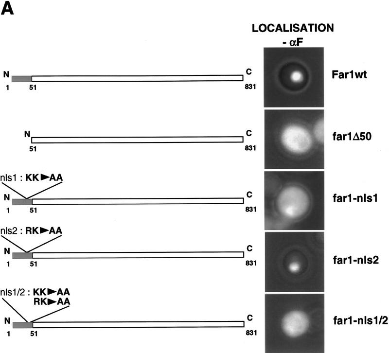
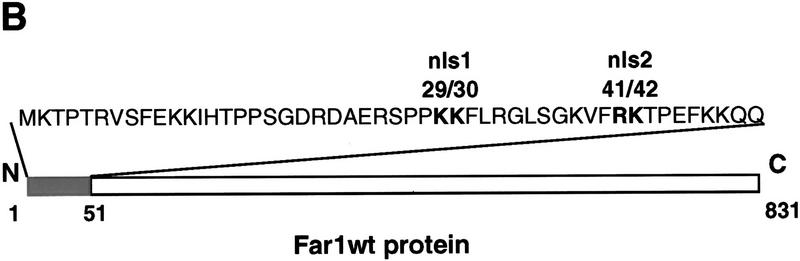
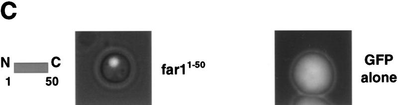
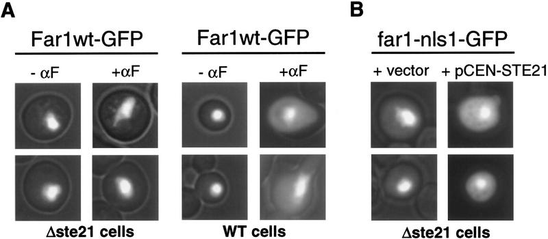
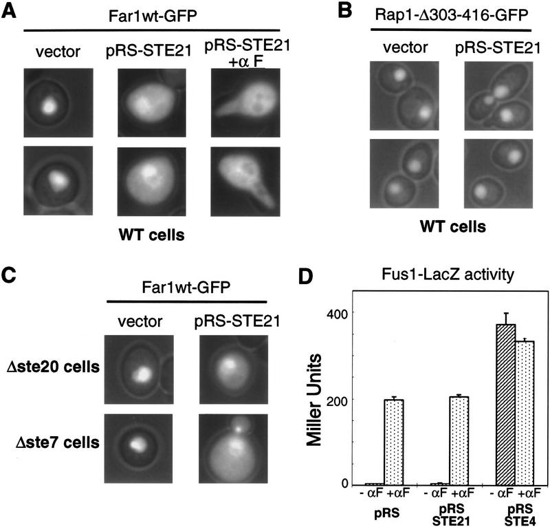
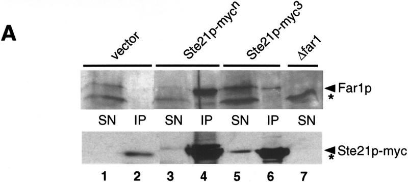
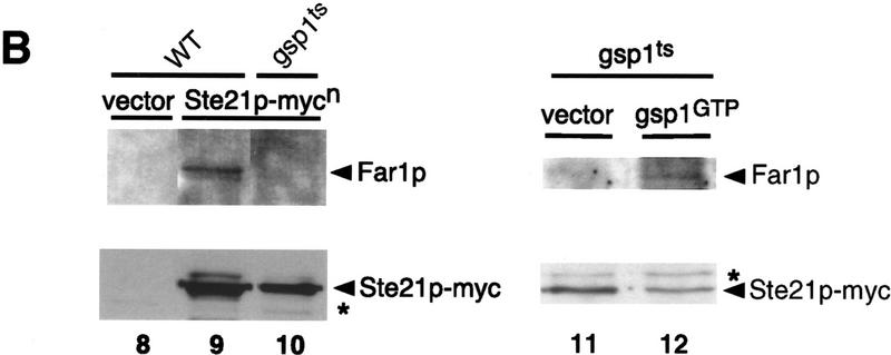
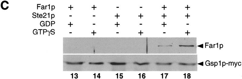
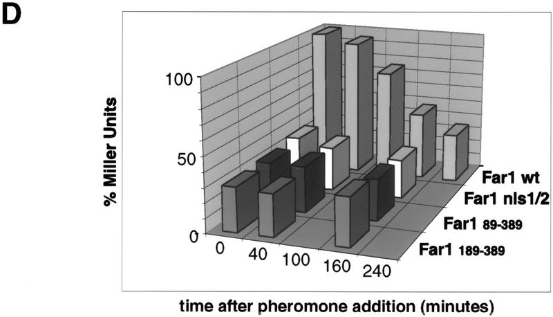
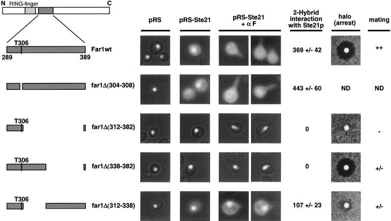
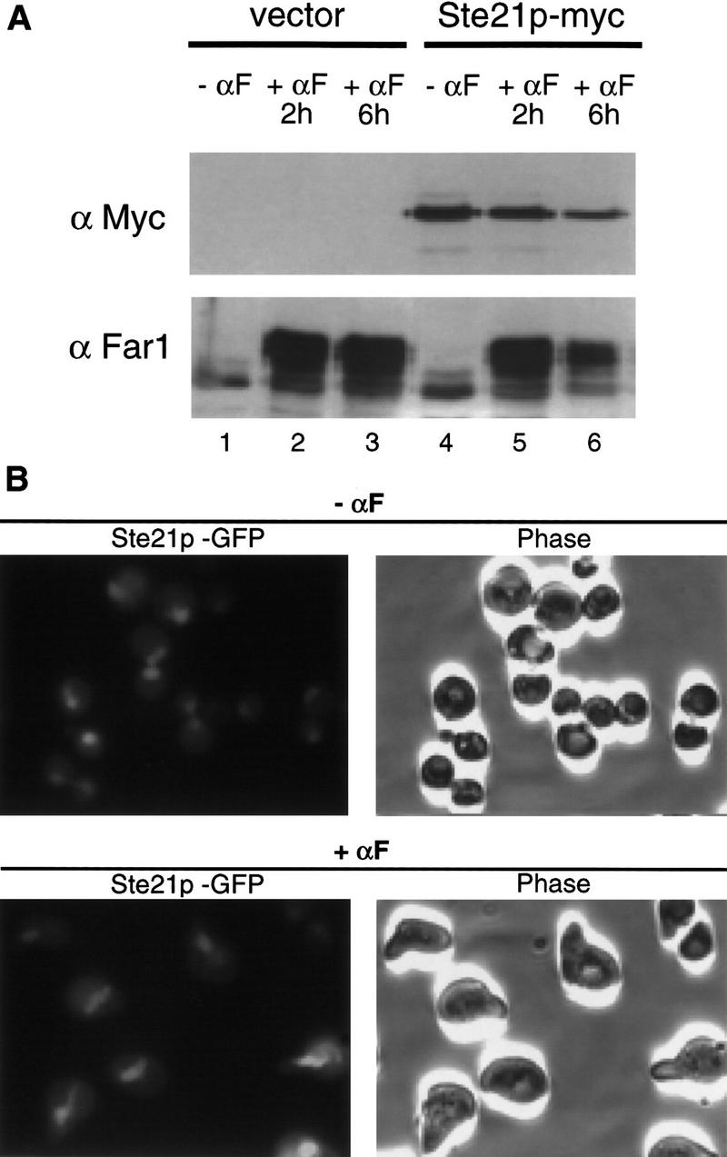
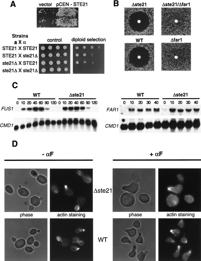
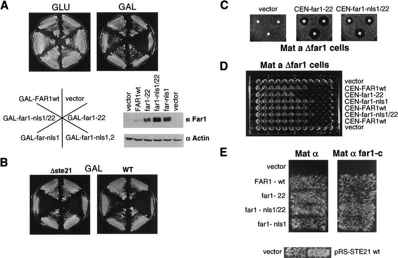
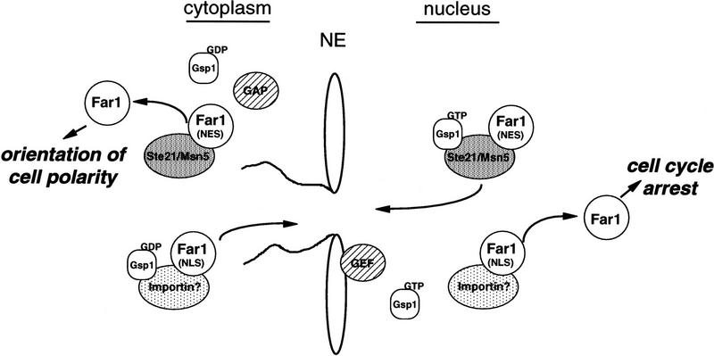
Far1p is a bifunctional protein that is required to arrest the cell cycle and to establish cell polarity during yeast mating. Far1p is localized predominantly in the nucleus but accumulates in the cytoplasm in cells exposed to pheromones. Here we show that Far1p functions in both subcellular compartments: nuclear Far1p is required to arrest the cell cycle, whereas cytoplasmic Far1p is involved in the establishment of cell polarity. The subcellular localization of Far1p is regulated by two mechanisms: (1) Far1p contains a functional bipartite nuclear localization signal (NLS), and (2) Far1p is exported from the nucleus by Msn5p/Ste21p, a member of the exportin family. Cells deleted for Msn5p/Ste21p failed to export Far1p in response to pheromones, whereas overexpression of Msn5p/Ste21p was sufficient to accumulate Far1p in the cytoplasm in the absence of pheromones. Msn5p/Ste21p was localized in the nucleus and interacted with Far1p in a manner dependent on GTP-bound Gsp1p. Two-hybrid analysis identified a small fragment within Far1p that is necessary and sufficient for binding to Msn5p/Ste21p, and is also required to export Far1p in vivo. Finally, similar to Deltamsn5/ste21 strains, cells expressing a mutant Far1p, which can no longer be exported, exhibit a mating defect, but are able to arrest their cell cycle in response to pheromones. Taken together, our results suggest that nuclear export of Far1p by Msn5p/Ste21p coordinates the two separable functions of Far1p during mating.


Figures

Figure 1
Figure 1
Far1p contains a functional NLS in the amino terminus. (A) Wild-type or various Far1p mutant proteins were expressed as fusions to GFP from the inducible GAL promoter and visualized by fluorescence microscopy. The introduced mutations are indicated schematically of left. Photographs show GFP fluorescence superimposed with phase contrast. (B) Schematic representation of Far1p and the 50 amino-terminal amino acids that are required for nuclear localization of Far1p. The mutated basic amino acids that are part of a bipartite nuclear localization signal are highlighted in bold. (C) The 50 amino-terminal amino acids of Far1p are sufficient to localize GFP in the nucleus. GFP fused to the 50 amino-terminal amino acids of Far1p (far11–50; left) or GFP alone (right) were expressed in cells and visualized as described above.
Figure 1
Figure 1
Far1p contains a functional NLS in the amino terminus. (A) Wild-type or various Far1p mutant proteins were expressed as fusions to GFP from the inducible GAL promoter and visualized by fluorescence microscopy. The introduced mutations are indicated schematically of left. Photographs show GFP fluorescence superimposed with phase contrast. (B) Schematic representation of Far1p and the 50 amino-terminal amino acids that are required for nuclear localization of Far1p. The mutated basic amino acids that are part of a bipartite nuclear localization signal are highlighted in bold. (C) The 50 amino-terminal amino acids of Far1p are sufficient to localize GFP in the nucleus. GFP fused to the 50 amino-terminal amino acids of Far1p (far11–50; left) or GFP alone (right) were expressed in cells and visualized as described above.
Figure 2
Figure 2
The exportin Msn5p/Ste21p is required to export Far1p. (A) Far1p–GFP was expressed in wild-type cells (right) or cells deleted for MSN5/STE21 (left) and treated (right row) or not treated (left row) with α-factor for 6 hr. Photographs show GFP fluorescence superimposed with phase contrast. Note that Far1p–GFP remains nuclear in Δmsn5/ste21 cells treated with α-factor. (B) Δmsn5/ste21 cells expressing Far1p–nls1–GFP were transformed with either an empty control plasmid (vector; left row) or a low copy number plasmid encoding MSN5/STE21 (pCEN-STE21; right row). Cells were analyzed as above. Note that Far1p–nls1–GFP is predominantly nuclear in Δmsn5/ste21.
Figure 3
Figure 3
Overexpression of Msn5p/Ste21p is sufficient to export Far1p. (A,B) Cells expressing Far1p–GFP (A) or for control Rap1p–Δ303–416-GFP (B) were transformed with a control plasmid (vector; left rows) or a plasmid allowing overexpression of Msn5p/Ste21p from the inducible GAL promoter (pRS–STE21; right rows). Cells were grown in the presence of galactose and analyzed by fluorescence microscopy. Where indicated α-factor was added for 3 hr. Note that complete cytoplasmic localization of Far1p requires overexpression of Msn5p/Ste21p and addition of α-factor. (C) Redistribution of Far1p to the cytoplasm does not require an intact mating pathway. Δste20 (top) or Δste7 cells (bottom) expressing Far1p–GFP were transformed with a control plasmid (vector; left row) or a plasmid allowing overexpression of Msn5p/Ste21p from the inducible GAL promoter (pRS–STE21; right row) and analyzed as described above. (D) The ability of cells carrying a control plasmid (pRS) or plasmids allowing overexpression of Msn5p/Ste21p (pRS–STE21) or Ste4p (pRS–STE4) to induce the reporter FUS1–lacZ was determined either in the absence (− αF) or presence of α-factor (+ αF). Bars show mean β-galactosidase activity ±s.d. for four independent transformants. Note that overexpression of Msn5p/Ste21p does not activate the pheromone response pathway.
Figure 4
Figure 4
Far1p interacts with Msn5p/Ste21p in a Gsp1p-GTP-dependent manner. (A,B) Msn5/Ste21p–myc was immunoprecipitated with 9E10 antibodies from extracts prepared from wild-type (K699; lanes 1–6, 8,9) or temperature-sensitive gsp1 cells (YSH80; lanes 10–12) expressing Far1p and either untagged Msn5p/Ste21p (lanes 1,2,8), Msn5p/Ste21p–mycn (lanes 3,4,9–12) or Msn5p/Ste21p–myc3 (lanes 5,6). The immunoprecipitates (IP; lanes 2, 4, 6, 8-12) and an aliquot of the supernatant before immunoprecipitation (SN; lanes 1,3,5,7) were analyzed for the presence of Far1p (top) or Msn5p/Ste21p–myc (bottom) by immunoblotting. Expression of Gsp1–GTP (YBM100; gsp1–G21V, lane 12) restored the ability of Msn5p/Ste21p to interact with Far1p. Cells lacking Far1p (YMP1054, lane 7) were included to control for the specificity of the antibodies. The arrowhead marks the position of Far1p (top) or Msn5p/Ste21p–myc (bottom); the asterisk points to the position of proteins that cross-reacts with the antibodies. Note that Far1p and Msn5p/Ste21p interact in a Gsp1p-dependent manner. (C) Sepharose beads containing immunoprecipitated Gsp1p–Myc expressed in E. coli and loaded with either GTPγS (lanes 14,16,18) or GDP (lanes 13,15,17) were incubated with yeast extracts containing as indicated Msn5p/Ste21p and Far1p expressed from the GAL promoter. Bound proteins were eluted and analyzed by immunoblotting for the presence of Far1p (top) and Gsp1p–Myc (bottom). Note that Far1p preferentially bound Gsp1p–GTP, but only in the presence of Msn5p/Ste21p. (D) Two-hybrid analysis of Msn5p/Ste21p and either wild-type (wt) or cytoplasmic Far1p mutant proteins in response to pheromones (times in minutes after addition of α-factor). The interaction was quantified as described and shown as percentage of Miller units relative to wild-type controls without pheromones. Note that the interaction between Msn5p/Ste21p and wild-type Far1p but not cytoplasmic mutant forms of Far1p decreases in an α-factor-dependent manner.
Figure 4
Figure 4
Far1p interacts with Msn5p/Ste21p in a Gsp1p-GTP-dependent manner. (A,B) Msn5/Ste21p–myc was immunoprecipitated with 9E10 antibodies from extracts prepared from wild-type (K699; lanes 1–6, 8,9) or temperature-sensitive gsp1 cells (YSH80; lanes 10–12) expressing Far1p and either untagged Msn5p/Ste21p (lanes 1,2,8), Msn5p/Ste21p–mycn (lanes 3,4,9–12) or Msn5p/Ste21p–myc3 (lanes 5,6). The immunoprecipitates (IP; lanes 2, 4, 6, 8-12) and an aliquot of the supernatant before immunoprecipitation (SN; lanes 1,3,5,7) were analyzed for the presence of Far1p (top) or Msn5p/Ste21p–myc (bottom) by immunoblotting. Expression of Gsp1–GTP (YBM100; gsp1–G21V, lane 12) restored the ability of Msn5p/Ste21p to interact with Far1p. Cells lacking Far1p (YMP1054, lane 7) were included to control for the specificity of the antibodies. The arrowhead marks the position of Far1p (top) or Msn5p/Ste21p–myc (bottom); the asterisk points to the position of proteins that cross-reacts with the antibodies. Note that Far1p and Msn5p/Ste21p interact in a Gsp1p-dependent manner. (C) Sepharose beads containing immunoprecipitated Gsp1p–Myc expressed in E. coli and loaded with either GTPγS (lanes 14,16,18) or GDP (lanes 13,15,17) were incubated with yeast extracts containing as indicated Msn5p/Ste21p and Far1p expressed from the GAL promoter. Bound proteins were eluted and analyzed by immunoblotting for the presence of Far1p (top) and Gsp1p–Myc (bottom). Note that Far1p preferentially bound Gsp1p–GTP, but only in the presence of Msn5p/Ste21p. (D) Two-hybrid analysis of Msn5p/Ste21p and either wild-type (wt) or cytoplasmic Far1p mutant proteins in response to pheromones (times in minutes after addition of α-factor). The interaction was quantified as described and shown as percentage of Miller units relative to wild-type controls without pheromones. Note that the interaction between Msn5p/Ste21p and wild-type Far1p but not cytoplasmic mutant forms of Far1p decreases in an α-factor-dependent manner.
Figure 5
Figure 5
Binding of Far1p and Msn5p/Ste21p is required for efficient mating but not cell cycle arrest. Cells expressing wild-type Far1p–GFP (Far1wt; top row) or mutant Far1p–GFP lacking various parts of the Msn5p/Ste21p binding domain (bottom rows) were transformed with a control plasmid (pRS; left row) or a plasmid expressing Msn5p/Ste21p from the inducible GAL promoter (pRS–STE21; right rows). Where indicated cells were treated with α-factor for 3 hr. Full-length or the various deletion mutants of Far1p are schematically represented on the left; deleted amino acids are indicated in parentheseis. The Far1p mutants were also tested for their ability to interact with Msn5p/Ste21p by two-hybrid assay; Miller units with standard deviations are shown. Cells deleted for FAR1 (YMP1054) transformed with low copy number plasmids expressing wild-type Far1p (top row) or the indicated Far1p mutant proteins (bottom rows) from the endogenous promoter were tested for their ability to arrest the cell cycle by halo assay, or for their ability to mate against the mating tester IH2625. (++) Wild-type mating; (+/−) strongly reduced mating; (−) sterile; (ND) not determined. Note that the Msn5p/Ste21p-binding domain of Far1p is required for nuclear export and efficient mating in vivo.
Figure 6
Figure 6
Msn5p/Ste21p is a nuclear protein that is not induced in response to pheromones. (A) Cells expressing epitope-tagged Msn5p/Ste21p (Ste21p–myc; lanes 4–6) or untagged Msn5p/Ste21p (vector; lanes 1–3) from the endogenous promoter were treated (lanes 2,3,5,6) or not treated with α-factor for the times indicated (lanes 1,4). Extracts were immunoblotted with 9E10 antibodies (top) or antibodies specific for Far1p (bottom). Note that in contrast to Far1p the levels of Msn5p/Ste21p do not increase in α-factor-treated cells. (B) Cells expressing Msn5p/Ste21p fused to GFP (Ste21p–GFP) from the endogenous promoter were treated (top) or not treated (bottom) with α-factor and analyzed by fluorescence microscopy (left). The corresponding phase contrast pictures are shown on the right. Note that Msn5p/Ste21p is nuclear in α-factor arrested cells and during all phases of the cell cycle.
Figure 7
Figure 7
Cells lacking Msn5p/Ste21p exhibit a bilateral mating defect but are able to arrest their cell cycle. (A; top) Mating assay with the mating tester IH2625 and Δmsn5/ste21 cells (PAY20) transformed with a control plasmid (vector) or a plasmid carrying MSN5/STE21 (pCEN–STE21). (Bottom) Mating assays with either MATa or MATα wild-type or Δmsn5/ste21 cells as indicated. Serial dilutions of the mating reactions were spotted on control plates (left) or selective plates where only diploid cells are able to grow (right). Note that Δmsn5/ste21 cells exhibit a bilateral mating defect. (B) The indicated strains were analyzed by halo assay for their ability to arrest the cell cycle in response to α-factor. Note that cells lacking Msn5p/Ste21p efficiently arrest their cell cycle in a manner dependent on Far1p. The following strains were analyzed: Δste21 (PAY20); Δste21 Δfar1 (YMP1067); wt (K699) and Δfar1 (YMP1054). (C) Δmsn5/ste21 cells (PAY20) were able to efficiently induce mating-specific transcription of FAR1 (right) and FUS1 (left). Northern analysis of total RNA isolated from wild-type (K699; left lanes) or Δmsn5/ste21 cells (PAY20, right lanes) treated with α-factor for the times indicated (in minutes). A probe against CMD1 RNA was used as total RNA loading control (bottom). (D) The morphology of cells deleted for Msn5p/Ste21p (PAY20; top row) or wild-type cells (K699; bottom row) in the absence (left rows) or presence of α-factor (right rows) was analyzed by phase contrast microscopy (left). Actin distribution is directed toward the growing bud or shmoo tip as visualized after staining with rhodamine–phalloidin (right).
Figure 8
Figure 8
Nuclear Far1p is required to arrest the cell cycle in response to pheromones, whereas cytoplasmic Far1p is necessary for efficient mating. (A) Wild-type cells (K699) were transformed with plasmids allowing overexpression of wild type or various mutant Far1p from the inducible GAL promoter as indicated, and grown on plates containing glucose (GLU; Far1p not expressed) or galactose (GAL; Far1p expressed). Extracts were prepared from cells and immunoblotted with specific antibodies against Far1p (top) or actin (bottom). Note that Far1p-22 is only toxic in the presence of a functional NLS. (B) Wild-type strains (K699, right) or strains deleted for MSN5/STE21 (Δste21; left) were transformed with the plasmids as indicated above and grown on media containing galactose. Note that in contrast to wild-type cells Far1–nls1/22 is toxic in Δmsn5/ste21 cells. (C) Cell deleted for FAR1 but harboring an empty plasmid (vector, left) or low copy number plasmids expressing Far1p-22 (middle) or Far1p–nls1/22 (right) from the endogenous promoter were analyzed by halo assay for their ability to arrest the cell cycle in response to pheromones. The circles contained 2, 10, and 20 μg of α-factor. (D) The minimal concentration of α-factor required to arrest cells expressing wild type or the indicated mutant Far1p from the endogenous promoter was determined by serial dilutions as described. Note that efficient cell cycle arrest in response to pheromones requires Far1p with a functional NLS. (E) Cells deleted for FAR1 but harboring an empty plasmid (vector) or low copy number plasmids expressing wild type or the indicated Far1p mutants from the endogenous promoter were analyzed for their ability to mate with wild-type (IH1793; left) or mating-reduced FAR1-C mating testers (IH2625, right). Note that cytoplasmic Far1p does not reduce the mating efficiency. Wild-type cells (K699) harboring an empty plasmid (vector; left patch) or a plasmid allowing overexpression of Msn5p/Ste21p (pRS–STE21; right patch) were mated to wild-type mating testers (IH1793). Note that overexpression of Msn5p/Ste21p slightly increases the mating efficiency.
Figure 9
Figure 9
A schematic representation of the Far1p functions. In the absence of pheromones Far1p is exclusively nuclear because of the presence of functional NLS sequences. Nuclear Far1p is inactive as a cyclin-dependent kinase inhibitor (CKI) because it needs to be phosphorylated by the MAPK Fus3p to be able to bind to the Cdc28p–Clnp kinase (Peter et al. 1993; Gartner et al. 1998). Nuclear Far1p interacts with Msn5p/Ste21p in a Gsp1p-dependent manner, which then transports Far1p into the cytoplasm. In the presence of pheromones the balance between import and export is shifted toward export presumably because of activation of Fus3p. Nuclear Far1p is required for cell cycle arrest, whereas cytoplasmic Far1p is needed for the establishment of oriented cell polarity.

References

    1. Akada R, Kallal L, Johnson DI, Kurjan J. Genetic relationships between the G-protein beta gamma complex, Ste5p, Ste20p and Cdc42p: Investigation of effector roles in the yeast pheromone response pathway. Genetics. 1996;143:103–117. - PMC - PubMed
    1. Alepuz, P.M., D. Matheos, K.W. Cunningham, and F. Estruch. 1999. The Saccharomyces cerevisiae RanGTP binding protein Msn5p is involved in different signal transduction pathways. Genetics (in press). - PMC - PubMed
    1. Arkowitz RA. Responding to attraction: Chemotaxis and chemotropism in Dictyostelium and yeast. Trends Cell Biol. 1999;9:20–27. - PubMed
    1. Ausubel FM, Brent R, Kingston RE, Moore DD, Seidman JG, Smith JA, Struhl K. Current protocols in molecular biology. New York, NY: Greene Publishing Associates and Wiley-Interscience; 1991.
    1. Bähler J, Peter M. Cell polarity in yeast. In: Drubin DG, editor. Frontiers in molecular biology: cell polarity. Oxford University Press; 1999. (In press).

Publication types

MeSH terms