. 1999 Sep 1;13(17):2315-27.
doi: 10.1101/gad.13.17.2315.

The balance between isoforms of the prickle LIM domain protein is critical for planar polarity in Drosophila imaginal discs

Affiliations

The balance between isoforms of the prickle LIM domain protein is critical for planar polarity in Drosophila imaginal discs

D Gubb et al. Genes Dev. .

Abstract

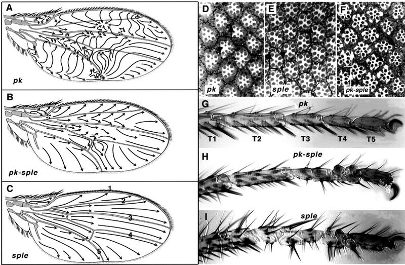
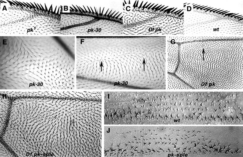
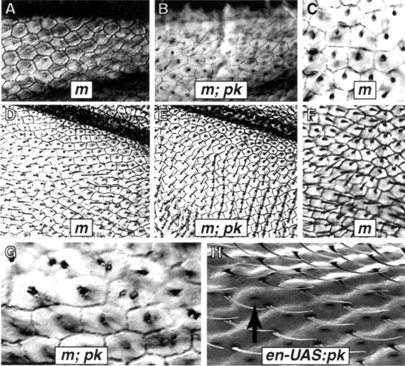
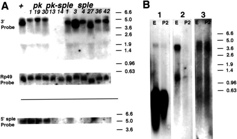
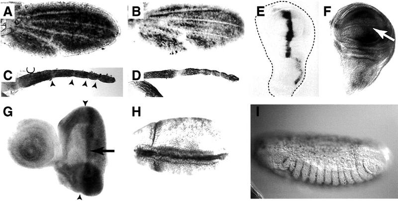
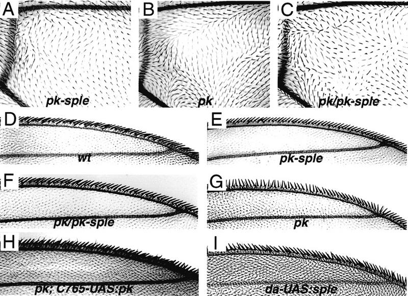
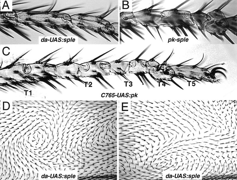
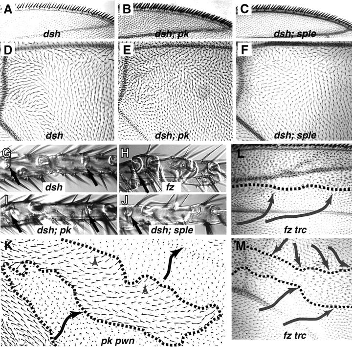
The tissue polarity mutants in Drosophila include a set of conserved gene products that appear to be involved in the control of cytoskeletal architecture. Here we show that the tissue polarity gene prickle (pk) encodes a protein with a triple LIM domain and a novel domain that is present in human, murine, and Caenorhabditis elegans homologs which we designate PET. Three transcripts have been identified, pk, pkM, and sple, encoding 93-, 100-, and 129-kD conceptual proteins, respectively. The three transcripts span 70 kb and share 6 exons that contain the conserved domains. The pk and sple transcripts are expressed with similar tissue-specific patterns but have qualitatively different activities. The phenotypes of pk mutants, and transgenic flies in which the different isoforms are overexpressed show that the balance between Pk and Sple is critical for the specification of planar polarity. In addition, these phenotypes suggest a tessellation model in which the alignment of wing hairs is dependent on cell shape and need not reflect fine-grained positional information. Lack of both pk and sple transcripts gives a phenotype affecting the whole body surface that is similar to those of dishevelled and frizzled (fz) suggesting a functional relationship between pk and fz signaling.


Figures

Figure 1
Figure 1
Pk mutant phenotypes. Complete lack of Pk function, in pkpk–sple alleles, gives a weak polarity phenotype in the wing, notum, abdomen, eye, and legs. pkpk alleles cause an extreme polarity phenotype in the wing and notum; pksple alleles affect eye, abdomen, and leg. The pkpk wing phenotype shows a characteristic reversal in the triple-row bristles along the anterior margin, a whorl in the wing hairs near the tip of vein 2, and abrupt discontinuities in hair polarity, e.g., pkpk1 (A). The weak PkPk–sple phenotype shows a slight effect on triple-row bristle orientation and gives gently curved hair polarity vectors, e.g., pkpk–sple13 (B); sple alleles are completely wild type, e.g., pksple1 (C). Arrows indicate the direction along which hairs are aligned; wing veins are designated 1–5 (C). The eye phenotype is wild-type in pkpk1 (D), showing a line of mirror symmetry along the equator (line). On both sides of the equator the R3 photoreceptor cell is aligned towards the pole, in the direction of the arrowhead. In addition to being rotated through 180° ommatidia show reversed chirality around the equator, so that both a rotation and a reflection in the plane of the epithelium is required to superimpose the ommatidial patterns. (E) pksple1 eyes contain a mixture of ommatidia with reversed polarity and chirality in both hemispheres of the eye. These ommatidia remain aligned along the polar axis, but with their R3 photoreceptors directed toward the equator rather than the pole giving rise to D/V mirror-image reversals of the normal rhabdomere pattern. In addition, all the pksple alleles show out ∼1% anteroposterior (A/P) reversed ommatidia (not shown). (F) pkpk–sple13 eyes contain a mixture of chiral forms of ommatidia. Some ommatidia fail to rotate properly, and the resulting imperfections in the hexagonal stacking give a slightly rough eye phenotype. Some ommatidia are aligned at 60° to the equator (black arrows) and some show A/P reversals (white arrows), with the R3 rhabdomere anterior to R4. (G) The tarsi of pkpk1 are wild type; tarsal segments are numbered T1–T5. (H) In pkpk–sple13, the T3 and T4 segments carry medial duplications of the proximal and distal joint structures, with the middle of each segment deleted. This results in alternating reversed-proximal and reversed-distal tarsal joint structures with half the length of a normal segment. (I) In pksple1 the tarsal duplications affect T2, T3, and T4 segments, with an occasional incipient ectopic joint in the distal T1. The distal T5 segment remains unaffected in all mutant alleles.
Figure 2
Figure 2
Comparison of wing polarity patterns between different alleles of pkpk. Junction of vein 1 and 2 at anterior margin in pkpk1 (A), Df(2R)pk-30 (B), Df(2R)nap-2/Df(2R)sple-J2 (C), and wild type (D). In pkpk mutant wings, the whorl close to the junction of vein 2 with the wing margin marks a discontinuity in polarity, or stacking flaw. Stacking flaws with different topologies are found in different regions of the wing. (E) Df(2R)pk-30, a cruciform discontinuity posterior to vein 5; note doubled hairs. (F) Df(2R)pk-30, radial and cruciform discontinuities (arrows) between veins 2 and 3. (G) Overlapping pkpk deletions, Df(2R)nap-2/Df(2R)sple-J2. Note triangular dislocation in polarity (arrow) distal to the posterior cross vein as in pkpk1 (Fig. 1A). (H) Overlapping pkpk–sple deletions removing the entire gene Df(2R)pk-N5/Df(2R)sple-J2 (cf. the pkpk–sple13 pattern; Fig. 1B). Embryonic denticle belt in wild type (I) and pkpk–sple13 (J). There is no detectable embryonic phenotype with alleles of pkpk, pksple, or pkpk–sple; in particular, the denticle belt morphology and denticle orientation remains normal.
Figure 3
Figure 3
Cell shapes and polarity. The m38c mutation was used to visualize cell boundaries in both wild-type and Df(2R)pk-30 backgrounds. In m38c wings the majority of cells are hexagonal, although occasional pentagonal cells are seen. (A) Distal vein 2 region in m38c wings. (B) The same region in m38c; Df(2R)pk-30 wings, with more irregular cell shapes. (C) A common packing defect consisting of a group of four irregular pentagons with one 90° corner fitted within the surrounding hexagonal array. (D) The region posterior to vein 5 in m38c shows a regular hexagonal array, although cells close to the vein tend to be trapezoidal or square. Wing hairs are aligned along the proximodistal axis. (E) The same region in m38c; Df(2R)pk-30 wings shows parallel rows of hexagons with hairs oriented anteriorly. (F) A small region between veins 3 and 4 in m38c wings shows cuboidal cells, with local polarity disruptions near the transition between cuboidal and hexagonal packing. (G) Cells near the anterior whorl in m38c; Df(2R)pk-30 wings tend to be roughly cuboidal and associated with duplicated hairs. (H) Hairs fail to migrate from the distal vertex of pupal wing cells in en–UAS:pk flies (gal4–en; P[UAS:pk+]). This s.e.m. micrograph of adult wings shows hairs remaining at the distal vertex; the pedicel in the middle of the mature cell (arrow) marks the position that the adult hair would normally occupy.
Figure 4
Figure 4
(A) Map showing the location of pk mutant breakpoints with respect to transcripts in the region. The zero coordinate is the EcoRI site 973 bp proximal to the pk transcription start site. Transcripts are indicated below the molecular coordinates. The 5′ start of the pk transcript lies within a cluster of three serpins (serine Proteinase inhibitors) (Spn43Aa, Spn43Ab, and Spn43Ac), one of which corresponds to the necrotic gene, and a transcript with homology to Adenosine kinases maps just distal to pk. Aberrations that break between coordinates 0 and 30 kb cause pk mutations, similar to the homozygous deletion Df(2R)pk-30, which deletes the pk 5′ start and the 3′ end of the medial serpin transcript Spn43Ab. Aberrations between coordinates 40 and 70 kb, which interrupt the common exons, give the weaker PkPk–Sple phenotype typical of pkpk–sple13. Overlapping deletions give an additional necrotic phenotype when the serpin cluster is removed, but deletion of the adenosine kinase transcript [Df(2R)sple–J2/Df(2R)pk-N5] gives no further phenotype. (B) The Pk protein isoforms encode putative 870 (pk), 936 (pkM), and 1206 (sple) amino acid peptides with the conserved PET and LIM domains mapping entirely within the common exons. The first pk exon contains an untranslated leader sequence of 0.8 kb, with the putative translation start site (Cavener and Ray 1991) being 39 bp 5′ to the large intron. The putative translation start sites of the pkM and sple transcripts are within their first and second introns, respectively.
Figure 5
Figure 5
Northern analysis. A probe homologous to sple 5′ sequences detects a 5.1-kb mRNA, but this band is no longer detected, or has an altered length, in the sple mutants. A shorter, 4.2-kb transcript is missing in pkpk mutants. Both 5.1- and 4.2-kb transcripts are detected by a common exon probe in wild-type RNA, and neither are detected in pkpk–sple mutant strains. (A) Total 2-day pupal RNA from homozygous wild-type (+) and mutant flies (pkpk1, pkpk19, Df(2R)pk-30, pkpk–sple13, pkpk–sple14, pksple1, pksple3, pksple4, pksple27, pksple36, pksple42) hybridized with a 3′ common exon probe (3′), an Rp49 loading control (Rp49) and a unique sple exon probe (5′). The 4.2-kb transcript is missing in pkpk and pkpk–splealleles but retained in pksple alleles. The 5.1-kb transcript is present in pkpk1, pkpk19 and Df(2R)pk-30; missing in pkpk–sple13, pkpk–sple14 and lost, or reduced in length, in pksple3, pksple4, pksple27, and pksple36. (B) Three strips cut from a filter carrying wild-type embryonic (E) and 2-day pupal (P2) poly(A)+ RNA at the same sample loading. Individual strips were hybridized with pk 5′ + Rp49 (1), pkM 5′ (2), and sple 5′ (3) probes. The pk and sple transcripts are expressed in embryo and 2-day pupae. The pkM transcript was only detected during the embryonic stage.
Figure 6
Figure 6
Tissue in situs. Wild-type pupal leg and wing discs (28–32 hr) probed with pk-specific (A,C) or sple-specific (B,D) 5′ probes. Hybridization is uniform with both probes, with the exception of the wing veins and the tarsal segment boundaries (arrowheads in C). The expression in larval imaginal discs is much weaker; no endogenous signal is detected with a common exon probe at a level at which dpp–UAS:pk (gal4–dpp P[UAS:pk+]) wing discs show strong hybridization within the dpp domain (E) (the dotted line indicates disc boundary). With increasing development times a signal is seen along both sides of the D/V compartment boundary (arrow) in the wing (F) and as a discrete band (arrow) behind the morphogenetic furrow (arrowheads) in the eye disc (G). These preparations show a high background of nonspecific signal (retained in pkpk–sple13 discs and with antisense probes; data not shown). In the embryo, pkM, sple, and common exon probes give similar patterns of expression. Stage 8 embryos (H) show a strong signal in the cephalic furrow and dorsal fold, pkM probe. (I) Stage 14 embryos show signal in the parasegmental folds, common exon probe.
Figure 7
Figure 7
The wing phenotypes associated with titrating the number of intact copies of pk and sple. (A) pk–sple, moderate phenotype resulting from homozygous deletion of the common exons, pkpk–sple13/pkpk–sple13. (B) pk, extreme phenotype resulting from deletion of the pk transcript, Df(2R)pk-30/Df(2R)pk-30. (C) pk/pk–sple, moderate phenotype resulting from deleting pk and halving the number of sple copies in Df(2R)pk-30/pkpk–sple13 flies. The polarity pattern shown here, distal to the posterior cross vein, is stronger than in the remainder of the wing blade, which is closer to pkpk–sple13. (D) polarity of the marginal triple row bristles in wild type. (E) pk–sple triple row (pkpk–sple13/pkpk–sple13). (F) pk/pk–sple triple row [Df(2R)pk-30/pkpk–sple13]; (G) pk triple row [Df(2R)pk-30 /Df(2R)pk-30]. The pk wing phenotype was rescued by driving UAS:pk+ expression with the gal4–C765 driver, which is expressed in the wing and leg disks. (H) The pkpk triple row bristle phenotype is completely rescued and the wing hair polarity is close to wild type in pk; C765–UAS:pk wings [Df(2R)pk-30; gal4–C765/P(UAS:pk+)]. Small regions of abnormal polarity remain within the wing blade (both in pk; C765–UAS:pk and C765–UAS:pk in a wild-type background). sple overexpression also gives a wing phenotype in da–UAS:sple+ wings (I); the wing shown is EP(2)2557/+; gal4–da/+ (EP(2)2557 inserts a UAS driver 80 bp 5′ to the sple first intron), but the same phenotype is shown with a P[UAS:sple+] transformant line, in P[UAS:sple+]/+; gal4–da/+ wings.
Figure 8
Figure 8
Overexpression phenotypes in leg and wing blade. Overexpression of sple in da–UAS:sple+ [EP(2)2557/+; gal4–da/+] legs gives no mutant phenotype; the wild-type morphology of T3 and T4 tarsal segments is shown in A. For comparison, the duplicated proximal (ball) and distal (socket) joint structures of the T3 and T4 segments resulting from lack of function in pkpk–sple-3 legs is shown in B; the T1, T2, and T5 segments (not shown) remain normal. A more extreme tarsal duplication phenotype affecting T1–T4 segments is given by Pk overexpression in 765–UAS:pk+ (P[UAS:pk]+/gal4–C765) flies (C). (D,E) The same region anterior to vein 4 in two different da–UAS:sple [EP(2)2557/+; gal4–da/+] wings. The polarity in any given region is unpredictable from wing to wing, but hair orientation changes gradually across large fields of cells. Whorls, cruciform, and radial stacking flaws are seen as in pkpk wings, but at variable positions within the wing blade.
Figure 9
Figure 9
Comparison of different planar polarity phenotypes in wing and tarsi. The triple row bristle phenotype and wing blade polarity patterns of dsh are similar to pkpk–sple13 (A,D). The extreme triple row phenotype of Df(2R)pk-30 is suppressed in dsh; Df(2R)pk-30 flies and becomes phenotypically Dsh in both dsh; Df(2R)pk-30 and dsh; pksple1 double mutants (B,C). In dsh; Df(2R)pk-30 double mutants, the wing blade phenotype is intermediate between the respective single mutants (E). A slight, but consistent modification in the Dsh polarity is also seen in dsh; pksple1 wings, despite the fact that pksple alleles show no wing phenotype (F). The Dsh tarsal phenotype is similar to pkpk–sple13 in giving complete duplications of the T3 and T4 segments; an incipient joint is present in T1 and T2 (arrows), but the external morphology of these segments remains normal (G). This phenotype is unaffected in dsh; Df(2R)pk-30 (I). The most extreme tarsal phenotypes, with complete duplications in T2–T4 and an ectopic joint structure in T1, are seen in fz (H), P[UAS:pk+] overexpression (Fig. 8C) and dsh; pksple (J) mutant strains. In somatic mosaics, the effects of pk and fz are distinct. Within pkpk pwn clones, the polarity pattern is autonomous. A pkpk pwn clone in cell E of the wing is shown in K which expresses pkpk polarity (black arrows) (cf. Fig. 1A); a short range alteration in polarity (gray arrowheads) is seen close to the clone boundary (broken line). This effect is clearest when a small peninsula of pkpk pwn tissue is surrounded by pwn+ tissue. In contrast, frz trc clones cause a long-range nonautonomous alteration in the polarity of surrounding cells, which tend to point toward the mutant clone (L,M).

References

    1. Adler PN. The genetic control of tissue polarity in Drosophila. BioEssays. 1992;14:735–741. - PubMed
    1. Adler PN, Vinson C, Park WJ, Conover S, Klein L. Molecular structure of frizzled, a Drosophila tissue polarity gene. Genetics. 1990;126:401–416. - PMC - PubMed
    1. Ashburner M. Drosophila, a laboratory manual. Cold Spring Harbor, NY: Cold Spring Harbor Laboratory Press; 1989.
    1. Axelrod JD, Matsuno K, Artavanis-Tsakonas S. Interaction between Wingless and Notch signaling pathways mediated by Dishevelled. Science. 1996;271:1826–1832. - PubMed
    1. Axelrod JD, Miller JR, Shulman JM, Moon RT, Perrimon N. Differential recruitment of Dishevelled provides signaling specificity in the planar cell polarity and Wingless signaling pathways. Genes & Dev. 1998;12:2610–2622. - PMC - PubMed

Publication types

MeSH terms

Associated data

LinkOut - more resources