

Detoxification of environmental mutagens and carcinogens: structure, mechanism, and evolution of liver epoxide hydrolase
- PMID: 10485878
- PMCID: PMC17935
- DOI: 10.1073/pnas.96.19.10637
Detoxification of environmental mutagens and carcinogens: structure, mechanism, and evolution of liver epoxide hydrolase
Abstract
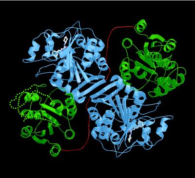
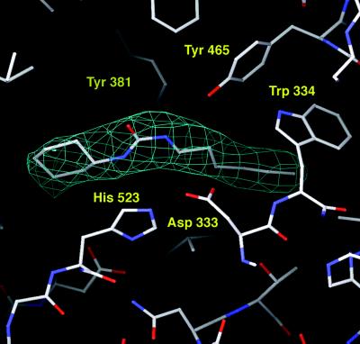
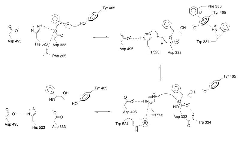
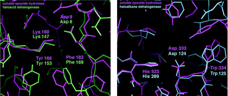
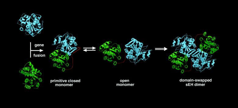
The crystal structure of recombinant murine liver cytosolic epoxide hydrolase (EC 3.3.2.3) has been determined at 2.8-A resolution. The binding of a nanomolar affinity inhibitor confirms the active site location in the C-terminal domain; this domain is similar to that of haloalkane dehalogenase and shares the alpha/beta hydrolase fold. A structure-based mechanism is proposed that illuminates the unique chemical strategy for the activation of endogenous and man-made epoxide substrates for hydrolysis and detoxification. Surprisingly, a vestigial active site is found in the N-terminal domain similar to that of another enzyme of halocarbon metabolism, haloacid dehalogenase. Although the vestigial active site does not participate in epoxide hydrolysis, the vestigial domain plays a critical structural role by stabilizing the dimer in a distinctive domain-swapped architecture. Given the genetic and structural relationships among these enzymes of xenobiotic metabolism, a structure-based evolutionary sequence is postulated.
Figures
References
-
- Wixtrom R N, Hammock B D. In: Biochemical Pharmacology and Toxicology. Zakim D, Vessey D A, editors. Vol. 1. New York: Wiley; 1985. pp. 1–93.
-
- Oesch F. Xenobiotica. 1973;3:305–340. - PubMed
-
- Ota K, Hammock B D. Science. 1980;207:1479–1481. - PubMed
-
- Arand M, Grant D F, Beetham J K, Friedberg T, Oesch F, Hammock B D. FEBS Lett. 1994;338:251–256. - PubMed
-
- Beetham J K, Grant D, Arand M, Garbarino J, Kiyosue T, Pinot F, Oesch F, Belknap W R, Shinozaki K, Hammock B D. DNA Cell Biol. 1995;14:61–71. - PubMed
Publication types
MeSH terms
Substances
Associated data
- Actions
- Actions
LinkOut - more resources
Full Text Sources
Other Literature Sources
Molecular Biology Databases
