. 1999 Sep 14;96(19):10655-60.
doi: 10.1073/pnas.96.19.10655.

Evidence for the function of an exonic splicing enhancer after the first catalytic step of pre-mRNA splicing

Affiliations

Evidence for the function of an exonic splicing enhancer after the first catalytic step of pre-mRNA splicing

S L Chew et al. Proc Natl Acad Sci U S A. .

Abstract

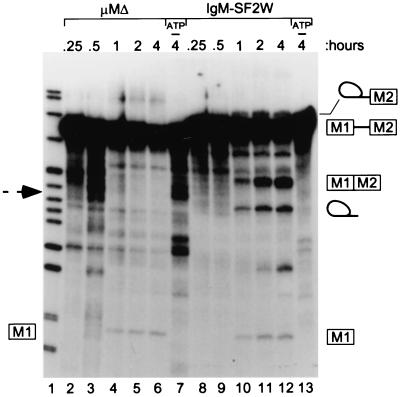
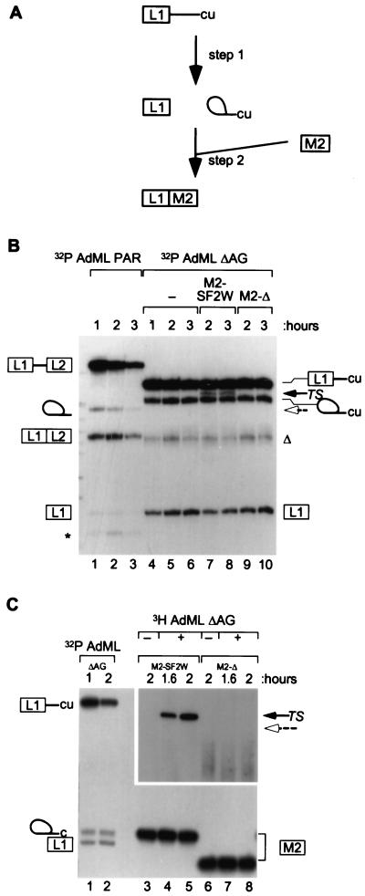
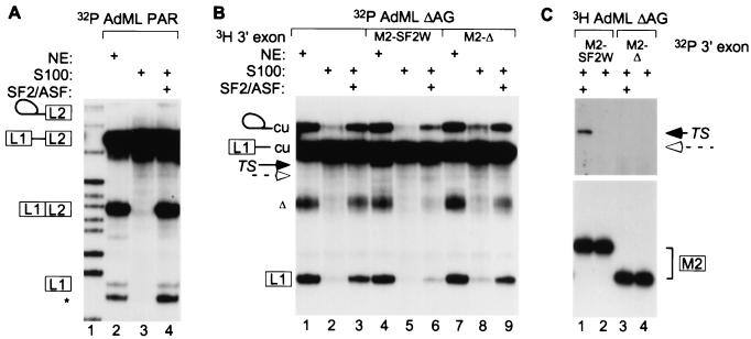
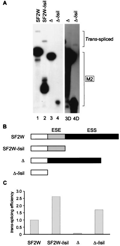
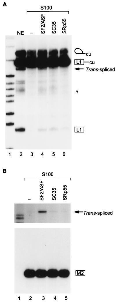
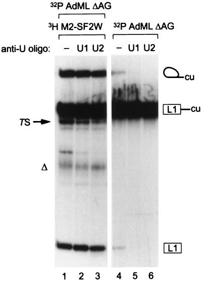
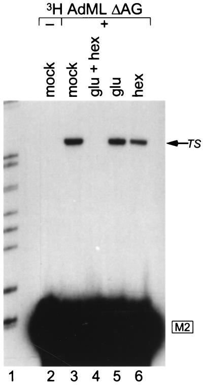
Exonic splicing enhancers (ESEs) activate pre-mRNA splicing by promoting the use of the flanking splice sites. They are recognized by members of the serine/arginine-rich (SR) family of proteins, such as splicing factor 2/alternative splicing factor (SF2/ASF), which recruit basal splicing factors to form the initial complexes during spliceosome assembly. The in vitro splicing kinetics of an ESE-dependent IgM pre-mRNA suggested that an SF2/ASF-specific ESE has additional functions later in the splicing reaction, after the completion of the first catalytic step. A bimolecular exon ligation assay, which physically uncouples the first and second catalytic steps of splicing in a trans-splicing reaction, was adapted to test the function of the ESE after the first step. A 3' exon containing the SF2/ASF-specific ESE underwent bimolecular exon ligation, whereas 3' exons without the ESE or with control sequences did not. The ESE-dependent trans-splicing reaction occurred after inactivation of U1 or U2 small nuclear ribonucleoprotein particles, compatible with a functional assay for events after the first step of splicing. The ESE-dependent step appears to take place before the ATP-independent part of the second catalytic step. Bimolecular exon ligation also occurred in an S100 cytosolic extract, requiring both the SF2/ASF-dependent ESE and complementation with SF2/ASF. These data suggest that some ESEs can act late in the splicing reaction, together with appropriate SR proteins, to enhance the second catalytic step of splicing.


Figures

Figure 1
Figure 1
In vitro splicing kinetics of IgM pre-mRNA. A time course of cis-splicing of IgM pre-mRNAs with (IgM-SF2W) or without (μMΔ) an SF2/ASF-specific ESE is shown. The positions of the pre-mRNA, 5′ exon M1 and lariat-exon M2 intermediates, and splicing products are shown by diagrams on the right. A dashed arrow shows the expected position of the μMΔ-spliced product. Lane 1, pBR322/MspI size markers; lanes 7 and 13, control reactions lacking ATP.
Figure 2
Figure 2
ESE dependence of bimolecular exon ligation. (A) A scheme of the trans-splicing assay is shown, with the 5′ leader exon denoted by the boxed L1 and the intron by a line ending with “cu” (17). After incubation, to allow the first step of splicing to occur, IgM M2 exon (with a 3′ splice site) was added as a 3′ substrate. (B) AdML ΔAG RNA was 32P-labeled and incubated in nuclear extract for the times shown (lanes 4–6). 3H-labeled 3′ exons were added for the second and third hours of the reactions (lanes 7–10). The trans-spliced mRNA (TS) is shown by a black arrow. The expected position of trans-splicing with M2-Δ is marked by a dashed open arrow. Control splicing in cis between AdML PAR exons L1 and L2 is shown in lanes 1–3. (C) In a reciprocal labeling experiment, the 3′ exons were 32P-labeled, and the 5′ fragment was made with [3H]GTP. Controls with 32P-labeled AdML ΔAG pre-mRNA are shown in lanes 1 and 2. Other controls omitting the 3H-labeled 5′ RNA are shown in lanes 3 and 6. A longer exposure of the trans-spliced products is shown. In B (and in subsequent figures), the asterisk indicates an artifactual band characteristic of the AdML PAR substrate (11, 17); the triangle indicates an artifactual product that appears to derive from the AdML ΔAG lariat intermediate or from use of an alternative branch site.
Figure 3
Figure 3
Trans-splicing requirement for an ESE and SF2/ASF. (A) Control AdML PAR pre-mRNA cis-splicing was carried out in nuclear extract (NE) or in S100 extract complemented with SF2/ASF. Lane 1, size markers. (B) Trans-splicing reactions were performed in nuclear extract or in S100 extract with or without SF2/ASF. 32P-labeled AdML ΔAG was incubated for 1 hr, and then excess 3H-labeled M2-SF2W, or M2-Δ, or water was added for the second hour. Trans-spliced mRNA (TS, lanes 4 and 6) is marked by a black arrow, and the expected position of the M2-Δ mRNA is marked with a dashed open arrow. (C) A reciprocal labeling experiment is shown. The top part of the autoradiogram is a longer exposure.
Figure 4
Figure 4
Interaction between enhancer and silencer elements in trans-splicing. (A) 3H-labeled AdML ΔAG pre-mRNA was incubated for 1 hr in nuclear extract. Then, the indicated 32P-labeled 3′ M2 exons with or without an ESE or a silencer were added. The lower and upper brackets on the right show the positions of the 3′ exons and of the trans-spliced mRNAs, respectively. A longer exposure of lanes 3 and 4 is shown (lanes 3D and 4D). (B) Schematic structure of the different 3′ M2 exons. The ESE is shaded gray, and the silencer segment (ESS) is shaded black. (C) Relative trans-splicing efficiencies of the four 3′ exons. The data from A were quantitated by densitometry and normalized for loading and differential labeling of the 3′ exons. The efficiency of the SF2W 3′ exon was arbitrarily set at 1.
Figure 5
Figure 5
SR protein specificity of second-step trans-splicing enhancement. (A) 32P-labeled AdML ΔAG pre-mRNA was incubated for 1 hr in S100 extract (with or without the indicated SR proteins), and 3H-labeled M2-SF2W 3′ exon was added for the second hour. Trans-spliced mRNA is shown by the black arrow. Lane 1: size markers. (B) A reciprocal labeling experiment is shown. The top part of the autoradiogram is a longer exposure. Lane 1, size markers.
Figure 6
Figure 6
Generation of trans-spliced products from accumulated first-step intermediates. 32P-labeled AdML ΔAG RNA was processed for 1 hr to give first-step intermediates, then incubated with buffer (lane 1), anti-U1 (lane 2), or anti-U2 (lane 3) oligonucleotides for 15 min, and finally, excess tritiated M2-SF2W 3′ exon RNA was added for another hour. Control reactions were preincubated without RNA for 1 hr, treated with buffer or oligonucleotides, and AdML ΔAG RNA was then added for a final hour of incubation.
Figure 7
Figure 7
ATP dependence of ESE-stimulated bimolecular exon ligation. Tritiated AdML ΔAG RNA was incubated for 1 hr (except in lane 2), followed by glucose (glu) and hexokinase (hex; lane 3), or control treatments (lanes 3, 5, and 6), with subsequent addition of radiolabeled M2-SF2W 3′ RNA. The trans-spliced product (TS) is marked by an arrow. Lane 1, size markers.

Similar articles

Cited by

References

    1. Cáceres J F, Krainer A R. In: Eukaryotic mRNA Processing. Krainer A R, editor. Oxford: IRL Press; 1997. pp. 174–212.
    1. Staknis D, Reed R. Mol Cell Biol. 1994;14:7670–7682. - PMC - PubMed
    1. Blencowe B J, Nickerson J A, Issner R, Penman S, Sharp P A. J Cell Biol. 1994;127:593–607. - PMC - PubMed
    1. Alzhanova-Ericsson A T, Sun X, Visa N, Kiseleva E, Wurtz T, Daneholt B. Genes Dev. 1996;10:2881–2893. - PubMed
    1. Cáceres J F, Screaton G R, Krainer A R. Genes Dev. 1998;12:55–66. - PMC - PubMed

Publication types