. 1999 Sep 14;96(19):10717-22.
doi: 10.1073/pnas.96.19.10717.

Targeted disruption of the beta adducin gene (Add2) causes red blood cell spherocytosis in mice

Affiliations

Targeted disruption of the beta adducin gene (Add2) causes red blood cell spherocytosis in mice

D M Gilligan et al. Proc Natl Acad Sci U S A. .

Abstract

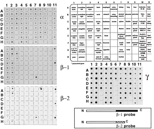
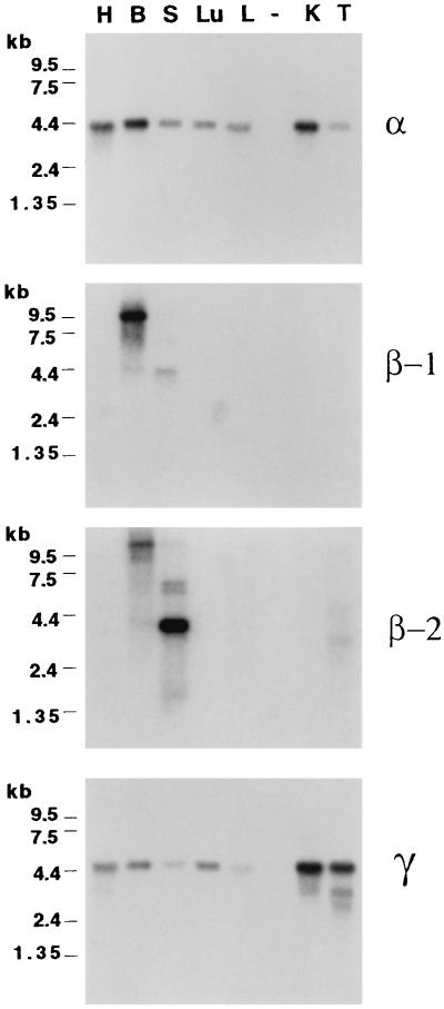
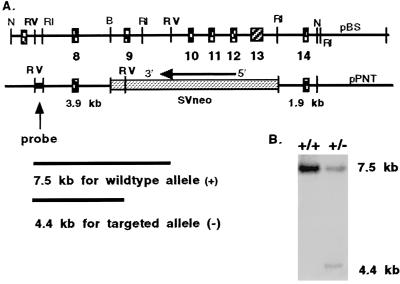
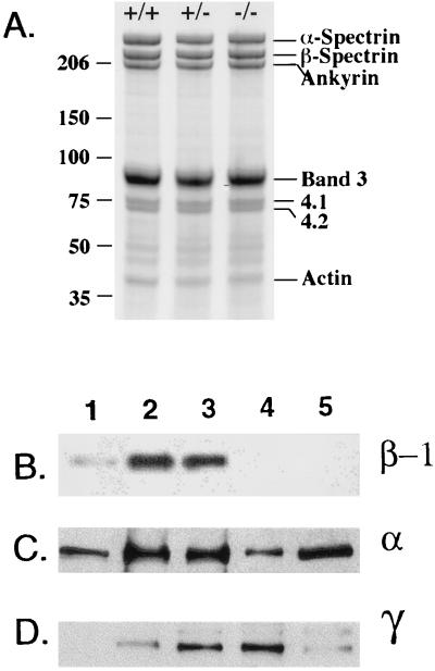
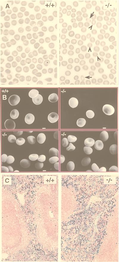
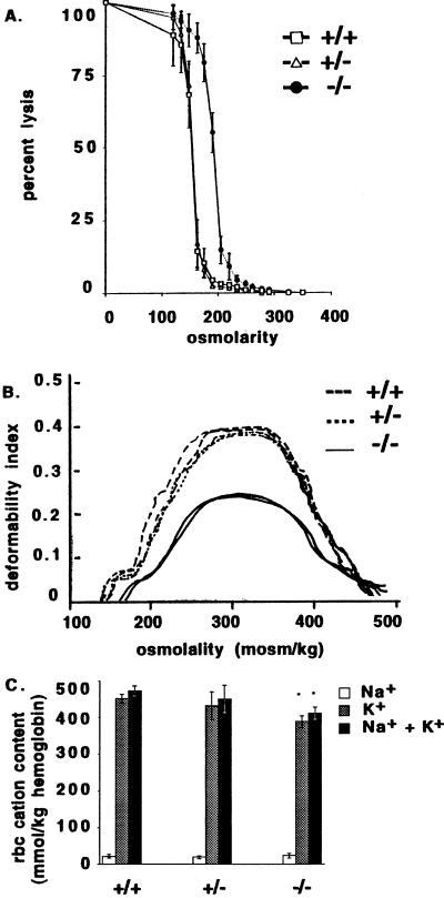
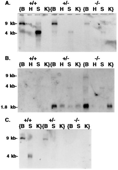
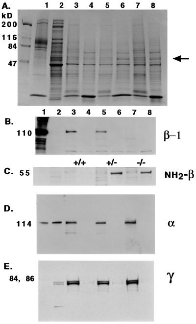
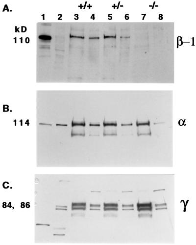
Adducins are a family of cytoskeleton proteins encoded by three genes (alpha, beta, gamma). In a comprehensive assay of gene expression, we show the ubiquitous expression of alpha- and gamma-adducins in contrast to the restricted expression of beta-adducin. beta-adducin is expressed at high levels in brain and hematopoietic tissues (bone marrow in humans, spleen in mice). To elucidate adducin's role in vivo, we created beta-adducin null mice by gene targeting, deleting exons 9-13. A 55-kDa chimeric polypeptide is produced from the first eight exons of beta-adducin and part of the neo cassette in spleen but is not detected in peripheral RBCs or brain. beta-adducin null RBCs are osmotically fragile, spherocytic, and dehydrated compared with the wild type, resembling RBCs from patients with hereditary spherocytosis. The lack of beta-adducin in RBCs leads to decreased membrane incorporation of alpha-adducin (30% of normal) and unexpectedly promotes a 5-fold increase in gamma-adducin incorporation into the RBC membrane skeleton. This study demonstrates adducin's importance to RBC membrane stability in vivo.


Figures

Figure 1
Figure 1
A multiple-tissue expression array (CLONTECH) of polyA RNA from 76 human tissues and cell lines probed with human cDNAs for α-, β-1, β-2, and γ-adducins. α- and γ-adducins show expression in all samples whereas β-1 and β-2 show restricted expression, mainly in brain and hematopoietic tissues. Probes for β-1 and β-2 were generated by PCR to encompass their distinct carboxy termini and 3′UTRs (β-1, exons 14–17; β2, exon 13b). Subcloned PCR products were labeled by random priming with 32P-dCTP.
Figure 2
Figure 2
A multiple-tissue expression Northern blot (CLONTECH) of polyA RNA from seven mouse tissues probed with mouse cDNAs for α, β-1, β-2, and γ-adducins. H, heart; B, brain; S, spleen; Lu, lung; L, liver; K, kidney; T, testis (- is a blank lane). α- and γ-adducin show expression in all lanes whereas β-1 and β-2 show restricted expression, mainly in brain and spleen.
Figure 3
Figure 3
(A) Strategy for targeted deletion of exons 9–13 from Add2, the gene encoding β-adducin. (B) Southern blot of EcoRV-restricted DNA from embryonic stem cell clones electroporated with the vector from A and selected for G418 resistance. The probe is indicated in A and detects the targeted allele in the clone on the right.
Figure 4
Figure 4
(A) Coomassie blue-stained Steck gel shows no difference in −/− RBC ghosts. (BD) Western blotting of RBC ghosts and platelets. Lanes: 1, human RBC ghosts; 2, +/+ RBC ghosts; 3, +/− RBC ghosts; 4, −/− RBC ghosts; 5, +/+ platelets. Antibodies used for detection are indicated to the right.
Figure 5
Figure 5
(A) Bright field light microscopy. Arrows indicate rounded elliptocytes; arrowheads indicate microspherocytes. (B) Scanning electron microscopy demonstrates that −/− RBCs are smaller and spherocytic compared with +/+ RBCs. (Bar = 1 μm.) (C) Increased iron deposition (dark blue color) in spleen from −/− mice compared with +/+ mice.
Figure 6
Figure 6
(A) Osmotic fragility curve demonstrates right shift for −/− RBCs. (B) Osmotic gradient deformability profiles demonstrate markedly reduced deformability for −/− RBCs; duplicate measurements are shown for each genotype. (C) Measurements of cation content show normal Na+ but decreased K+ and Na+ plus K+ in −/− RBCs.
Figure 7
Figure 7
Northern blotting of total RNA from mouse tissues. B, brain; H, heart; S, spleen; K, kidney. (A) Using a full-length β-1 cDNA as probe, 9- and 4-kb mRNAs are detected in brain and spleen. (B) Using a probe from the neoR cassette, a 1.8-kb mRNA is seen in all tissues from targeted mice and also hybridizes to the 9- and 4-kb mRNAs from targeted mice. (C) Using a probe for the deleted exons 9–12, no mRNA is detected in −/− mice, indicating that the 9- and 4-kb mRNAs are aberrant transcripts inserting antisense neoR sequence in the place of deleted β-adducin exons.
Figure 8
Figure 8
Western blotting of spleen homogenates. Lanes: 1, human RBC ghosts; 2, human platelets; 3–8, spleen homogenates, separated into 30,000 × g pellet (P) and supernatant (S) fractions: 3, +/+ P; 4, +/+ S; 5, +/− P; 6, +/− S; 7, −/− P; 8, −/− S. Gel in A is Coomassie blue-stained to demonstrate protein loading. Gels in BE are stained with the antibodies indicated on the right of the panels. The arrow marks the 55-kDa band in lanes 6 and 8 that is immunoreactive to NH2-β antibody. β-1 is not detected in −/− samples, but α and γ are detected in all pellet samples.
Figure 9
Figure 9
Western blotting of brain homogenates. Lanes: 1, human RBC ghosts; 2, human platelets; 3–8, brain homogenates, separated into 30,000 × g pellet (P) and supernatant (S) fractions: 3, +/+ P; 4, +/+ S; 5, +/− P; 6, +/− S; 7, −/− P; 8, −/− S. β-1 is not detected in −/− samples, but α and γ are detected in all samples.

References

    1. Palfrey H C, Waseem A. J Biol Chem. 1985;260:16021–16029. - PubMed
    1. Gardner K, Bennett V. J Biol Chem. 1986;261:1339–1348. - PubMed
    1. Gardner K, Bennett V. Nature (London) 1987;328:359–362. - PubMed
    1. Mische S M, Mooseker M S, Morrow J S. J Cell Biol. 1987;105:2837–2845. - PMC - PubMed
    1. Kuhlman P A, Hughes C A, Bennett V, Fowler V M. J Biol Chem. 1996;271:7986–7991. - PubMed

Publication types

MeSH terms

LinkOut - more resources