. 1999 Sep 14;96(19):10723-8.
doi: 10.1073/pnas.96.19.10723.

Muscle degeneration without mechanical injury in sarcoglycan deficiency

Affiliations

Muscle degeneration without mechanical injury in sarcoglycan deficiency

A A Hack et al. Proc Natl Acad Sci U S A. .

Abstract

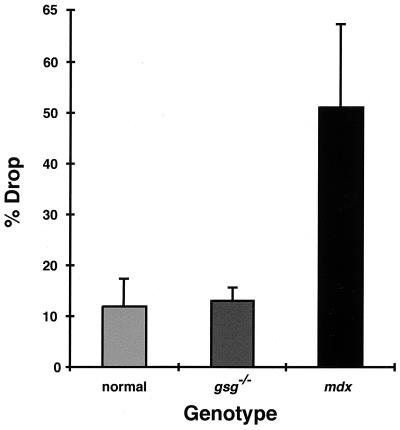
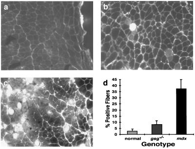
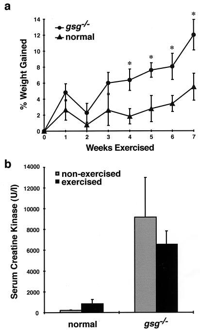
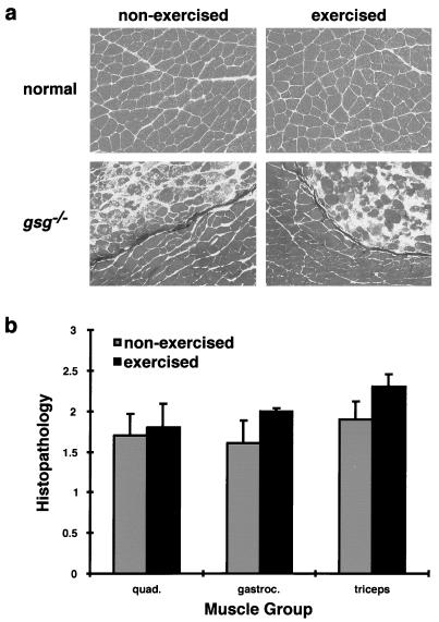
In humans, mutations in the genes encoding components of the dystrophin-glycoprotein complex cause muscular dystrophy. Specifically, primary mutations in the genes encoding alpha-, beta-, gamma-, and delta-sarcoglycan have been identified in humans with limb-girdle muscular dystrophy. Mice lacking gamma-sarcoglycan develop progressive muscular dystrophy similar to human muscular dystrophy. Without gamma-sarcoglycan, beta- and delta-sarcoglycan are unstable at the muscle membrane and alpha-sarcoglycan is severely reduced. The expression and localization of dystrophin, dystroglycan, and laminin-alpha2, a mechanical link between the actin cytoskeleton and the extracellular matrix, appears unaffected by the loss of sarcoglycan. We assessed the functional integrity of this mechanical link and found that isolated muscles lacking gamma-sarcoglycan showed normal resistance to mechanical strain induced by eccentric muscle contraction. Sarcoglycan-deficient muscles also showed normal peak isometric and tetanic force generation. Furthermore, there was no evidence for contraction-induced injury in mice lacking gamma-sarcoglycan that were subjected to an extended, rigorous exercise regimen. These data demonstrate that mechanical weakness and contraction-induced muscle injury are not required for muscle degeneration and the dystrophic process. Thus, a nonmechanical mechanism, perhaps involving some unknown signaling function, likely is responsible for muscular dystrophy where sarcoglycan is deficient.


Figures

Figure 1
Figure 1
ECC and sarcolemmal damage in normal, gsg−/−, and mdx animals. The mean and SD of the percent drop in tetanic force generation between the first and fifth ECC is shown for mature normal (n = 4), gsg−/− (n = 9), and mdx (n = 7) EDLs. There is no difference in percent drop between the normal and gsg−/−, in contrast to the mdx muscles.
Figure 2
Figure 2
Eccentric contraction-induced Procion orange uptake in normal (n = 5), gsg−/− (n = 4), and mdx (n = 6) EDL. Representative sections from normal (a), gsg−/− (b), and mdx EDL muscles (c) after the five ECC protocol are shown. (d) The percentage of fibers showing dye uptake (±SD), indicative of sarcolemmal rupture, is similar between normal and gsg−/− EDL muscles. The mdx EDL shows a substantial increase in dye uptake after ECC indicative of sarcolemmal rupture.
Figure 3
Figure 3
The effect of exercise on weight gain (a) and serum creatine kinase levels (b) in γ-sarcoglycan-deficient muscular dystrophy. (a) gsg−/− animals were weighed at the end of each week of exercise. The percent increase in weight compared with the starting weight for each genotype was calculated. The mean ± SEM is shown. gsg−/− animals gained significantly more weight than normal controls (∗, P < 0.05 for weeks 4–7) over the course of the protocol. (b) Serum creatine kinase levels were measured 2 days after completion of the 7-week swimming protocol. The mean ± SEM is shown. Animals lacking γ-sarcoglycan failed to show an increase in creatine kinase that was consistent with a change in the overall amount of degeneration occurring in skeletal muscle.
Figure 4
Figure 4
The effect of exercise on histopathology in normal and gsg−/− animals. (a) Representative sections of normal and gsg−/− quadriceps taken from age-matched animals that did (exercised, Right) and did not (non-exercised, Left) undergo the swimming protocol. Normal muscle showed no evidence of exercise-induced degeneration. gsg−/− muscles also showed no increase in histopathology as a result of exercise. (b) Quantitation of histopathology in age-matched exercised and nonexercised gsg−/− muscles. Histologic sections from femoral quadriceps (quad.), gastrocnemius (gastroc.), and triceps bracheii (triceps) were scored (blinded) for the presence of adipose tissue, degeneration, and fibrosis on a scale of 0–3. The mean ± SEM is shown. There was no significant difference in histopathology between exercised and nonexercised gsg−/− animals (P values of 0.80, 0.24, and 0.23, respectively).

Similar articles

Cited by

References

    1. Campbell K P. Cell. 1995;80:675–679. - PubMed
    1. Bonnemann C G, McNally E M, Kunkel L M. Curr Opin Peds. 1996;8:569–582. - PubMed
    1. Straub V, Campbell K P. Curr Opin Neurol. 1997;10:168–175. - PubMed
    1. Lim L E, Campbell K P. Curr Opin Neurol. 1998;11:443–452. - PubMed
    1. Burghes A H, Logan C, Hu X, Belfall B, Worton R G, Ray P N. Nature (London) 1987;328:434–437. - PubMed

Publication types

MeSH terms

LinkOut - more resources