. 1999 Sep 14;96(19):10812-7.
doi: 10.1073/pnas.96.19.10812.

Targeted gene disruption reveals an essential role for ceruloplasmin in cellular iron efflux

Affiliations

Targeted gene disruption reveals an essential role for ceruloplasmin in cellular iron efflux

Z L Harris et al. Proc Natl Acad Sci U S A. .

Abstract

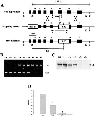
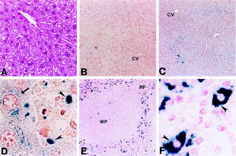
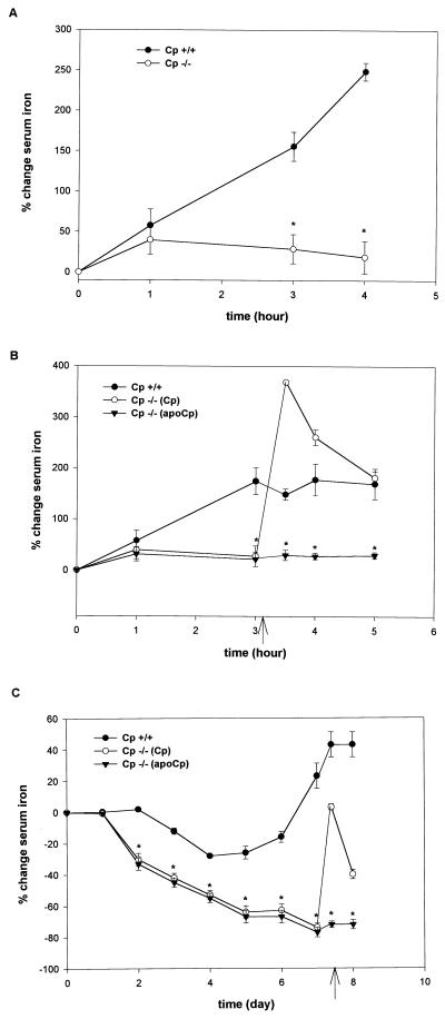
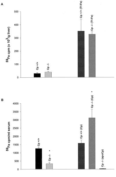
Aceruloplasminemia is an autosomal recessive disorder of iron metabolism. Affected individuals evidence iron accumulation in tissue parenchyma in association with absent serum ceruloplasmin. Genetic studies of such patients reveal inherited mutations in the ceruloplasmin gene. To elucidate the role of ceruloplasmin in iron homeostasis, we created an animal model of aceruloplasminemia by disrupting the murine ceruloplasmin (Cp) gene. Although normal at birth, Cp(-/-) mice demonstrate progressive accumulation of iron such that by one year of age all animals have a prominent elevation in serum ferritin and a 3- to 6-fold increase in the iron content of the liver and spleen. Histological analysis of affected tissues in these mice shows abundant iron stores within reticuloendothelial cells and hepatocytes. Ferrokinetic studies in Cp(+/+) and Cp(-/-) mice reveal equivalent rates of iron absorption and plasma iron turnover, suggesting that iron accumulation results from altered compartmentalization within the iron cycle. Consistent with this concept, Cp(-/-) mice showed no abnormalities in cellular iron uptake but a striking impairment in the movement of iron out of reticuloendothelial cells and hepatocytes. Our findings reveal an essential physiologic role for ceruloplasmin in determining the rate of iron efflux from cells with mobilizable iron stores.


Figures

Figure 1
Figure 1
Targeted disruption of the ceruloplasmin gene. (A) Cp locus, targeting vector and predicted recombinant allele. The 5′ flanking probe used for Southern analysis is shown. Restriction sites: E, EcoRV; B, BglII. (B) PCR products from tail genomic DNA corresponding to a 0.5-kb fragment (made by using primers a and b) from exon 16 to exon 17 in Cp+/+ alleles and a 1.0-kb fragment (made by using primers c and d) from exon 16 to the Neo cassette in Cp−/− mice. (C) Immunoblot analysis of ceruloplasmin in serum from Cp+/+, Cp+/−, and Cp−/− mice. (D) Ferroxidase activity in serum of Cp+/+, Cp+/−, and Cp−/− mice. Results are expressed as means ± standard deviations; ∗, P < 0.001.
Figure 2
Figure 2
(A) Hematoxylin/eosin stain of liver from a representative 1-year-old Cp−/− animal (×17). (B) Perls’ stain of liver section from a representative 1-year-old Cp+/+ mouse (×8.5). CV, central vein. (C) Perls’ stain of liver section from Cp−/− littermate (×8.5). (D) High-power view from C (×100). Arrow indicates iron accumulation in hepatocyte; arrowheads indicate Kupffer cells. (E) Perls’ stain of Cp−/− spleen from 1-year-old mouse (×8.5). RP, red pulp; WP, white pulp. (F) High-power view from E (×100). Arrowheads indicate iron within splenic reticuloendothelial cells.
Figure 3
Figure 3
(A) Change in serum iron concentration after the i.v. infusion of damaged red blood cells in 10-wk-old Cp+/+ and Cp−/− mice. Results are expressed as means ± standard deviations, n = 8 per time point (∗, P < 0.001). (B) Change in serum iron concentration after i.v. infusion of damaged red blood cells. Arrow indicates time of infusion of ceruloplasmin (Cp) or apoceruloplasmin (apoCp) as 6 μg/100 μl of circulating blood volume. Results expressed as means ± standard deviations, n = 8 per time point (∗, P < 0.001). (C) Change in serum iron concentration after serial phlebotomy in 10-wk-old Cp+/+ and Cp−/− mice. Results are expressed as means ± standard deviations, n = 6 per time point (∗, P < 0.001). Arrow indicates timing of infusion of ceruloplasmin (Cp) or apoceruloplasmin (apoCp) as 6 μg/100 μl of circulating blood volume.
Figure 4
Figure 4
(A) 59Fe uptake in the livers of control and transferrin-saturated (Tf-Fe) 16-wk-old Cp+/+ and Cp−/− mice. Results are expressed as means ± standard deviations, n = 6 per group. (B) Analysis of serum 59Fe in transferrin saturated Cp+/+ and Cp−/− mice revealed a statistically significant difference 96 hr after 59Fe injection (∗, P < 0.001). Results are expressed as means ± standard deviations, n = 4 per group. Subsequent infusion of ceruloplasmin (Cp) or apoceruloplasmin (apoCp) as 6 μg/100 μl of circulating blood volume in these mice reveals a statistically significant increase in 59Fe in the serum of Cp−/− mice (∗, P < 0.001).

References

    1. Ponka P, Beaumont C, Richardson D R. Semin Hematol. 1998;35:35–54. - PubMed
    1. Brittenham G M. In: Iron Metabolism in Health and Disease. Brock J H, Halliday J W, Pippard M J, Powell L W, editors. Philadelphia: Saunders; 1994. pp. 31–62.
    1. Andrews N C, Levy J E. Blood. 1998;92:1845–1851. - PubMed
    1. Bacon B R, Schilsky M L. Adv Intern Med. 1999;44:91–116. - PubMed
    1. Gitlin J D. Pediatr Res. 1998;44:271–276. - PubMed

Publication types

MeSH terms