. 1999 Sep 14;96(19):10911-6.
doi: 10.1073/pnas.96.19.10911.

Orexin A activates locus coeruleus cell firing and increases arousal in the rat

Affiliations

Orexin A activates locus coeruleus cell firing and increases arousal in the rat

J J Hagan et al. Proc Natl Acad Sci U S A. .

Abstract

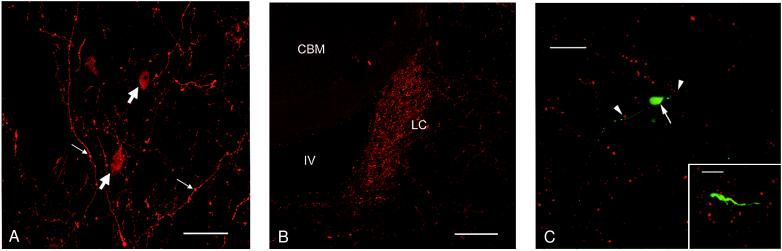
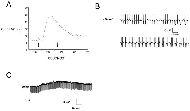
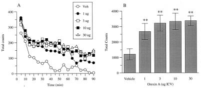
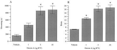
The localization of orexin neuropeptides in the lateral hypothalamus has focused interest on their role in ingestion. The orexigenic neurones in the lateral hypothalamus, however, project widely in the brain, and thus the physiological role of orexins is likely to be complex. Here we describe an investigation of the action of orexin A in modulating the arousal state of rats by using a combination of tissue localization and electrophysiological and behavioral techniques. We show that the brain region receiving the densest innervation from orexinergic nerves is the locus coeruleus, a key modulator of attentional state, where application of orexin A increases cell firing of intrinsic noradrenergic neurones. Orexin A increases arousal and locomotor activity and modulates neuroendocrine function. The data suggest that orexin A plays an important role in orchestrating the sleep-wake cycle.


Figures

Figure 1
Figure 1
Immunofluorescence images of orexin A immunoreactive profiles in the rat brain. All of these micrographs were generated by using the polyclonal antiserum against orexin A. (A) Coronal section through rat lateral hypothalamus showing orexin A immunofluorescence labeling in cell bodies (block arrows) and varicose axonal fibers (small arrows). (Bar = 50 μm.) (B) Coronal section through rat locus coeruleus (LC) showing dense plexus of immunolabeled axon fibers and terminals. CBM, cerebellum; IV, fourth ventricle. (Bar = 200 μm.) (C) An orexin A-responsive cell in a locus coeruleus brain slice, filled with Neurobiotin via the recording electrode. The red signal illustrates orexin A immunoreactivity, and the green signal indicates intracellular Neurobiotin. The large arrow indicates a neuronal soma bearing two main dendrites. The dendrites extend throughout the field of dense orexin A-positive varicosities. The arrowheads indicate varicosities that are closely apposed to a dendritic structure. The image is a brightest point projection of 64 confocal optical sections taken at 2-μm intervals. (Bar = 35 μm.) (Inset) Higher-power micrograph of a similarly treated section showing detail of proximity of orexin A-immunoreactive fiber varicosities to a process of an intrinsic locus coeruleus neurone. (Bar = 10 μm.)
Figure 2
Figure 2
Orexin A excitation in the locus coeruleus. (A) Extracellular recording of a single unit. Vertical axis is the number of suprathreshold spikes per 10-s period. Orexin A (1 μM) was applied between the arrows. (B) Intracellular recording of identified locus coeruleus neurone. (Upper) Baseline firing at the resting potential in normal artificial cerebrospinal fluid. (Lower) Increased action potential frequency at the peak of the response to 1 μM orexin A. (C) Intracellular recording of a locus coeruleus cell hyperpolarized to −80 mV by steady current injection. In the absence of action potentials, a clear depolarization is observed as a response to 1 μM orexin A (applied at arrow). Downward deflections in the trace are responses to 200-ms current pulses to monitor cell resistance. Note the absence of a clear change of resistance at the peak of the orexin A response.
Figure 3
Figure 3
Effects of orexin A on LMA. Data are shown as (A) mean total beam breaks during 5-min time bins for a 90-min recording period and (B) mean (±SEM) cumulative beam breaks for total period. Significant differences from vehicle-treated (Veh) rats are shown by **P < 0.01 (n = 7–8 per treatment group).
Figure 4
Figure 4
The effects of orexin A on (A) grooming duration (s) and (B) number of grooming bouts recorded for a 60-min period immediately after peptide administration. Significant differences from vehicle-treated (Veh) rats are shown by *P < 0.05 (n = 6 per treatment group).

References

    1. Sakurai T, Amemiya A, Ishii M, Matsuzaki I, Chemilli R M, Tanaka H, Clay Williams S, Richardson J A, Kozlowski G P, Wilson S, et al. Cell. 1998;92:573–585. - PubMed
    1. De Lecea L, Kilduff T S, Peyron C, Gao X-B, Foye P E, Danielson P E, Fukuhara C, Battenberg E L F, Gautvik V T, Bartlett II F S, et al. Proc Natl Acad Sci USA. 1998;95:322–327. - PMC - PubMed
    1. van den Pol A N, Gao X B, Obrietan K, Kilduff T S, Belousov A B. J Neurosci. 1998;18:7962–7971. - PMC - PubMed
    1. Elias C F, Saper C B, Maratos-Flier E, Tritos N A, Lee C, Kelly J, Tatro J B, Hoffman G E, Ollman M M, Barsh G S, et al. J Comp Neurol. 1998;402:442–459. - PubMed
    1. Peyron C, Tighe D K, van den Pol A N, de Lecea L, Heller H C, Sutcliffe J G, Kilduff T S. J Neurosci. 1998;18:9996–10015. - PMC - PubMed

MeSH terms

LinkOut - more resources