Comparison of three dimensional magnetic resonance imaging in conjunction with a blood pool contrast agent and nuclear scintigraphy for the detection of experimentally induced gastrointestinal bleeding
- PMID: 10486369
- PMCID: PMC1727663
- DOI: 10.1136/gut.45.4.581
Comparison of three dimensional magnetic resonance imaging in conjunction with a blood pool contrast agent and nuclear scintigraphy for the detection of experimentally induced gastrointestinal bleeding
Abstract
Background and aims: To compare the performance of 3D magnetic resonance imaging (MRI) in conjunction with an intravascular contrast agent with that of scintigraphy, with respect to detection and localisation of gastrointestinal haemorrhage in vivo in pigs.
Methods: Intraluminal bleeding sites were surgically created in the small bowel and colon of six pigs. The animals underwent scintigraphy with (99m)Tc labelled red blood cells and 3D MRI following administration of an intravascular contrast agent (NC100150) at five minute intervals over 30 minutes. For analysis, the intestinal tract was divided into six segments. Based on the two evaluated methods, each segment was characterised on a five point scale regarding the presence of a bleed. At autopsy, the surgically manipulated bowel segments were inspected for the presence of haemorrhage.
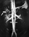
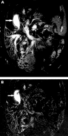
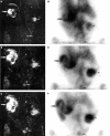
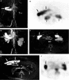
Results: Bleeding was confirmed at autopsy in 18/36 segments. Contrast extravasation with subsequent movement through the bowel could be documented on MRI data sets. All segments were correctly characterised, resulting in 100% sensitivity and specificity for MRI. Based on scintigraphy, interpretation of seven segments (19%) was false (sensitivity/specificity of 78%/72%). Differences in diagnostic performance were evident in the receiver operator characteristic (ROC) analysis, with an area under the MRI curve of 0.99 and under the scintigraphy curve of 0.85.
Conclusion: In conjunction with an intravascular contrast agent, 3D MRI permits accurate detection and localisation of gastrointestinal bleeding. The extent and evolution of intestinal bleeding can be determined with repeated data acquisition.
Figures


Similar articles
-
Intestinal and peritoneal bleeding: detection with an intravascular contrast agent and fast three-dimensional MR imaging--preliminary experience from an experimental study.Radiology. 1998 Dec;209(3):769-74. doi: 10.1148/radiology.209.3.9844672. Radiology. 1998. PMID: 9844672
-
Single-photon emission computed tomography enhanced Tc-99m-pertechnetate disodium-labelled red blood cell scintigraphy in the localization of small intestine bleeding: a single-centre twelve-year study.Digestion. 2011;84(3):207-11. doi: 10.1159/000328389. Epub 2011 Jul 12. Digestion. 2011. PMID: 21757912 Clinical Trial.
-
[Nuclear Medicine in the diagnosis of lower gastrointestinal bleeding].Hell J Nucl Med. 2007 Sep-Dec;10(3):197-204. Hell J Nucl Med. 2007. PMID: 18084667 Greek, Modern.
-
A role for subtraction scintigraphy in the evaluation of lower gastrointestinal bleeding in the athlete.Sports Med. 2007;37(10):923-8. doi: 10.2165/00007256-200737100-00007. Sports Med. 2007. PMID: 17887815 Review.
-
Gastrointestinal Bleeding Scintigraphy in the Early 21st Century.J Nucl Med. 2016 Feb;57(2):252-9. doi: 10.2967/jnumed.115.157289. Epub 2015 Dec 17. J Nucl Med. 2016. PMID: 26678616 Review.
Cited by
-
Comparison of capsule endoscopy and magnetic resonance (MR) enteroclysis in suspected small bowel disease.Int J Colorectal Dis. 2006 Mar;21(2):97-104. doi: 10.1007/s00384-005-0755-0. Epub 2005 Apr 22. Int J Colorectal Dis. 2006. PMID: 15846497
-
Lower gastrointestinal bleeding in chronic hemodialysis patients.Int J Nephrol. 2011;2011:272535. doi: 10.4061/2011/272535. Epub 2011 Oct 5. Int J Nephrol. 2011. PMID: 22007297 Free PMC article.
-
[Gastrointestinal bleeding. Diagnostics and therapy by interventional radiology].Chirurg. 2006 Feb;77(2):117-25. doi: 10.1007/s00104-005-1140-9. Chirurg. 2006. PMID: 16411076 Review. German.
-
[Negative endoscopy and MSCT findings in patients with acute lower gastrointestinal hemorrhage. Value of (99m)Tc erythrocyte scintigraphy].Radiologe. 2007 Jan;47(1):64-70. doi: 10.1007/s00117-006-1431-2. Radiologe. 2007. PMID: 17096110 German.
References
Publication types
MeSH terms
Substances
LinkOut - more resources
Full Text Sources
Medical
