. 1999 Sep;104(5):541-9.
doi: 10.1172/JCI6974.

Inhibition of apoptosis induced by ischemia-reperfusion prevents inflammation

Affiliations

Inhibition of apoptosis induced by ischemia-reperfusion prevents inflammation

M A Daemen et al. J Clin Invest. 1999 Sep.

Abstract

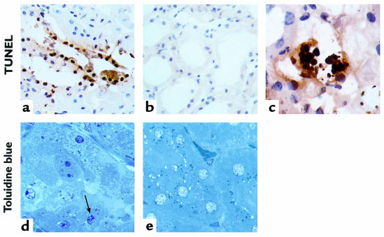
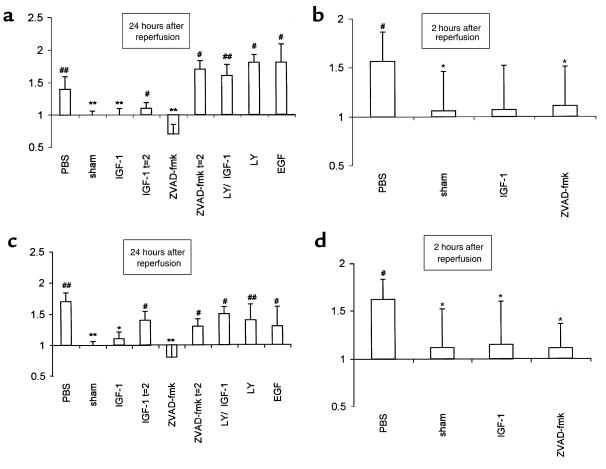
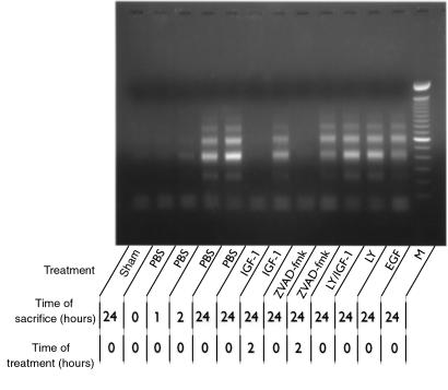
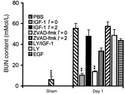
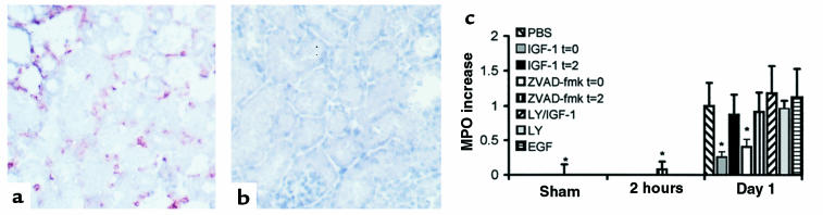
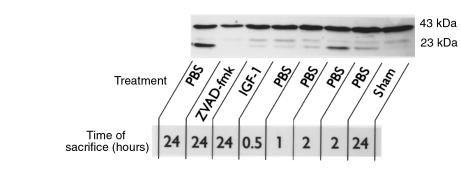
Ischemia followed by reperfusion leads to severe organ injury and dysfunction. Inflammation is considered to be the most important cause of tissue injury in organs subjected to ischemia. The mechanism that triggers inflammation and organ injury after ischemia remains to be elucidated, although different causes have been postulated. We investigated the role of apoptosis in the induction of inflammation and organ damage after renal ischemia. Using a murine model, we demonstrate a relationship between apoptosis and subsequent inflammation. At the time of reperfusion, administration of the antiapoptotic agents IGF-1 and ZVAD-fmk (a caspase inactivator) prevented the early onset of not only renal apoptosis, but also inflammation and tissue injury. Conversely, when the antiapoptotic agents were administered after onset of apoptosis, these protective effects were completely abrogated. The presence of apoptosis was directly correlated with posttranslational processing of the endothelial monocyte-activating polypeptide II (EMAP-II), which may explain apoptosis-induced influx and sequestration of leukocytes in the reperfused kidney. These results strongly suggest that apoptosis is a crucial event that can initiate reperfusion-induced inflammation and subsequent tissue injury. The newly described pathophysiological insights provide important opportunities to effectively prevent clinical manifestations of reperfusion injury in the kidney, and potentially in other organs.


Figures

Figure 1
Figure 1
Representative light micrographs showing evident in situ detection of DNA nick ends by TUNEL histology (ac). The tubular epithelial cells of the outer medulla from biopsies obtained after 2 hours of reperfusion showed positive staining (a), in contrast to kidneys obtained from sham-operated mice (b) (×250). The presence of apoptotic bodies could be detected extracellularly and extruded into the tubular lumen after 2 hours of reperfusion (c) (×1,000). Toluidine blue stains of 1-μm sections, obtained from kidneys that were fixed in situ after 3 hours of reperfusion and subsequently embedded in plastic (d and e), revealed the presence of dense condensation of nuclear chromatin and nuclear fragmentation in apoptotic tubular epithelial cells (arrow) in the outer medulla (d), whereas morphological attributes of tubular epithelial cell apoptosis were generally absent in sham-operated mice (e) (×1,000).
Figure 2
Figure 2
Renal caspase-like activities were determined kinetically in homogenates of tissue obtained after 24 hours (a and c) and 2 hours (b and d) of renal reperfusion in a fluorogenic substrate assay in which Ac-YVAD-amc (caspase-1–like) (a and b) or Ac-DEVD-amc (caspase-3–like) (c and d) served as substrates. Note that these data may represent a more generalized form of caspase activation, as outlined in the main text. Data are expressed as the increase in fluorescence as a function of time, normalized against data obtained from the sham-operated group. The groups that indicate t = 2 on the x-axis received the indicated treatment after 2 hours of reperfusion. All other groups were treated at the time of reperfusion. *P < 0.05, **P < 0.01 vs. control-treated animals. #P < 0.05, ##P < 0.01 vs. contralateral control kidney. The data shown are mean ± SEM.
Figure 3
Figure 3
The extent of renal apoptosis in different treatment groups is also reflected by the presence of fragmented DNA (as a result of internucleosomal DNA cleavage), amplified by LM-PCR and then viewed on ethidium bromide–stained gel. M, molecular weight markers (range: 100–2,000 bp).
Figure 4
Figure 4
Renal function in the different experimental groups as reflected by BUN content. **P < 0.01, ***P < 0.001 vs. control-treated animals. The data shown are mean ± SEM.
Figure 5
Figure 5
Representative light micrographs (a and b) showing infiltrating neutrophils in areas with impaired renal morphological integrity in biopsies obtained from mice subjected to renal I/R after 1 day of reperfusion (a). Neutrophils were stained with the anti-murine neutrophil mAb Gr-1. Neither loss of morphological integrity nor infiltrating neutrophils were observed in kidneys obtained from sham-operated mice (b) (×200). Neutrophil influx was assessed quantitatively by determination of MPO increase at 2 hours and 1 day of reperfusion (c). Values are presented relative to the amount of MPO present in the contralateral kidney harvested immediately after reperfusion, and then normalized with respect to the MPO increase at day 1 in PBS-treated mice subjected to I/R. *P < 0.05 vs. control-treated animals. The data shown are mean ± SEM.
Figure 6
Figure 6
Posttranslational processing of the EMAP-II protein is induced in parallel with apoptosis after as little as 2 hours of reperfusion. Western blots were performed with protein isolated from kidneys in mice that were sacrificed after different periods of reperfusion. Incubation with the EMAP-II antiserum SA 2846 revealed constitutive expression of inactive pro–EMAP-II, which resulted in a 43-kDa band. After 2 hours of reperfusion, the presence of 23-kDa mature EMAP-II was observed, which was even more apparent after 24 hours of reperfusion. Antiapoptotic treatment at the time of reperfusion with IGF-1 or ZVAD-fmk evidently inhibited I/R-induced EMAP-II maturation.

Similar articles

Cited by

References

    1. Vedder NB, et al. Inhibition of leukocyte adherence by anti-CD18 monoclonal antibody attenuates reperfusion injury in the rabbit ear. Proc Natl Acad Sci USA. 1990;87:2643–2646. - PMC - PubMed
    1. Shreeniwas R, et al. Hypoxia-mediated induction of endothelial cell interleukin-1 alpha. An autocrine mechanism promoting expression of leukocyte adhesion molecules on the vessel surface. J Clin Invest. 1992;90:2333–2339. - PMC - PubMed
    1. Metinko AP, Kunkel SL, Standiford TJ, Strieter RM. Anoxia-hyperoxia induces monocyte-derived interleukin-8. J Clin Invest. 1992;90:791–798. - PMC - PubMed
    1. Nogae S, et al. Induction of apoptosis in ischemia-reperfusion model of mouse kidney: possible involvement of Fas. J Am Soc Nephrol. 1998;9:620–631. - PubMed
    1. Fadok VA, et al. Macrophages that have ingested apoptotic cells in vitro inhibit proinflammatory cytokine production through autocrine/paracrine mechanisms involving TGF-beta, PGE2, and PAF. J Clin Invest. 1998;101:890–898. - PMC - PubMed

Publication types

MeSH terms