. 1999 Sep;104(5):567-76.
doi: 10.1172/JCI6713.

RGS4 causes increased mortality and reduced cardiac hypertrophy in response to pressure overload

Affiliations

RGS4 causes increased mortality and reduced cardiac hypertrophy in response to pressure overload

J H Rogers et al. J Clin Invest. 1999 Sep.

Abstract

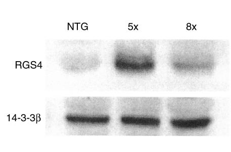
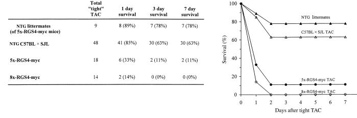
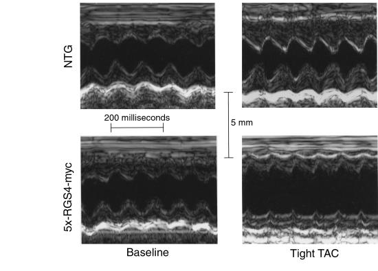
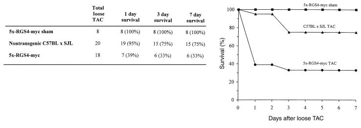
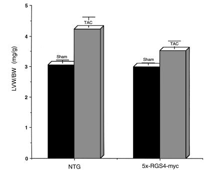
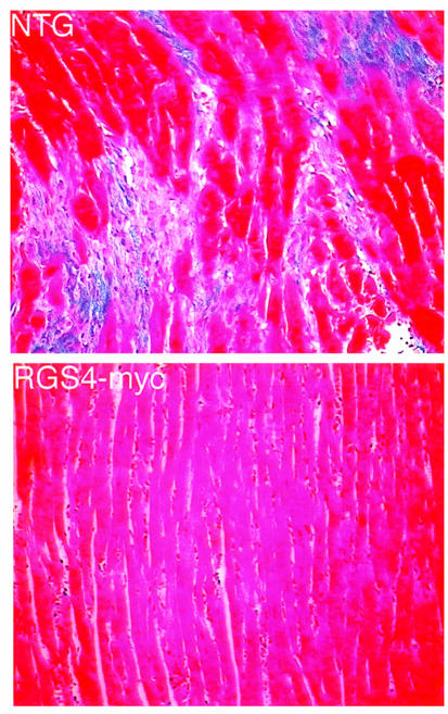
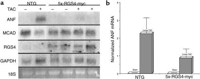
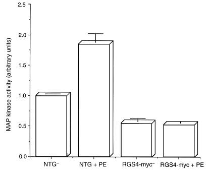
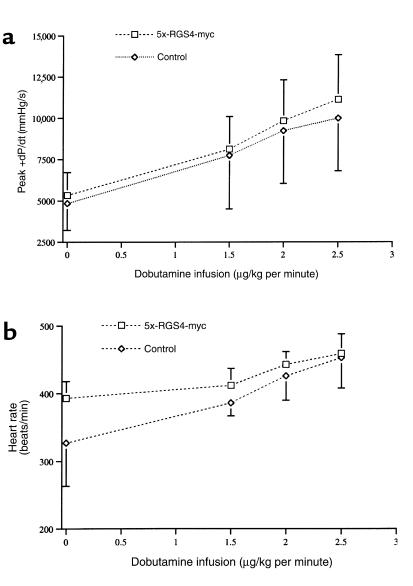
RGS family members are GTPase-activating proteins (GAPs) for heterotrimeric G proteins. There is evidence that altered RGS gene expression may contribute to the pathogenesis of cardiac hypertrophy and failure. We investigated the ability of RGS4 to modulate cardiac physiology using a transgenic mouse model. Overexpression of RGS4 in postnatal ventricular tissue did not affect cardiac morphology or basal cardiac function, but markedly compromised the ability of the heart to adapt to transverse aortic constriction (TAC). In contrast to wild-type mice, the transgenic animals developed significantly reduced ventricular hypertrophy in response to pressure overload and also did not exhibit induction of the cardiac "fetal" gene program. TAC of the transgenic mice caused a rapid decompensation in most animals characterized by left ventricular dilatation, depressed systolic function, and increased postoperative mortality when compared with nontransgenic littermates. These results implicate RGS proteins as a crucial component of the signaling pathway involved in both the cardiac response to acute ventricular pressure overload and the cardiac hypertrophic program.


Figures

Figure 1
Figure 1
Characteristics of RGS4-myc cardiac tissue. Increased ventricular RGS4 protein levels in 5x-RGS4-myc (5x) and 8x-RGS4-myc (8x) mice compared with nontransgenic littermate mice (NTG). The 5x-RGS4-myc mice express 4- to 5-fold excess protein, whereas the 8x-RGS4-myc mice express only 2- to 3-fold excess protein. A hamster monoclonal anti-RGS4 antibody and a rabbit polyclonal anti–14-3-3β antibody (to confirm equal loading) were used. Similar results were obtained in 6 hearts in each group.
Figure 2
Figure 2
Decreased survival of RGS4-myc transgenic mice after tight TAC. Survival rates after tight TAC in 8x-RGS4-myc mice, 5x-RGS4-myc mice, nontransgenic littermates of 5x-RGS4-myc mice (NTG littermates), and nontransgenic congenic mice (NTG C57BL × SJL TAC).
Figure 3
Figure 3
Analysis of cardiac function in 5x-RGS4-myc transgenic mice by M-mode echocardiography. Representative transthoracic M-mode echocardiographic tracings in a 5x-RGS4-myc mouse and a nontransgenic littermate (NTG) at baseline. TAC images shown for nontransgenic (1 week after tight TAC) and 5x-RGS4-myc (premorbid, 1 day after TAC) mice.
Figure 4
Figure 4
Decreased survival of RGS4-myc transgenic mice after loose TAC. Survival rates after loose TAC in 5x-RGS4-myc mice (5x-RGS4-myc TAC) and nontransgenic congenic C57BL × SJL mice (C57BL × SJL TAC) and after a sham operation in 5x-RGS4-myc mice (5x-RGS4-myc sham).
Figure 5
Figure 5
Reduced hypertrophic response to TAC in 5x-RGS4-myc mice. The LV weight/body weight ratio (LVW/BW), an index of LV mass, was determined 7 days after loose TAC or after a sham operation in nontransgenic congenic C57BL × SJL mice (NTG) or in 5x-RGS4-myc mice. Mice were excluded from LVW/BW analysis after TAC if the ascending aortic SBP was less than 2 SDs greater than the mean ascending aortic SBP obtained in sham-operated animals. The error bars represent the SEM.
Figure 6
Figure 6
Histologic analysis of cardiac morphology after TAC. Ventricular tissue sections from 5x-RGS4-myc (RGS4-myc) and nontransgenic littermates (NTG) of 5x-RGS4-myc mice were stained with Masson’s trichrome 1 week after loose TAC (original magnification ×200). Note the increased extracellular matrix content (blue color), cardiomyocyte enlargement, and disarray in the nontransgenic cardiac tissue.
Figure 7
Figure 7
Northern blot analysis of cardiac gene expression after TAC. (a) Northern blot analysis of ANF, MCAD, RGS4, and GAPDH gene expression after loose TAC. Loose TAC (+) or a sham operation (–) was performed on 5x-RGS4-myc or nontransgenic (NTG) congenic C57BL × SJL mice. (b) Quantitative analysis of cardiac ANF gene expression 7 days after loose TAC. The relative intensities of the resultant bands were quantified in their linear range by automated 2-dimensional computer densitometry. The graph depicts normalized ANF mRNA levels in ventricular tissue obtained from 5x-RGS4-myc or nontransgenic congenic C57BL × SJL mice (NTG). RGS4 mRNA levels were normalized by GAPDH mRNA. Data are presented in arbitrary units and error bars reflect the SE of 4 determinations.
Figure 8
Figure 8
Reduced RGS4-myc cardiac response to the Gi/Gq-coupled ligand phenylephrine. The p44 MAP kinase activity was assessed in the ventricular tissue of RGS4-myc mice or their nontransgenic littermates 90 seconds after intracardiac infusion of phenylephrine (or control buffer). Immunoblots of cytosolic extracts were analyzed using an anti–active ERK-1 MAP kinase mAb. Equal amounts of total protein were loaded in each lane. Densitometric analysis of 3 separate experiments was performed using NIH Image software, and data are expressed as the mean signal intensity ± SEM.
Figure 9
Figure 9
Preserved inotropic and chronotropic response of 5x-RGS4-myc mice to dobutamine. Peak LV +dP/dt (a) and heart rate (b) are shown at baseline and after progressive infusion of dobutamine in 5x-RGS4-myc mice (squares; n = 3) or nontransgenic C57BL × SJL mice (diamonds; n = 3). Peak +dP/dt, maximal first derivative of LV pressure. P = NS between 5x-RGS4-myc mice and nontransgenic mice at any level of dobutamine infusion.
Figure 10
Figure 10
Apoptotic indices are not increased in 5x-RGS4-myc mice after TAC. All figures are of LV myocardium (original magnification ×400), and are representative of TdT assays performed. (a) Positive control: tissue incubated in DNase demonstrating pan staining of nucleic DNA by TdT-labeling assay. (b) 5x-RGS4-myc 1 week after loose TAC. Note paucity of apoptotic nuclei shown by the arrow. (c) 5x-RGS4-myc, death less than 24 hours after TAC. Note absence of apoptotic nuclei. Nonapoptotic nuclei are counterstained with methyl green.

Similar articles

Cited by

References

    1. Sadoshima J, Izumo S. The cellular and molecular response of cardiac myocytes to mechanical stress. Annu Rev Physiol. 1997;59:551–571. - PubMed
    1. Thorburn J, Frost JA, Thorburn A. Mitogen-activated protein kinases mediate changes in gene expression, but not cytoskeletal organization associated with cardiac muscle cell hypertrophy. J Cell Biol. 1994;126:1565–1572. - PMC - PubMed
    1. Knowlton KU, et al. Co-regulation of the atrial natriuretic factor and cardiac myosin light chain-2 genes during α-adrenergic stimulation of neonatal rat ventricular cells. J Biol Chem. 1991;266:7759–7768. - PubMed
    1. Izumo S, Nadal-Ginard B, Mahdavi V. Proto-oncogene induction and reprogramming of cardiac gene expression produced by pressure overload. Proc Natl Acad Sci USA. 1988;85:339–343. - PMC - PubMed
    1. Lee H, et al. α1-adrenergic stimulation of cardiac gene transcription in neonatal rat myocardial cells: effects on myosin light chain-2 gene expression. J Biol Chem. 1988;263:7352–7358. - PubMed

Publication types

MeSH terms