. 1999 Sep;104(5):577-88.
doi: 10.1172/JCI7291.

O(2) deprivation inhibits Ca(2+)-activated K(+) channels via cytosolic factors in mice neocortical neurons

Affiliations

O(2) deprivation inhibits Ca(2+)-activated K(+) channels via cytosolic factors in mice neocortical neurons

H Liu et al. J Clin Invest. 1999 Sep.

Abstract

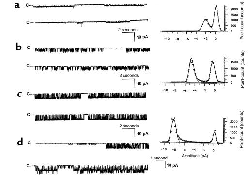
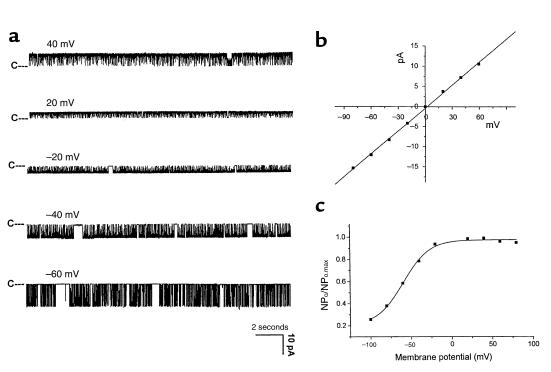
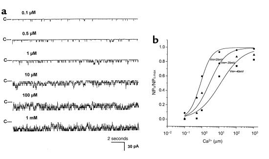
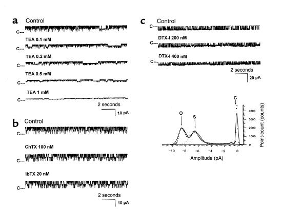
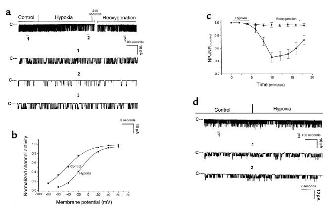
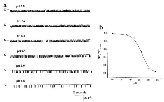
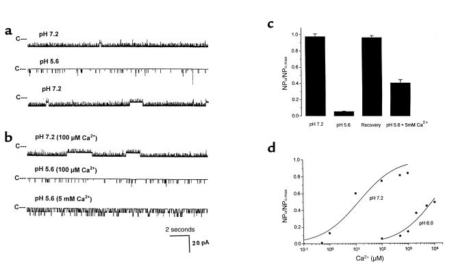
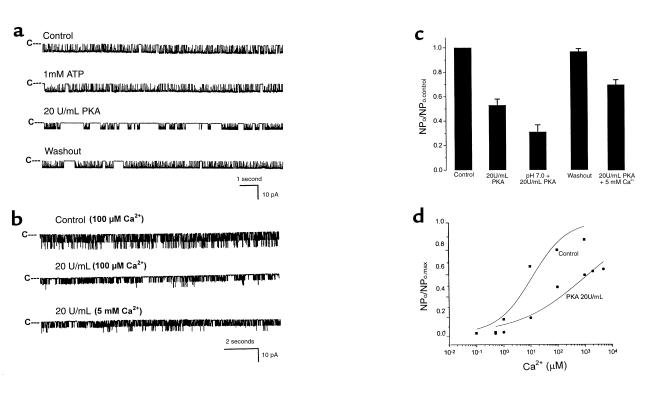
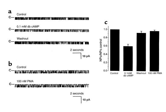
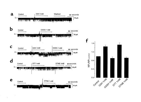
O(2) deprivation induces membrane depolarization in mammalian central neurons. It is possible that this anoxia-induced depolarization is partly mediated by an inhibition of K(+) channels. We therefore performed experiments using patch-clamp techniques and dissociated neurons from mice neocortex. Three types of K(+) channels were observed in both cell-attached and inside-out configurations, but only one of them was sensitive to lack of O(2). This O(2)-sensitive K(+) channel was identified as a large-conductance Ca(2+)-activated K(+) channel (BK(Ca)), as it exhibited a large conductance of 210 pS under symmetrical K(+) (140 mM) conditions, a strong voltage-dependence of activation, and a marked sensitivity to Ca(2+). A low-O(2) medium (PO(2) = 10-20 mmHg) markedly inhibited this BK(Ca) channel open probability in a voltage-dependent manner in cell-attached patches, but not in inside-out patches, indicating that the effect of O(2) deprivation on BK(Ca) channels of mice neocortical neurons was mediated via cytosol-dependent processes. Lowering intracellular pH (pH(i)), or cytosolic addition of the catalytic subunit of a cAMP-dependent protein kinase A in the presence of Mg-ATP, caused a decrease in BK(Ca) channel activity by reducing the sensitivity of this channel to Ca(2+). In contrast, the reducing agents glutathione and DTT increased single BK(Ca) channel open probability without affecting unitary conductance. We suggest that in neocortical neurons, (a) BK(Ca) is modulated by O(2) deprivation via cytosolic factors and cytosol-dependent processes, and (b) the reduction in channel activity during hypoxia is likely due to reduced Ca(2+) sensitivity resulting from cytosolic alternations such as in pH(i) and phosphorylation. Because of their large conductance and prevalence in the neocortex, BK(Ca) channels may be considered as a target for pharmacological intervention in conditions of acute anoxia or ischemia.


Figures

Figure 1
Figure 1
Different types of K+ channels in isolated neocortical neurons of mice. Currents were recorded from inside-out patches of a neocortical neuron using symmetrical 140 mM KCl on both sides of the membrane at a Vm of –40mV. The channel closed level is indicated by “C”. (a) Example traces of small-conductance K+ channel. The amplitude histogram to the left was compiled from 40 seconds of current record digitized at 500 Hz. (b) Example traces and the amplitude histogram of the medium conductance K+ channel. (c) Example traces and the amplitude histogram of the large-conductance K+ channel. (d) Example traces containing multiple K+ channel types.
Figure 2
Figure 2
(a) Single large-conductance K+ currents recorded from an inside-out patch, recorded at various holding potentials in the presence of symmetrical 140 mM KCl across the neuronal membrane. (b) The current-voltage relationship of the large-conductance K+ channel under conditions already described. The line drawn through these points is a linear regression fit with a conductance of 210 ± 6.9 pS (n = 15). (c) Relationship between open probability and voltage for this large-conductance channel. The line is fitted to a Boltzmann distribution: y = 1/{1 + exp [(V0.5 – Vm)/k]}, where y is normalized open probability (NPo/NPo.max), Vm is membrane potential, V0.5 is the membrane potential at which NPo is half of NPo.max, and k is a constant related to the slope of curve. V0.5 = –46 mV.
Figure 3
Figure 3
(a) Single-channel recordings of BKCa channel activity in the presence of varying concentration of Ca2+i at a Vm of –40 mV. (b) Relationship between Ca2+i and channel open probability at various membrane potentials. Data were fitted to Hill’s equation: y = 1/{1 + ([Ca2+]i/k)h}, where y is the normalized open probability (NPo/NPo.max), [Ca2+]i is intracellular free Ca2+ concentration, k is the free Ca2+ concentration required for maximum channel activation, and h is the Hill coefficient.
Figure 4
Figure 4
(a) Currents recorded under symmetrical 140 mM KCl condition in an outside-out patch from a neocortical neuron at a Vm of 30 mV. TEA (0–1 mM), from the extracellular side, blocks the BKCa channel. (b) Effects of ChTX (100 nM) and IbTX (20 nM), also on the extracellular side of the BKCa channels. Currents were also recorded in an outside-out patch at a Vm of 30 mV. (c) Effect of DTX-I on single BKCa channels. Currents were recorded in an inside-out patch under symmetrical 140 mM KCl condition at a Vm of –40 mV. DTX-I (200–400 nM) induced a subconductance state with 72% of normal open-state current. Amplitude histogram to the upper trace was compiled from 60 seconds (DTX-I 200 nM) of current record digitized at 500 Hz. Amplitude peaks are identified as closed state (c), substate (s), or open state (o).
Figure 5
Figure 5
(a) Effect of hypoxia on single BKCa channel in a cell-attached patch from a neocortical neuron. Current was recorded with high-KCl (140 mM) solution in the pipette and physiological solution in the bath at a Vm of –30 mV. The channel closed level is indicated by “C”. Three parts of the compressed trace are shown as indicated by numbers 1–3 (fast time resolution). (b) Effect of hypoxia on the voltage activation of BKCa channel under ionic condition just described. The line is fitted to a Boltzmann distribution. V0.5 shifted from –42.4 ± 4.8 mV to –18.6 ± 3.5 mV after exposure of hypoxia for 10 minutes. (c) Time course of the hypoxia-induced effect on NPo. In cell-attached recordings (filled squares), channel inhibition started about 5 minutes after the onset of hypoxia, and a maximum inhibition was reached in about 10 minutes. After that time, NPo was markedly reduced to about 43% of control level. Reoxygenation led to partial recovery. In inside-out recordings (filled circles), NPo was not significantly affected during hypoxia. (d) Continuous recording of a single BKCa channel current from an inside-out patch of a neocortical neuron during hypoxia, using a symmetrical 140 mM KCl on both sides of the membrane, with a Vm of –30 mV. The channel closed level is indicated by “C”. Two parts of the compressed trace (indicated by the numbers 1 and 2) are shown below at fast time resolution.
Figure 6
Figure 6
Cytosolic pH modulates NPo of BKCa channels in inside-out membrane patches from neocortical neurons. (a) Example from a patch. Currents were recorded at a Vm of –40mV. Changing the pH of the bath solution (intracellular side of the membrane) leads to marked changes in NPo. (b) Relationship between pHi and channel NPo at a Vm of –40 mV. Data were fitted to Hill’s equation. The half-inhibition pH value was 6.42.
Figure 7
Figure 7
Reversibility of the pH effect on the BKCa channels. Currents were recorded from an inside-out patch of a neocortical neuron, using symmetrical 140 mM KCl on both sides of the membrane, Vm of –30 mV. (a) Single-channel trace showing inhibition by pH 5.6 and complete reactivation at pH 7.2. (b) Similar experiment, but where the reactivation was done by the addition of 5 mM Ca2+. (c) Mean change of NPo, measured at different conditions. (d) Effect of pH (6.0 and 7.2) on the relation of Ca2+ and NPo. Data were acquired from inside-out patches of neocortical neurons using symmetrical 140 mM KCl on both sides of the membrane, Vm of –40 mV, and fitted to Hill’s equation.
Figure 8
Figure 8
Effect of the catalytic subunit of PKA on the BKCa channel. Currents were recorded from inside-out patches of neocortical neurons, using symmetrical 140 mM KCl on both sides of the membrane, Vm of –30 mV. (a) Single-channel traces showing the inhibition by PKA 20 U/mL and reactivation upon washing out. (b) Similar experiment, where the reactivation was done by the addition of 5 mM Ca2+. (c) Mean change in NPo, measured at different conditions. (d) Effect of 20 U/mL PKA on the relation between Ca2+ and NPo. Data were acquired from inside-out patches of neocortical neurons using symmetrical 140 mM KCl on both sides of the membrane, Vm of –40 mV and fitted to Hill’s equation.
Figure 9
Figure 9
Effects of PKA and PKC activators on BKCa channel currents. Currents were recorded from a cell-attached patch of a neocortical neuron with high-KCl (140 mM) solution in the pipette and bathing outside solution, at a Vm of –20 mV. (a) Application of 0.1 mM db cAMP markedly decreased the single BKCa channel activity and recovered after washout. (b) Application of 100 nM PMA had no apparent effect on BKCa channel activity. (c) Mean change in NPo, measured in different conditions.
Figure 10
Figure 10
Effect of the redox agents on the BKCa channel. Currents were recorded from inside-out patches of neocortical neurons, using symmetrical 140 mM KCl on both sides of the membrane, Vm of –30 mV. (a) Application of 1 mM GSH markedly increased the single BKCa channel activity, and this activity persisted after washout. (b) Application of 1 mM GSSG had no apparent effect on BKCa channel activity. (c) The increase in channel activity caused by GSH (1 mM) recovered to control level when GSSG was applied. (d) Reducing agent DTT (1 mM) markedly augmented BKCa channel activity, which was reversed by the oxidizing agent DTNB (1 mM). (e) DTNB (1 mM) had no significant effect on the BKCa channel. (f) Mean change in NPo, measured at different conditions.

Similar articles

Cited by

References

    1. Haddad GG, Jiang C. O2 deprivation in the central nervous system: on mechanisms of neuronal response, different sensitivity and injury. Prog Neurobiol. 1993;40:277–318. - PubMed
    1. Cummins TR, Jiang C, Haddad GG. Decrease in human neocortical excitability during hypoxia via Na+ channel modulation. J Clin Invest. 1993;91:608–615. - PMC - PubMed
    1. O’Reilly J, Cummins TR, Haddad GG. Oxygen deprivation inhibits Na+ current in rat hippocampal neurons via protein kinase C. J Physiol. 1997;503:479–488. - PMC - PubMed
    1. Haddad GG, Jiang C. O2-sensing mechanisms in excitable cells: role of plasma membrane K+ channel. Annu Rev Physiol. 1997;59:23–43. - PubMed
    1. Lopez-Barneo J, Lopez-Lopez JR, Urena J, Gonzalez C. Chemotransduction in the carotid body: K+ current modulated by PO2 in type I chemoreceptor cells. Science. 1988;241:580–582. - PubMed

Publication types

MeSH terms