. 1999 Sep;155(3):765-74.
doi: 10.1016/S0002-9440(10)65175-9.

Formation of new vasa vasorum in vasculitis. Production of angiogenic cytokines by multinucleated giant cells

Affiliations

Formation of new vasa vasorum in vasculitis. Production of angiogenic cytokines by multinucleated giant cells

M Kaiser et al. Am J Pathol. 1999 Sep.

Abstract

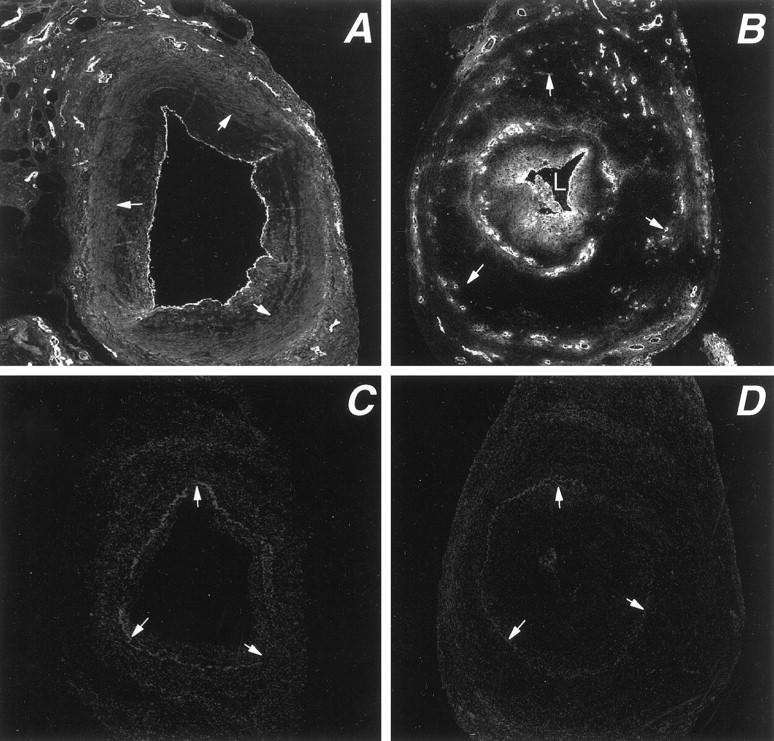
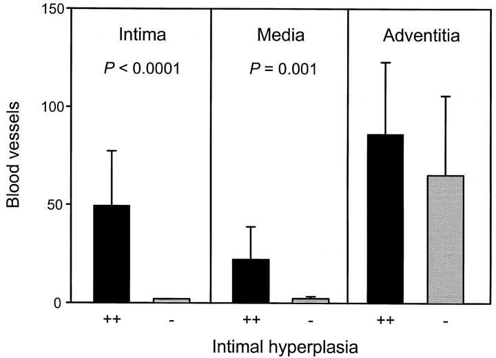
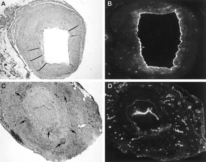
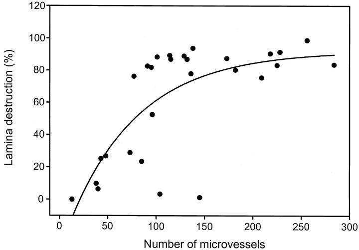
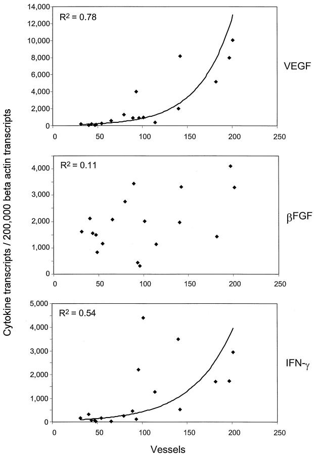
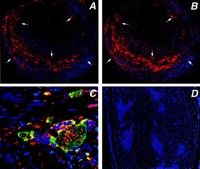
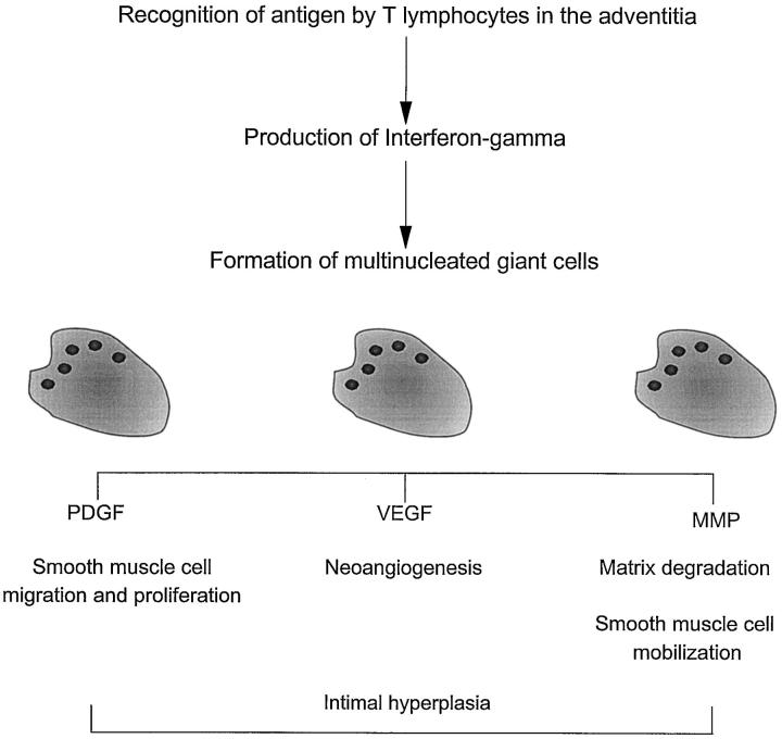
Inflammation of the arterial wall in giant cell arteritis induces a series of structural changes, including the formation of new vasa vasorum. To study the regulation of neoangiogenesis in giant cell arteritis, temporal arteries were examined for the extent and localization of microvessel generation and for the production of angiogenic factors. In normal arteries, vasa vasorum were restricted to the adventitia, but in inflamed arteries, capillaries emerged in the media and the intima. These capillaries displayed a distinct topography with a circumferential arrangement in the external one-third of the intima. Neovascularization was closely correlated with the formation of lumen-obstructing intima, the fragmentation of the internal elastic lamina, and the presence of multinucleated giant cells. Comparison of tissue cytokine transcription in temporal arteries of giant cell arteritis patients with and without up-regulated neoangiogenesis identified interferon-gamma and vascular endothelial growth factor but not fibroblast growth factor-2 as mediators associated with vasa vasorum proliferation. Giant cells and CD68-positive macrophages at the media-intima junction were found to be the major cellular sources of vascular endothelial growth factor. These data demonstrate that formation of new vasa vasorum in vasculitis is regulated by inflammatory cells and not by arterial wall cells, raising the possibility that it represents a primary disease mechanism and not a secondary hypoxia-induced event. Increased neovascularization in interferon-gamma-rich arteries suggests that the formation of new vasa vasorum is determined by the nature of the immune response in the arterial wall, possibly resulting from the generation and functional activity of multinucleated giant cells.


Figures

Figure 1.
Figure 1.
Distribution of vasa vasorum in temporal arteries affected by GCA. Tissue sections from temporal arteries of GCA patients were stained with antibodies specific for von Willebrand’s factor and a Cy-2-labeled secondary antibody to visualize capillary lumina. Stained sections were scanned by confocal microscopy. In temporal arteries with minimal intimal thickening, vasa vasorum are essentially restricted to the adventitial layer (A). In temporal arteries with marked luminal stenosis due to intimal hyperplasia, microvessels emerge in the media and form a circumferential ring in the outer intima (B). Arrows delineate the internal elastic lamina. Negative control stains are shown as C and D. Original magnification, ×100.
Figure 2.
Figure 2.
Layer-specific distribution of vasa vasorum in temporal arteries with minimal and marked intimal hyperplasia. Temporal arteries from a cohort of 28 patients with GCA were stained for von Willebrand’s factor to visualize microvessels. Based upon the thickening of the intimal layer, patients were divided into two categories with minimal to moderate intimal hyperplasia (<50%) or marked luminal stenosis (>50%), designated − and ++, respectively. Capillary lumen were counted in the entire arterial circumference and assigned to the adventitia, media, or intima. Results are given as means ± SD. Neovascularization in the media and intima occurred only in cases with intimal hyperplasia. The number of microvessels in the adventitia did not correlate with luminal stenosis.
Figure 3.
Figure 3.
Giant cell formation, fragmentation of the internal elastic lamina, and neoangiogenesis in GCA. Temporal arteries from GCA patients with and without marked neoangiogenesis were compared. Hematoxylin/eosin (A, C) and von Willebrand’s factor staining (B, D) of two representative specimens are shown. Original magnification, ×100. B and C: In the temporal artery with vasa vasorum confined to the adventitia, multinucleated giant cells were absent and the internal elastic lamina (IEL), was well maintained. C and D: In the temporal artery with numerous microvessels in the media and the hyperplastic intima, a garland of multinucleated giant cells (arrows) was arranged along the disrupted IEL.
Figure 4.
Figure 4.
Correlation of the destruction of the internal elastic lamina (IEL) and the formation of new vasa vasorum in GCA. Cross-sections of temporal arteries from 28 patients with GCA were stained for von Willebrand’s factor to identify vasa vasorum. The number of capillary lumina in all layers of the arterial wall were counted. The perimeter of the IEL and the fraction of disrupted IEL were determined by digital image analysis. The percentage of fragmented IEL is shown in correlation to the number of microvessels in the arterial wall. Vasa vasorum formation was low in patients with a large proportion of the IEL intact but was high in patients with almost complete disruption of the IEL (R2 = 0.53).
Figure 5.
Figure 5.
Expression of cytokine gene transcripts in temporal arteries with minimal and marked neoangiogenesis. Total RNA was obtained from 18 consecutive temporal artery biopsies from patients with GCA. cDNAs were adjusted for the number of β-actin transcripts and amplified with primer sets specific for IFN-γ, FGF-2, and VEGF. Results are given as the number of gene transcripts per 2 × 10 copies of β-actin. In parallel, sections of the temporal artery specimens were stained with antibodies to von Willebrand’s factor and microvessels per section were enumerated as described in Figure 1 ▶ . Transcription of IFN-γ and VEGF, but not of FGF-2 (βFGF), correlated with neoangiogenesis.
Figure 6.
Figure 6.
Production of vascular endothelial growth factor (VEGF) in temporal arteries with GCA. Tissue sections from temporal arteries were stained with anti-VEGF and anti-CD68 Abs. Anti-CD68 was visualized by using an FITC-labeled secondary antibody, anti-VEGF was developed by using a secondary biotinylated antibody, streptavidine-alkaline phosphatase and vector red. Stained sections were scanned by confocal microscopy and composite pictures were generated. Stainings for VEGF (A) and CD68 (B) are shown at an original magnification of ×100. VEGF production in the arterial wall was unevenly distributed with VEGF+ cells (red) accumulating at the media-intima junction. A small population of VEGF-producing cells was detected along the external elastic lamina. Cell nuclei are stained blue. More than 90% of the VEGF-producing cells expressed the CD68 marker. C: Strong expression of VEGF was typical for multinucleated giant cells. Blue, cell nuclei; red, VEGF; green, CD68; yellow, co-expression of VEGF and CD68. Original magnification, ×630. D: Control stainings without primary antibodies only showed minimal autofluorescence.
Figure 7.
Figure 7.
Schematic model summarizing the role of multinucleated giant cells in vasculitis. A model is proposed that the generation, differentiation, and functional activity of multinucleated giant cells and macrophages at the media intima junction are regulated through T lymphocytes stimulated by antigen in the adventitia. Giant cells and specialized macrophages have a role in several pathways, including regulation of neoangiogenesis, intimal hyperplasia, and matrix degradation.

References

    1. Hunder GG: Giant cell arteritis and polymyalgia rheumatica. Med Clin North Am 1997, 81:195-219 - PubMed
    1. Nordborg E, Nordborg C, Malmvall BE, Andersson R, Bengtsson BA: Giant cell arteritis. Rheum Dis Clin North Am 1995, 21:1013-1026 - PubMed
    1. Kaiser M, Weyand CM, Bjornsson J, Goronzy JJ: Platelet-derived growth factor, intimal hyperplasia, and ischemic complications in giant cell arteritis. Arthritis Rheum 1998, 41:623-633 - PubMed
    1. Evans JM, O’Fallon WM, Hunder GG: Increased incidence of aortic aneurysm and dissection in giant cell (temporal) arteritis: a population-based study. Ann Intern Med 1995, 122:502-507 - PubMed
    1. Weyand CM, Goronzy JJ: Giant cell arteritis as an antigen-driven disease. Rheum Dis Clin North Am 1995, 21:1027-1039 - PubMed

Publication types

MeSH terms