

Id-1 and Id-2 are overexpressed in pancreatic cancer and in dysplastic lesions in chronic pancreatitis
- PMID: 10487839
- PMCID: PMC1866883
- DOI: 10.1016/S0002-9440(10)65180-2
Id-1 and Id-2 are overexpressed in pancreatic cancer and in dysplastic lesions in chronic pancreatitis
Abstract
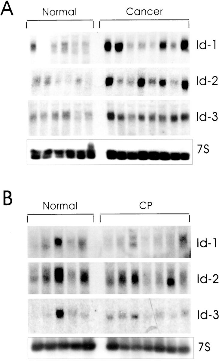
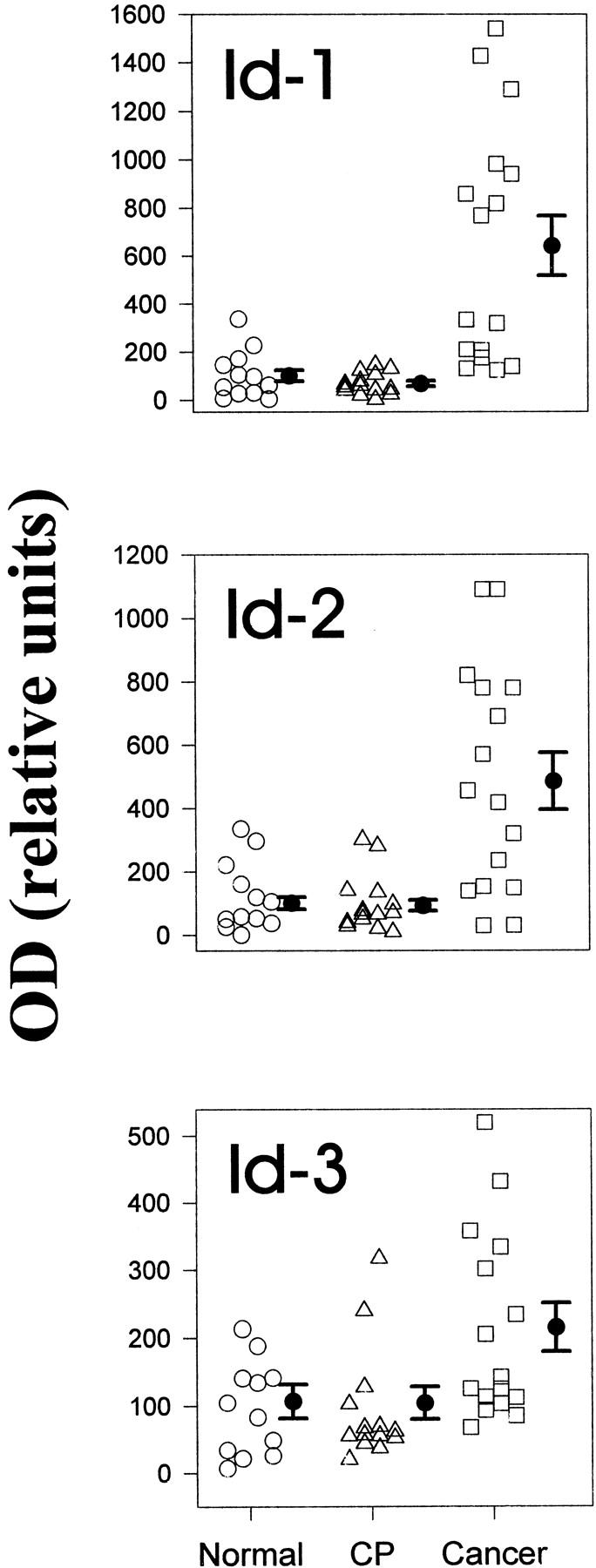
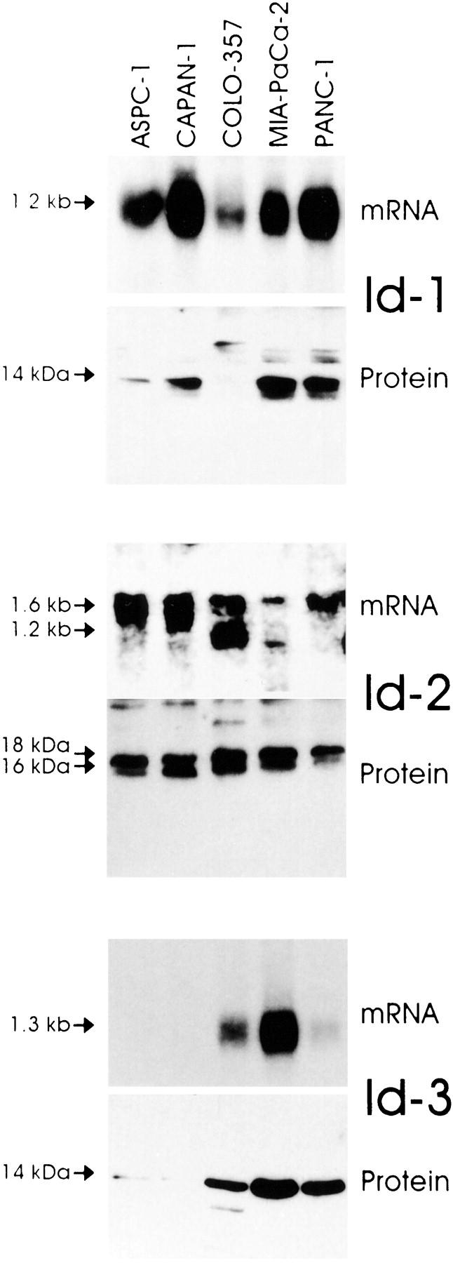
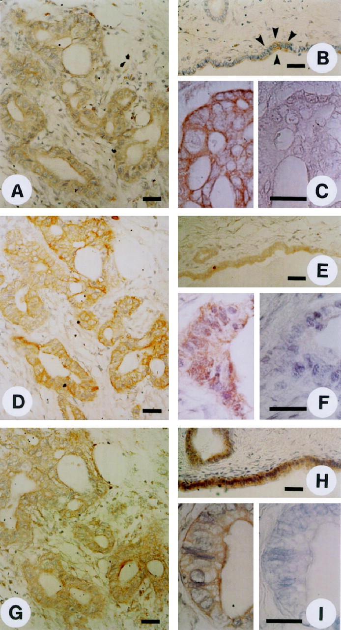
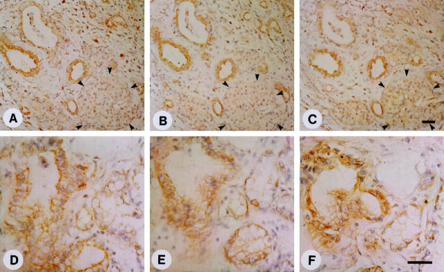
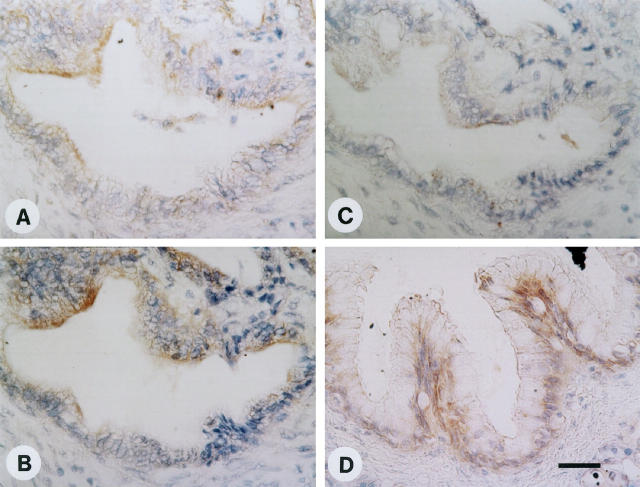
Id proteins antagonize basic helix-loop-helix proteins, inhibit differentiation, and enhance cell proliferation. In this study we compared the expression of Id-1, Id-2, and Id-3 in the normal pancreas, in pancreatic cancer, and in chronic pancreatitis (CP). Northern blot analysis demonstrated that all three Id mRNA species were expressed at high levels in pancreatic cancer samples by comparison with normal or CP samples. Pancreatic cancer cell lines frequently coexpressed all three Ids, exhibiting a good correlation between Id mRNA and protein levels, as determined by immunoblotting with highly specific anti-Id antibodies. Immunohistochemistry using these antibodies demonstrated the presence of faint Id-1 and Id-2 immunostaining in pancreatic ductal cells in the normal pancreas, whereas Id-3 immunoreactivity ranged from weak to strong. In the cancer tissues, many of the cancer cells exhibited abundant Id-1, Id-2, and Id-3 immunoreactivity. Scoring on the basis of percentage of positive cells and intensity of immunostaining indicated that Id-1 and Id-2 were increased significantly in the cancer cells by comparison with the respective controls. Mild to moderate Id immunoreactivity was also seen in the ductal cells in the CP-like areas adjacent to these cells and in the ductal cells of small and interlobular ducts in CP. In contrast, in dysplastic and atypical papillary ducts in CP, Id-1 and Id-2 immunoreactivity was as significantly elevated as in the cancer cells. These findings suggest that increased Id expression may be associated with enhanced proliferative potential of pancreatic cancer cells and of proliferating or dysplastic ductal cells in CP.
Figures
Similar articles
-
Preferential expression of cystein-rich secretory protein-3 (CRISP-3) in chronic pancreatitis.Histol Histopathol. 2003 Apr;18(2):425-33. doi: 10.14670/HH-18.425. Histol Histopathol. 2003. PMID: 12647793
-
The helix-loop-helix protein Id2 is overexpressed in human pancreatic cancer.Cancer Res. 1998 Sep 1;58(17):3769-72. Cancer Res. 1998. PMID: 9731481
-
COMP is selectively up-regulated in degenerating acinar cells in chronic pancreatitis and in chronic-pancreatitis-like lesions in pancreatic cancer.Scand J Gastroenterol. 2003 Feb;38(2):207-15. doi: 10.1080/00365520310000717. Scand J Gastroenterol. 2003. PMID: 12678339
-
Id genes in nervous system development.Histol Histopathol. 2000 Apr;15(2):603-18. doi: 10.14670/HH-15.603. Histol Histopathol. 2000. PMID: 10809382 Review.
-
Id family of helix-loop-helix proteins in cancer.Nat Rev Cancer. 2005 Aug;5(8):603-14. doi: 10.1038/nrc1673. Nat Rev Cancer. 2005. PMID: 16034366 Review.
Cited by
-
Physical and functional interaction between the ID1 and p65 for activation of NF-κB.Am J Physiol Cell Physiol. 2012 Aug 1;303(3):C267-77. doi: 10.1152/ajpcell.00365.2011. Epub 2012 May 16. Am J Physiol Cell Physiol. 2012. PMID: 22592405 Free PMC article.
-
Structural insights into interacting mechanism of ID1 protein with an antagonist ID1/3-PA7 and agonist ETS-1 in treatment of ovarian cancer: molecular docking and dynamics studies.J Mol Model. 2012 Nov;18(11):4865-84. doi: 10.1007/s00894-012-1489-x. Epub 2012 Jun 20. J Mol Model. 2012. PMID: 22714536
-
Role of inhibitor of DNA binding-1 protein is related to angiogenesis in the tumor advancement of uterine endometrial cancers.Exp Ther Med. 2010 Mar;1(2):351-356. doi: 10.3892/etm_00000055. Epub 2010 Mar 1. Exp Ther Med. 2010. PMID: 22993548 Free PMC article.
-
Overexpression of Id-1 is significantly associated with tumour angiogenesis in human pancreas cancers.Br J Cancer. 2004 Mar 22;90(6):1198-203. doi: 10.1038/sj.bjc.6601684. Br J Cancer. 2004. PMID: 15026801 Free PMC article.
-
The ID proteins: master regulators of cancer stem cells and tumour aggressiveness.Nat Rev Cancer. 2014 Feb;14(2):77-91. doi: 10.1038/nrc3638. Epub 2014 Jan 20. Nat Rev Cancer. 2014. PMID: 24442143 Review.
References
-
- Jan YN, Jan LY: HLH proteins, fly neurogenesis, and vertebrate myogenesis. Cell 1993, 75:827-830 - PubMed
-
- Olson EN, Klein WH: bHLH factors in muscle development: dead lines and commitments, what to leave in and what to leave out. Genes Dev 1994, 8:1-8 - PubMed
-
- Johnson JE, Birren SJ, Anderson DJ: Two rat homologues of Drosophila achaete-scute specifically expressed in neuronal precursors. Nature 1990, 346:858-861 - PubMed
-
- Weintraub H: The MyoD family and myogenesis: redundancy, networks, and thresholds. Cell 1993, 75:1241-1244 - PubMed
Publication types
MeSH terms
Substances
Grants and funding
LinkOut - more resources
Full Text Sources
Other Literature Sources
Medical
Miscellaneous
