. 1999 Sep;155(3):915-25.
doi: 10.1016/S0002-9440(10)65191-7.

Involvement of soluble CD95 in Churg-Strauss syndrome

Affiliations

Involvement of soluble CD95 in Churg-Strauss syndrome

M Müschen et al. Am J Pathol. 1999 Sep.

Abstract

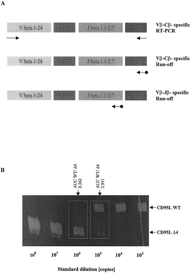
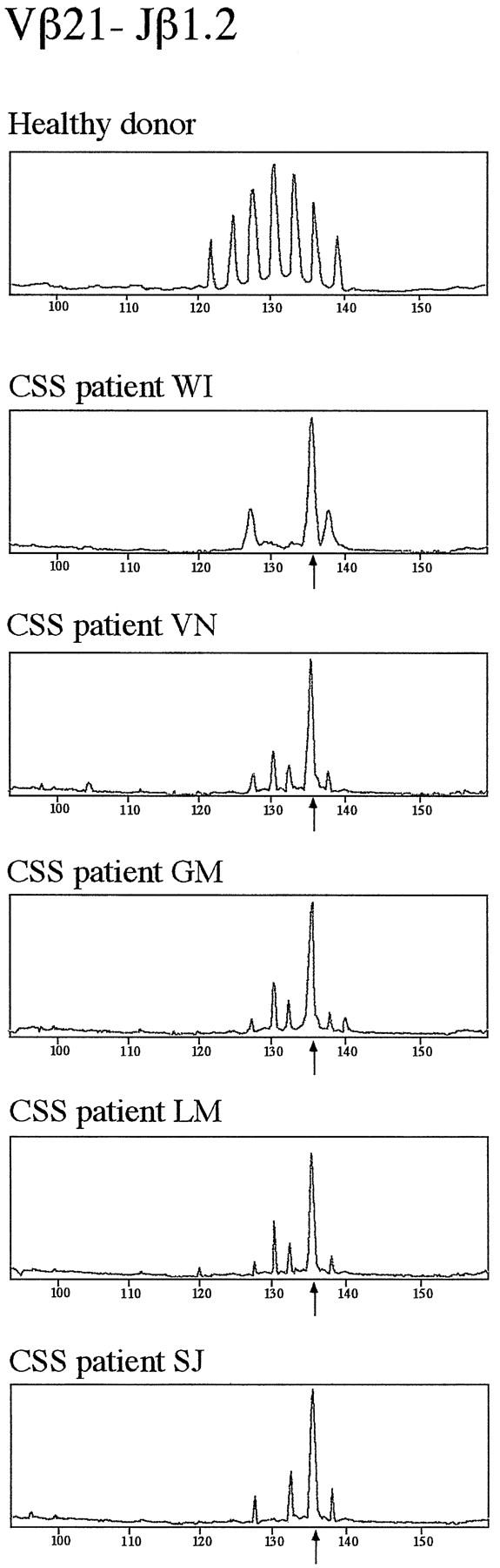
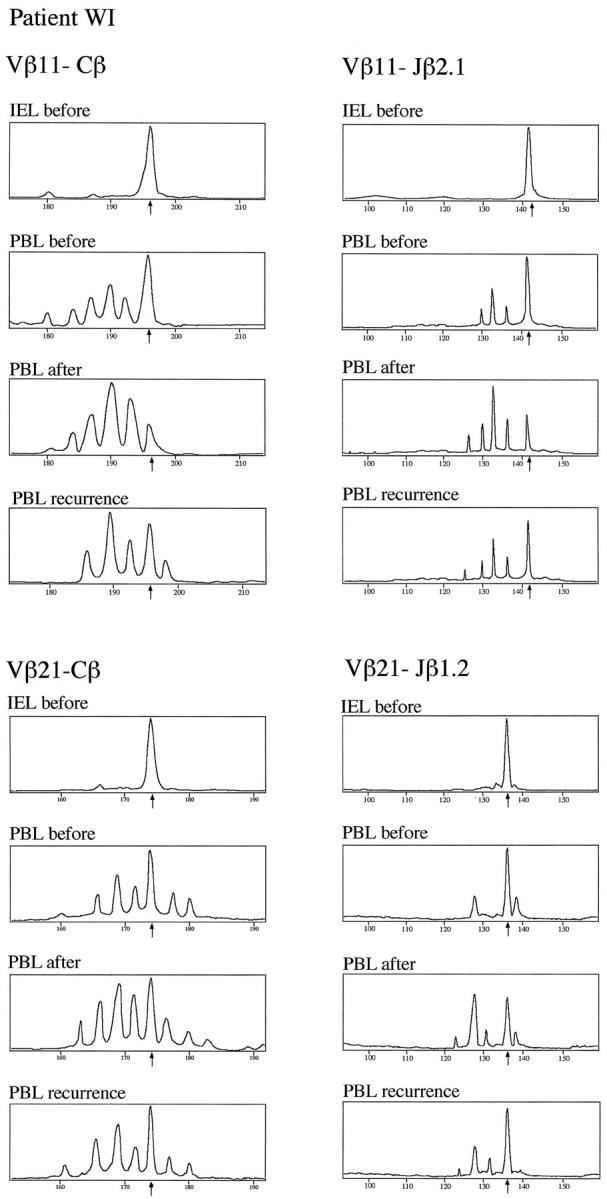
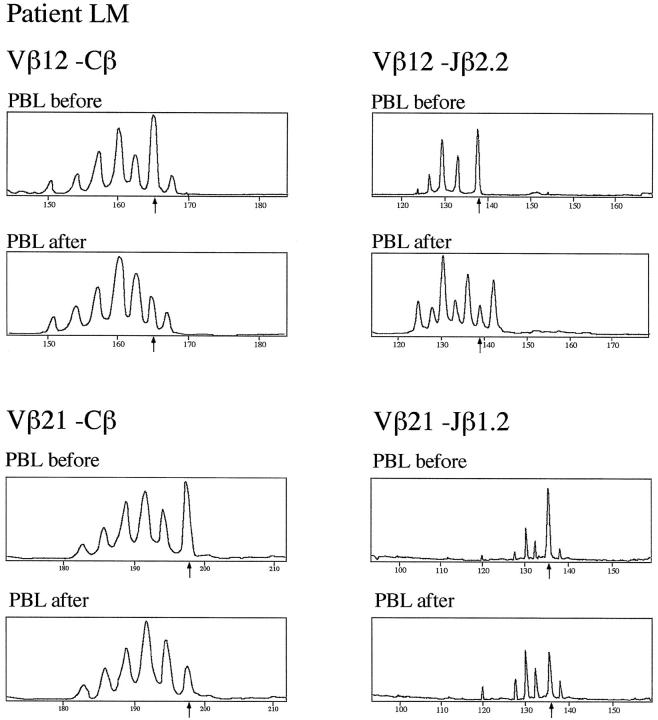
Deficiency of CD95 (Apo-1/Fas)-mediated apoptosis has recently been found in some autoimmune lymphoproliferative disorders due to inherited mutations of the CD95 gene. In this study, impairment of CD95 ligand-mediated killing of lymphocytes and eosinophils in Churg-Strauss Syndrome (CSS), which was a result of variation of CD95 receptor isoform expression, is demonstrated. Compared to those from healthy individuals, peripheral blood lymphocytes from eight CSS patients exhibit a switch from the membrane-bound CD95 receptor expression to its soluble splice variant, which protects from CD95L-mediated apoptosis. In five out of seven CSS patients recurrent oligoclonal T cell expansions were found, all using a Vbeta-gene from the Vbeta21 family associated with similar CDR3 motifs, indicating the predominance of T cell clones of a similar specificity in the CSS patients. In two of them, the effect of immunosuppressive therapy was studied. In both cases aberrant overexpression of the soluble CD95 receptor isoform and deviations from normal TCR Vbeta-gene usage normalized in parallel with the clinical improvement. Furthermore, soluble CD95 was identified as a survival factor for eosinophils rescuing eosinophils from apoptosis in the absence of growth factors in vitro. Given the role of eosinophils as effector cells in CSS, these findings suggest that soluble CD95 may be mechanistically involved in the disease.


Figures

Figure 1.
Figure 1.
A: Immunoscope-based analysis of the clonality of TCRβ VDJ-transcripts. T cell receptor β (TCRβ) transcripts are reverse transcribed and amplified using a panel of 24 Vβ- and Cβ-specific primers. Thus, the first step of the Immunoscope technique involves 24 RT-PCR reactions run to saturation (top). In an initial low-resolution analysis, a dye labeled Cβ-specific primer is used to visualize the amplified products in run-off reactions (middle). If higher resolution is required, run-off experiments are carried out using 13 dye-labeled Jβ-specific primers in theoretically 24 × 13 = 312 run-off reactions (bottom). After electrophoresis on an automated sequencer and subsequent analysis, the different size peaks are separated and their CDR3 size in amino acids (aa) and area are calculated. B: Quantification of CD95 ligand and receptor isoform mRNA levels by quantitative competitive RT-PCR. The cDNA to be assayed (WT) was co-amplified with known amounts (108, 107, … 10 copies) of an internal DNA standard (Δ4), which was apart from a deletion of four nucleotides identical to the corresponding fragment of the assayed cDNA. PCR products were specifically labeled in run-off reactions, loaded on an acrylamide gel, and analyzed by an automated sequencer. The fluorescent profiles were recorded and the profile areas were analyzed. For co-amplifications with 10 and 10 copies of the CD95L standard, respectively, the peak area ratios for CD95L wild-type (CD95L WT) and standard (CD95L Δ4) were calculated. The number of CD95L WT copies in the cDNA sample was calculated as the mean of CD95L WT/CD95L Δ4 peak area ratios at two standard dilutions (eg, for the sample shown here: (0.262 × 10⁶ + 2.931 × 10⁵)/2, or 277,550 copies).
Figure 2.
Figure 2.
Preferential usage of distinct TCR-Vβ and Jβ genes in T cells from five CSS patients. Fluorescent profiles of the Vβ21-Jβ1.2 rearrangements are shown in samples from the peripheral blood of one healthy donor and five CSS patients (WI, VN, GM, LM, and SJ). The peripheral blood samples of these five CSS patients contained clonally expanded T cells carrying a gene from the Vβ21 gene family rearranged to the Jβ1.2 gene with a dominant CDR3-size peak corresponding to a CDR3 of 11 aa in length (arrows).
Figure 3.
Figure 3.
T cell repertoire diversity and clonal expansions in gastric IEL and PBL from Churg-Strauss Syndrome patient WI. Using the Immunoscope RT-PCR technique, TCRVβ chain segments in IEL and PBL from patient WI were amplified using a panel of 24 Vβ family-specific PCR primers. Clonal expansions were detected in several Vβ-Cβ profiles. Clonality of Vβ11- and Vβ21-specific transcripts is shown here. Samples were analyzed from infiltrated ulcerative gastric mucosa (IEL before) and the peripheral blood before (PBL before) and after immunosuppressive treatment (PBL after) as well as after recurrence of CSS (PBL recurrence). The peak sizes of clonal expansions are indicated by arrows. Clonality of the Vβ-Cβ profiles was further analyzed using Jβ-specific primers. For both Vβ-Cβ profiles, each one of 13 Vβ-Jβ recombinations was selected to demonstrate the strongest deviation from normal polyclonal gaussian-like peak size distribution, ie, Vβ11-Jβ2.1 for the Vβ11 gene family and Vβ21-Jβ1.2 for the Vβ21 gene family.
Figure 4.
Figure 4.
T-cell repertoire diversity and clonal expansions in PBL from Churg-Strauss syndrome patient LM. Using the Immunoscope RT-PCR technique, TCRVβ chain segments in PBL from patient LM was amplified using a panel of 24 Vβ subfamily-specific PCR primers. Clonal expansions was detected in several Vβ-Cβ profiles. Clonality of Vβ12- and Vβ21-specific transcripts is shown here. Samples were analyzed from the peripheral blood prior to (PBL before) and after immunosuppressive treatment (PBL after). Clonal expansions are indicated by arrows. Clonality of the Vβ-Cβ profiles was confirmed using Jβ-specific primers. For both Vβ-Cβ profiles, each of 13 Vβ-Jβ recombinations was selected to demonstrate the strongest deviation from normal polyclonal gaussian-like peak size distribution, ie, Vβ12-Jβ2.2 for the Vβ12 family and Vβ21-Jβ1.2 for the Vβ21 family.
Figure 5.
Figure 5.
Sensitivity of PBL to CD95-mediated apoptosis. Lymphocytes were isolated from the peripheral blood from four healthy donors (filled circles) and from five CSS-patients (open circles). After treatment with PHA (2.4 μg/ml) for 24 hours, the lymphocytes were incubated with an agonistic anti-CD95 antibody at various concentrations. After another 24 hours, the lymphocytes were subjected to TUNEL analysis as described in Materials and Methods and percentages of apoptotic lymphocytes were counted. Data are given as means ± SE.
Figure 6.
Figure 6.
Distribution of CD95 receptor isoform mRNA expression in peripheral blood T cells (PBL), gastric intraepithelial T cells (IEL) and eosinophils. A: cDNAs derived from PBL of one healthy donor, patient WI 1 day prior to (CSS, day −1) and 14 days after gastrectomy and immunosuppressive treatment (CSS, day 14) were amplified using CD95 isoform- and HPRT-specific primers as described in Materials and Methods in 30 PCR cycles. Amplification products for membrane-bound CD95 (CD95Tm, 392 bp), soluble CD95 (CD95Sol, 330 bp), and HPRT (237 bp) were separated on a 2% agarose gel. B: CD95 membrane-bound and soluble isoform mRNA expression was analyzed in eosinophils from healthy donors (representative for three donors), one patient with infectious eosinophilia (patient DA, 25% eosinophilic counts), and one CSS patient (patient PF, 32% eosinophilic counts). C: CD95 isoform mRNA expression in ulcerative and non-ulcerative gastric mucosa was studied in samples from gastric antrum and corpus of patient WI at the time of gastrectomy.
Figure 7.
Figure 7.
Identification of T cells as CD95L-expressing cells in ulcerative gastric mucosa in patient WI. Cryosections from ulcerative gastric mucosa from patient WI were double-stained using CD3-specific and CD95L-specific antibodies as described in Materials and Methods. In A and B, one cryosection from infiltrated muscularis propria and in C and D one from infiltrated mucosa is shown. Double fluorescence staining for CD3 and CD95L was performed on the same cryosection. In A and C, signals are specific for CD3 (FITC-labeled), in B and D, for CD95L expression (CY3-labeled). CD3-expressing cells (ie, T lymphocytes) and CD95L expression were colocalized (arrows).

References

    1. Churg J, Strauss L: Allergic granulomatosis. Allergic angiitis, and periarteriitis nodosa. Am J Pathol 1951, 27:277-301 - PMC - PubMed
    1. Modigliani R, Muschart JM, Galian A, Clauvel JP, Desruisseaux JL: Allergic granulomatous vasculitis: report of a case with digestive involvement. Dig Dis Sci 1981, 26:264-270 - PubMed
    1. Masi AT, Hunder GG, Lie JT, Michel BA, Bloch DA, Arend WP, Calabrese LH, Edworthy SM, Fauci AS, Leavitt RY: The American College of Rheumatology 1990 criteria for the classification of Churg-Strauss syndrome. Arthritis Rheum 1990, 33:1094-1102 - PubMed
    1. Lanham GJ, Elkon KB, Pusey CD, Hughes GR: System vasculitis with asthma and eosinophilia: a clinical approach to the Churg-Strauss syndrome. Medicine 1984, 63:65-81 - PubMed
    1. Schmitt WH, Csernok E, Kobayashi S, Klinkenborg A, Reinhold-Keller E, Gross WL: Churg-Strauss syndrome: serum markers of lymphocyte activation and endothelial damage. Arthritis Rheum 1998, 41:445-452 - PubMed

MeSH terms