. 1999 Sep;155(3):841-51.
doi: 10.1016/s0002-9440(10)65183-8.

Activation of rat alveolar macrophage-derived latent transforming growth factor beta-1 by plasmin requires interaction with thrombospondin-1 and its cell surface receptor, CD36

Affiliations

Activation of rat alveolar macrophage-derived latent transforming growth factor beta-1 by plasmin requires interaction with thrombospondin-1 and its cell surface receptor, CD36

T Yehualaeshet et al. Am J Pathol. 1999 Sep.

Abstract

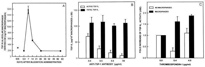
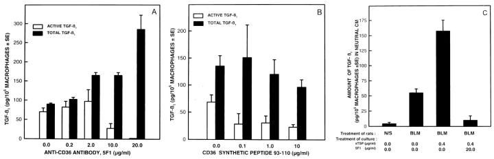
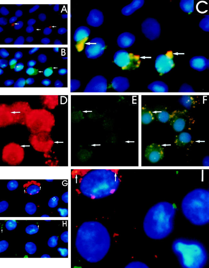
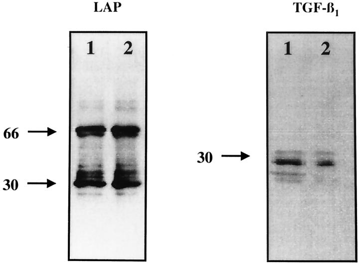
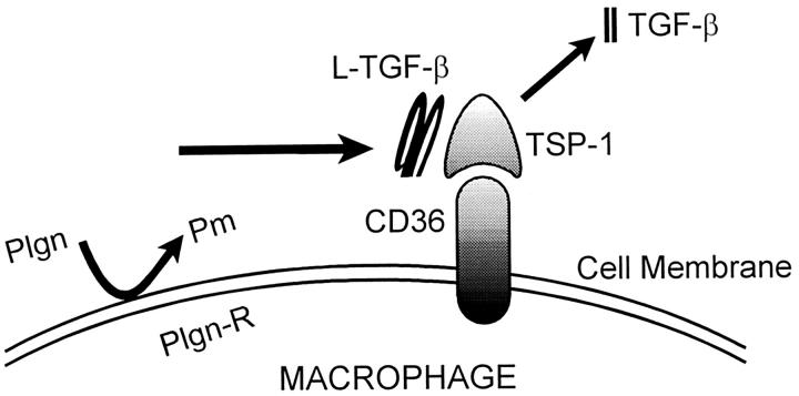
Transforming growth factor-beta-1 (TGF-beta1) is secreted by cells in a latent form (L-TGF-beta1) noncovalently bound to a latency-associated peptide. Activated alveolar macrophages obtained from rat lungs after bleomycin-induced pulmonary injury released increased amounts of active TGF-beta1 as well as plasmin, a protease, and thrombospondin-1 (TSP-1), a trimeric glycoprotein. Previously we had demonstrated that plasmin was critical to the activation of L-TGF- beta1. In the present study we demonstrated that TSP-1 is also important for the activation of L-TGF- beta1 because the activation can be inhibited by anti-TSP-1 monoclonal antibody. Proteins obtained from alveolar macrophage cell lysates immunoprecipitated with antibodies specific for TSP-1 were identified on immunoblots as LAP and TGF-beta1, indicating that TSP-1/L-TGF-beta1 complexes are present on alveolar macrophages. However, in the presence of plasmin both latency-associated peptide and TGF-beta1 were decreased in the same cell lysates, indicating that L-TGF-beta1 associated with TSP-1 is released by plasmin. Using immunofluorescence and antibodies to TGF-beta1 and CD36, a receptor for TSP-1, there was colocalization of TGF-beta1 with CD36. Because TSP-1 but not TGF-beta1 is a natural ligand for CD36, these findings suggest that the L-TGF-beta1 in a complex with TSP-1 localizes to the macrophage cell surface when TSP-1 interacts with its receptor, CD36. Furthermore, the association of TSP-1/L-TGF-beta1 complex with CD36 is necessary to the activation of L-TGF-beta1 because antibodies to CD36 prevent the colocalization of TGF-beta1 with CD36 as observed by immunofluorescence and inhibit activation of the L-TGF-beta1 by explanted alveolar macrophages. These findings suggest that activation of L-TGF-beta1 by plasmin occurs at the cell surface of activated alveolar macrophages and requires a TSP-1/CD36 interaction.


Figures

Figure 1.
Figure 1.
Activation of alveolar macrophage derived L-TGF-β1 by sTSP-1. A: Quantity of TSP-1 secreted by explanted alveolar macrophages obtained from rats at varying lengths of time after bleomycin administration. Each point is the mean of samples from 3 to 6 rats. The P value is <0.0003 when the quantity of TSP-1 produced by alveolar macrophages from normal saline-treated rats is compared to TSP-1 produced by alveolar macrophages from rats treated with bleomycin 7 days earlier. B: The effects of anti-TSP-1 antibody on the activation of alveolar macrophage-derived L-TGF-β1. Alveolar macrophages obtained 7 days after bleomycin administration were cultured in the absence of anti-TSP-1 antibody or in the presence of varying concentrations. The TGF-β1 present in neutral CM (□) represents bioactive TGF-β1; that present in acidified then neutralized CM (▪) represents the total TGF-β1 in the same sample. Each point is the mean of samples from 6 rats. The quantity of TGF-β1 in neutral CM compared to the quantity of TGF-β1 in acidified CM has a P ≤ 0.001 when the anti-TSP-1 antibody used was 50 μg/ml/10 macrophages, and P ≤ 0.003 when the anti-TSP-1 antibody used was 5.0 or 0.5 μg/ml/10 macrophages. There was no statistical difference in the quantity of TGF-β1 in neutral and acidified CM when no anti-TSP-1 antibody was present. C: Induction of activation of L-TGF-β1 by addition of sTSP-1. Alveolar macrophages were obtained 7 days after bleomycin administration and cultured for 20 hours before the addition of sTSP-1 (0–4 μg/ml) to alveolar macrophages (▪) or the CM (□) from parallel cultures of alveolar macrophages. After a 2-hour incubation with sTSP-1, the TGF- β1 in neutral and acidified CM (not shown) of the same sample was determined. The -fold increase in the quantity of TGF-β1 present was calculated by using the quantity of active TGF-β1 present in the absence of sTSP-1 as the denominator and the quantity of TGF-β1 present after the addition of sTSP-1 as the numerator. Each point is the mean of samples from 3 rats. The -fold increase in the quantity of TGF-β1 in neutral CM when alveolar macrophages were treated with 0.4 or 4.0 μg/ml of sTSP-1, compared to TGF-β1 in CM when no sTSP-1 was present, has P value ≤ 0.05.
Figure 2.
Figure 2.
Effects of anti-CD36 antibody on the activation of L-TGF-β1. Alveolar macrophages obtained 7 days after bleomycin administration were cultured in the absence or presence of different concentrations of anti-CD36 antibody or the CD36 synthetic peptide 93–110. A: The TGF-β1 present in neutral CM □ and acidified then neutralized CM ▪ from the same sample was quantitated. Each point is the mean of samples from 4 rats. The quantity of TGF-β1 present in neutral CM compared to TGF-β1 in acidified CM has a P value ≤0.0001 when the antibody was 20 μg/ml/10 macrophages, and P ≤ 0.01 when the antibody was 10 μg/ml/10 macrophages. The other comparisons between TGF-β1 in neutral and acidified CM were not significant. B: The effects of CD36 synthetic peptide aa 93–110 on the activation of L-TGF-β1. Alveolar macrophages were cultured in the absence or presence of several concentrations of the synthetic peptide of CD36 aa 93–110. TGF-β1 in neutral □ and acidified, then neutralized CM ▪ was quantitated. All points are the mean of 4 to 6 animals. The quantity of TGF-β1 in neutral CM, compared to the TGF-β1 in acidified CM of the same sample, had a P value ≤0.0006 only when CD36 peptide 93–110 was present. C: Effects of anti-CD36 antibody on the activation of L-TGF-β1 in the presence of sTSP-1. Alveolar macrophages were cultured in the absence or presence of 20 μg/ml/10 macrophages of anti-CD36 antibody, and in the absence or presence of 0.4 μg/ml/10 macrophages of sTSP-1. The TGF- β1 present in neutral CM was quantitated. Each point is the mean of samples from 4–6 rats. TGF-β1 in neutral CM of alveolar macrophages from bleomycin (BLM)-treated rats compared to normal saline (N/S)-treated rats has a P value ≤0.07. TGF-β1 in neutral CM of alveolar macrophages from BLM-treated rats cultured in the absence of sTSP-1, compared to when sTSP-1 was present, has a P value ≤0.02. TGF-β1 in neutral CM of alveolar macrophages from BLM treated rats cultured with sTSP-1, compared to when sTSP-1 and anti-CD36 antibody was also present, has a P value ≤0.006.
Figure 3.
Figure 3.
Localization of TGF-β1 and CD36 on the alveolar macrophage using immunofluoresence. Alveolar macrophages obtained 7 days after normal saline administration treated with (A) polyclonal anti-rabbit lgG antibody conjugated with TRITC reacts with cell surface-associated anti-TGF-β1 primary antibody, and the location is seen as a red coloration (arrow). B: Monoclonal anti-mouse lgM antibody conjugated with FITC reacts with cell surface-associated anti-CD36 primary antibody; the location is seen as a green coloration (arrow). C: Immunofluorescent images obtained from the above observations were used to generate a pixel overlay of the images resulting in a yellow coloration (arrow) and demonstrating the regions of colocalization of TGF-β1 and CD36. Alveolar macrophages obtained 7 after bleomycin administration and treated as described above identify (D) TGF-β1 seen as a red coloration (arrow); (E) CD36 seen as a green coloration (arrow). F: Immunofluorescent images obtained from the above observations were used to generate a pixel overlay of images resulting in a yellow coloration (arrow). Alveolar macrophages obtained 7 days after bleomycin administration treated in the same manner as described above but incubated with anti-CD36 antibody before cell surface localization of (G) TGF-β1 and (H) CD36. A pixel overlay of the images demonstrates either no areas of colocalization of TGF-β1 and CD36 or markedly decreased areas of colocalization (arrows) on a few cells (I).
Figure 4.
Figure 4.
Immunoblot analysis of LAP and TGF-β1 antigens obtained from immunoprecipitates using anti-sTSP-1 antibody (mAb 133) and lysates of alveolar macrophages. Alveolar macrophages obtained 7 days after treatment with either normal saline (lane 1) or bleomycin (lane 2) were immunoblotted using anti-LAP antibodies and demonstrate protein bands compatible with the dimer (68 kd) and monomer (34 kd) of LAP. The same nitrocellulose filter was reprobed for immunoblotting with anti-TGF-β1 antibodies and demonstrate the presence of TGF-β1 protein as a dimer (25 kd) and monomer in samples obtained after normal saline (lane 1) and primarily as a dimer after bleomycin treatment (lane 2). The immunoblots are representative of experiments done 6 times. The numbers on the left denote the molecular weight of the bands in kilodaltons.
Figure 5.
Figure 5.
The effects of plasmin inhibitors on the expression of LAP, TGF-β1 and activation of L-TGF-β1. Immunoblot analysis of LAP and TGF-β1 antigens obtained from immunoprecipitates using anti-TSP-1 antibody (mAb133) and lysates of alveolar macrophages cultured in the absence or presence of α2-antiplasmin or aprotinin. A, top: Alveolar macrophages obtained 7 days after bleomycin administration cultured in the absence (lane 1) or presence of α2-antiplasmin at 10−4 units/ml (lane 2), α2-antiplasmin 10−1 unit/ml (lane 3), or aprotinin 0.01 μg/μm l (lane 4) were immunoblotted with anti-LAP antibodies and demonstrate protein bands compatible with L-TGF-β (100 kd) and the dimer (68 kd) of LAP. A, bottom: The same nitrocellulose filter was reprobed for immunoblotting with anti-TGF-β1 antibodies and demonstrates the dimer (25 kd) of TGF-β1. The culture conditions of alveolar macrophages used for lanes 1–4 are described above. The immunoblots are representative of experiments done 3 times. The numbers on the left denote the molecular weight of the bands in kilodaltons. B: Effects of aprotinin on the activation of L-TGF-β1 in the presence of sTSP-1. Alveolar macrophages obtained 7 days after bleomycin administration were cultured in the absence or presence of 0.01 μg/μm l/10 macrophages of aprotinin and/or in the absence or presence of 0.4 μg/ml/10 macrophages of sTSP-1. The TGF-β1 present in neutral and acidified then neutralized CM was quantitated and the percentage of active TGF-β1 was calculated. Each point is the mean of 3 to 7 rats. Percentage of active TGF-β1 in CM of alveolar macrophages from BLM-treated rats compared to normal saline N/S-treated rats or the CM of alveolar macrophages obtained after BLM treatment cultured with aprotinin have a P value ≤0.0001. Percentage of active TGF-β1 in CM of alveolar macrophages cultured in the presence of sTSP-1 compared to when no TSP-1 or aprotinin were present has a P value ≤0.0004. Percentage of active TGF-β1 in CM of alveolar macrophages obtained after BLM treatment and no in vitro treatment, compared to when sTSP-1 and aprotinin was also present, has a P value ≤0.0976. Percentage of active TGF-β1 in CM of alveolar macrophages cultured with sTSP-1 compared to when sTSP-1 and aprotinin were present has a P value <0.0001.
Figure 6.
Figure 6.
Proposed model for the activation of alveolar macrophage-derived L-TGF-β1. Resting alveolar macrophages secrete small amounts of L-TGF-β1 and TSP-1 but no plasmin. CD36 and TGF-β1 are present in small quantities on the cell surface, but no active TGF-β1 is produced. After bleomycin-induced lung injury, the alveolar macrophages are activated to secrete increased quantities of L-TGF-β1 and TSP-1 and generate increased quantities of plasmin. TSP-1 associates with the alveolar macrophage-derived L-TGF-β1 released in the immediate vicinity of the macrophage. The TSP-1/L-TGF-β1 complex then associates with the cell surface of the alveolar macrophage by the CD36 receptor. After association of CD36 with the TSP-1/L-TGF-β1 complex, the plasmin generated by the macrophages releases the TGF-β1 from the LAP. TGF-β1 is then available to react with its receptor and have a biological effect. Plgn, plasminogen; pm, plasmin; Plgn-R, plasminogen receptor.

Similar articles

Cited by

References

    1. Rappolee DA, Werb Z: Macrophage derived growth factors. Curr Topics Microbiol Immumol 1992, 181:87-140 - PubMed
    1. Stein M, Keshav A: The versatility of macrophages. Clin Exp Allergy 1992, 22:19-27 - PubMed
    1. Wahl SM: Transforming growth factor β: the good, the bad and the ugly. J Exp Med 1994, 108:1587-1590 - PMC - PubMed
    1. Gleizes PE, Munger JS, Nunes I, Harpel JG, Mazzieri R, Noguera S, Rifkin DB: TGF-β latency: biological significance and mechanism of activation. Stem Cells 1997, 15:190-197 - PubMed
    1. Khalil N, Corne S, Whitman C, Yacyshyn H: Plasmin regulates the activation of cell associated latent TGF-β1 secreted by rat alveolar macrophages after in vivo bleomycin injury. Am J Resp Mol Cell Biol 1996, 15:252-259 - PubMed

Publication types

MeSH terms