

Detection of prosthetic hip infection at revision arthroplasty by immunofluorescence microscopy and PCR amplification of the bacterial 16S rRNA gene
- PMID: 10488193
- PMCID: PMC85548
- DOI: 10.1128/JCM.37.10.3281-3290.1999
Detection of prosthetic hip infection at revision arthroplasty by immunofluorescence microscopy and PCR amplification of the bacterial 16S rRNA gene
Abstract
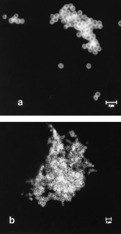
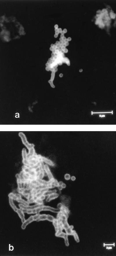
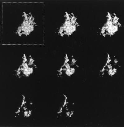
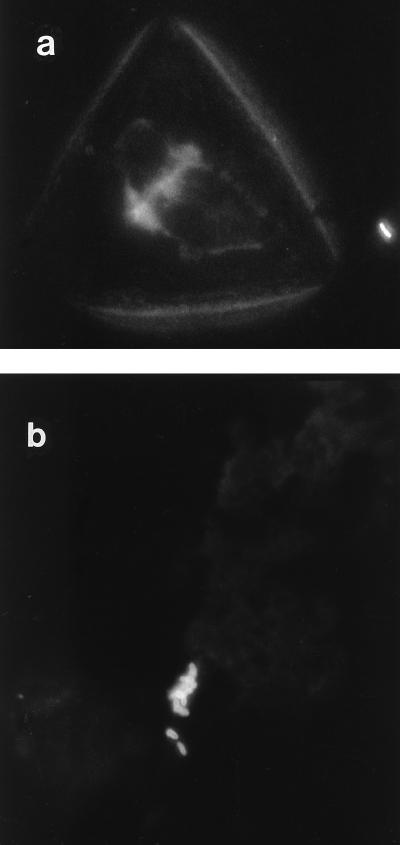
In this study the detection rates of bacterial infection of hip prostheses by culture and nonculture methods were compared for 120 patients with total hip revision surgery. By use of strict anaerobic bacteriological practice during the processing of samples and without enrichment, the incidence of infection by culture of material dislodged from retrieved prostheses after ultrasonication (sonicate) was 22%. Bacteria were observed by immunofluorescence microscopy in 63% of sonicate samples with a monoclonal antibody specific for Propionibacterium acnes and polyclonal antiserum specific for Staphylococcus spp. The bacteria were present either as single cells or in aggregates of up to 300 bacterial cells. These aggregates were not observed without sonication to dislodge the biofilm. Bacteria were observed in all of the culture-positive samples, and in some cases in which only one type of bacterium was identified by culture, both coccoid and coryneform bacteria were observed by immunofluorescence microscopy. Bacteria from skin-flake contamination were readily distinguishable from infecting bacteria by immunofluorescence microscopy. Examination of skin scrapings did not reveal large aggregates of bacteria but did reveal skin cells. These were not observed in the sonicates. Bacterial DNA was detected in 72% of sonicate samples by PCR amplification of a region of the bacterial 16S rRNA gene with universal primers. All of the culture-positive samples were also positive for bacterial DNA. Evidence of high-level infiltration either of neutrophils or of lymphocytes or macrophages into associated tissue was observed in 73% of patients. Our results indicate that the incidence of prosthetic joint infection is grossly underestimated by current culture detection methods. It is therefore imperative that current clinical practice with regard to the detection and subsequent treatment of prosthetic joint infection be reassessed in the light of these results.
Figures
References
-
- Athanasou N A, Pandey R, DeSteiger R, Crook D, McLardy Smith P. Diagnosis of infection by frozen section during revision arthroplasty. J Bone Jt Surg Br Vol. 1995;77:28–33. - PubMed
-
- Atkins B L, Athanasou N A, Deeks J J, Crook D W M, Simpson H, Peto T E A, McLardy-Smith P, Berendt A R The Osiris Collaborative Study Group. Prospective evaluation of criteria for microbiological diagnosis of prosthetic-joint infection at revision arthroplasty. J Clin Microbiol. 1998;36:2932–2939. - PMC - PubMed
-
- Barrack R L, Harris W H. The value of aspiration of the hip joint before revision total hip arthroplasty. J Bone Jt Surg Am Vol. 1993;75:66–76. - PubMed
-
- Canvin J M G, Goutcher S C, Hagig M, Gemmell C G, Sturrock R D. Persistence of Staphylococcus aureus as detected by polymerase chain reaction in the synovial fluid of a patient with septic arthritis. Br J Rheumatol. 1997;36:203–206. - PubMed
Publication types
MeSH terms
Substances
LinkOut - more resources
Full Text Sources
Other Literature Sources
Medical
Molecular Biology Databases
Miscellaneous
