. 1999 Oct;37(10):3338-47.
doi: 10.1128/JCM.37.10.3338-3347.1999.

Two sensitive PCR-based methods for detection of hepatitis B virus variants associated with reduced susceptibility to lamivudine

Affiliations

Two sensitive PCR-based methods for detection of hepatitis B virus variants associated with reduced susceptibility to lamivudine

M I Allen et al. J Clin Microbiol. 1999 Oct.

Abstract

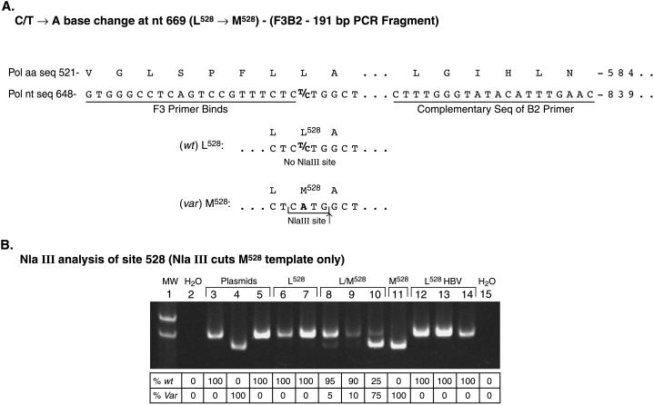
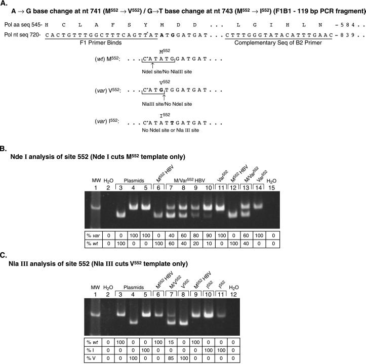
Two novel assays, a restriction fragment length polymorphism (RFLP) assay and an assay based on the 5'-nuclease activity of Taq DNA polymerase, were developed for screening viral variants in lamivudine-treated patients' sera containing <1,000 copies of the hepatitis B virus (HBV) genome per ml. Both assays were designed to detect single-nucleotide changes within the HBV DNA polymerase gene that are associated with lamivudine resistance in vitro and have been used to screen a number of patients' sera for variant virus. Results obtained with these assays and standard sequencing technology were compared with regard to throughput, ability to detect individual virus species present at low concentrations, and ability to detect, distinguish, and quantitate wild-type (wt) and HBV tyrosine methionine(552) aspartate aspartate motif variants in mixed viral populations. Unlike DNA sequencing, both assays are amenable to high-throughput screening and were shown to be able to quantitatively detect variant virus in the presence of a background of wt virus. As with DNA sequencing, both new assays incorporate a PCR amplification step and are able to detect the relatively low amounts of virus found in lamivudine-treated patients' sera. However, these assays are far less labor intensive than the DNA-sequencing techniques presently in use. Overall, the RFLP assay was more sensitive than DNA sequencing in detecting and determining the ratios of wt to variant virus. Furthermore, the RFLP assay and 5'-nuclease assay were equally sensitive in the detection of mixed viral species, but the RFLP assay was superior to the 5'-nuclease assay in the quantitation of mixed viral species. These assays should prove useful for further understanding of virological response to therapy and disease progression.


Figures

FIG. 1
FIG. 1
Design and gel image of an RFLP assay for rapid detection of variant HBV at codon 528. (A) PCR primers designed to amplify the region of codon 528 (primers F3 and B2) have been used to detect the presence of the methionine codon at 528. A methionine at codon 528 creates an NlaIII restriction site (CATG↓), whereas the NlaIII site is absent in the wt sequence. (B) Result of sample analysis at codon 528 after NlaIII enzyme digestion. HBV DNA containing either the wt (Leu), variant (Met), or both sequences at codon 528 can be determined by PCR amplification with primers F3 and B2, digestion of PCR products with NlaIII, and resolution of DNA band size on a 6% polyacrylamide gel. The faster migrating band indicates the variant sequence, whereas the slower migrating band indicates the wt sequence. Lane 1, molecular weight markers; lane 2, water (negative control); lane 3, wt pCMVHBV plasmid; lane 4, L528M/M552V pCMVHBV plasmid; lane 5, M552I pCMVHBV plasmid; lanes 6 to 14, HBV DNAs from sera of different patients on lamivudine therapy that are either wt at codon 528, L528M, or both; lane 15, water. The amount of each DNA present in lanes (expressed as a percentage) is shown below the image and was quantitated as described in Materials and Methods. Var, variant.
FIG. 2
FIG. 2
Design and gel image of an RFLP assay for rapid detection of HBV variant at codon 552. (A) For detecting variations at codon 552 in the HBV polymerase, the YMDD nucleotide sequence TATATGGATGAT was used, and a 5′-PCR primer (primer F1) was designed that contains a CAT sequence at the 3′ end (changing the first tyrosine codon from a TAT to a CAT) in order to introduce an NdeI restriction site (CA↓TATG) upon amplification of part of the HBV polymerase gene with the 5′-PCR primer and a 3′-PCR primer located 120 bases downstream. The creation of an NdeI site occurs only if the template HBV has the wt DNA sequence and is absent if the template HBV is mutated, indicating the loss of the NdeI site only. (B) Result of sample analysis at codon 552 after NdeI enzyme digestion. Resolution of PCR products on a polyacrylamide gel after amplification of template HBV DNA using primers F1 and B2 with subsequent digestion using NdeI indicates either wt sequence (faster moving band) or variant sequence (slower moving band). Lane 1, molecular weight markers; lane 2, water (negative control); lane 3, wt pCMVHBV plasmid; lane 4, L528M/M552V pCMVHBV plasmid; lane 5, M552I pCMVHBV plasmid; lanes 6 to 11, HBV DNA from sera of different patients undergoing lamivudine therapy that are either wt at codon 552, M552V, or both; lanes 12 to 14, HBV DNA from sera of different patients on lamivudine therapy that are wt at codon 552, M552I, or both; lane 15, water. The amount of each DNA present in lanes (expressed as a percentage) is shown below the image and was quantitated as described in Materials and Methods. var, variant. (C) After determining the sequence (wt or variant) of a sample from results of the NdeI analysis shown in Fig. 2B, the sequence of the variant template (Val or Ile) can be determined by NlaIII enzyme digestion of the PCR products. Resolution of the PCR products on a polyacrylamide gel after amplification of template HBV DNA using primers F1 and B2 with subsequent digestion using NlaIII indicates either a valine sequence (faster moving band) or methionine or isoleucine sequence (slower moving band) at codon 552. Lane 1, molecular weight markers; lane 2, water (negative control); lane 3, wt pCMVHBV plasmid; lane 4, L528M/M552V pCMVHBV plasmid; lane 5, M552I pCMVHBV plasmid; lanes 6 to 8, HBV DNA from sera of different patients undergoing lamivudine therapy that are either wt at codon 552, M552V, or both; lanes 9 to 11, HBV DNA from sera of different patients undergoing lamivudine therapy that are wt at codon 552, M552I, or both; lane 15, water. The amount of each DNA present in lanes (expressed as a percentage) is shown below the image and was quantitated as described in Materials and Methods.

References

    1. Allen M I, DesLauriers M, Andrews C W, Tipples G A, Walters K-A, Tyrrell D L J, Brown N, Condreay L D. Identification and characterization of mutations in hepatitis B virus resistant to lamivudine. Hepatology. 1998;27:1670–1677. - PubMed
    1. Atkins M, Hunt C M, Brown N, Gray F, Sanathanan L, Woessner M, Lai C L, Dusheiko G, Dienstag J, Wright T, Barnard J, Bourne E, Condreay L. Clinical significance of YMDD mutant hepatitis B virus (HBV) in a large cohort of lamivudine-treated hepatitis B patients, abstr. 625. Hepatology. 1998;28:319.
    1. Bartholomeusz A, Locarnini S A. Mutations in the hepatitis B virus polymerase gene that are associated with resistance to famciclovir and lamivudine. Int Antiviral News. 1997;5:123–124.
    1. Bartholomew M M, Jansen R W, Jeffers L J, Reddy K R, Johnson L C, Bunzendahl H, Condreay L D, Tzakis A G, Schiff E R, Brown N A. Hepatitis B virus resistant to lamivudine given for recurrent infection after orthotopic liver transplantation. Lancet. 1997;349:20–22. - PubMed
    1. Bassler H A, Flood S J A, Livak K J, Marmaro J, Knorr R, Batt C A. Use of a fluorogenic probe in a PCR-based assay for the detection of Listeria monocytogenes. Appl Environ Microbiol. 1995;61:3724–3728. - PMC - PubMed

Publication types

MeSH terms