. 1999 Oct;19(10):6471-8.
doi: 10.1128/MCB.19.10.6471.

Identification of a novel dexamethasone-sensitive RNA-destabilizing region on rat monocyte chemoattractant protein 1 mRNA

Affiliations

Identification of a novel dexamethasone-sensitive RNA-destabilizing region on rat monocyte chemoattractant protein 1 mRNA

M Poon et al. Mol Cell Biol. 1999 Oct.

Abstract

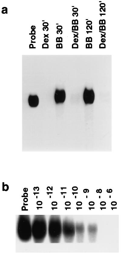
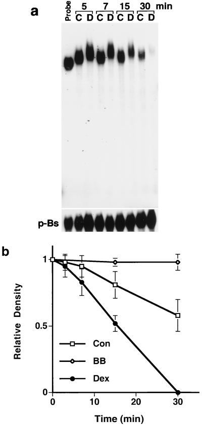
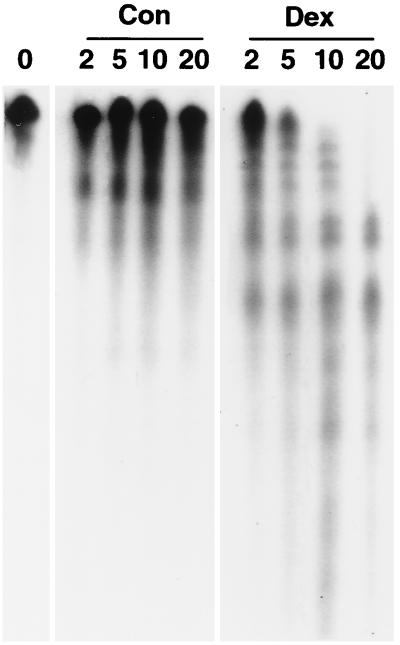
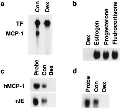
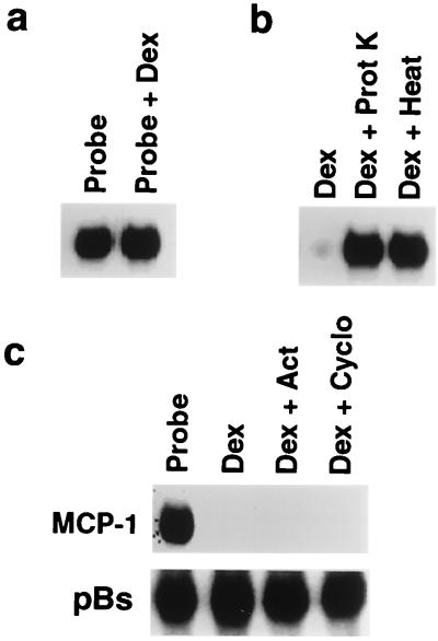
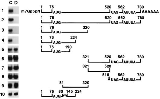
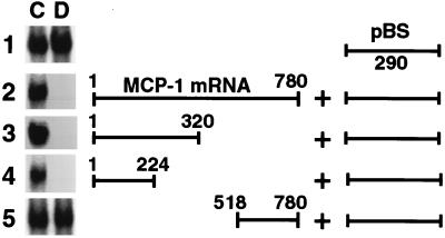
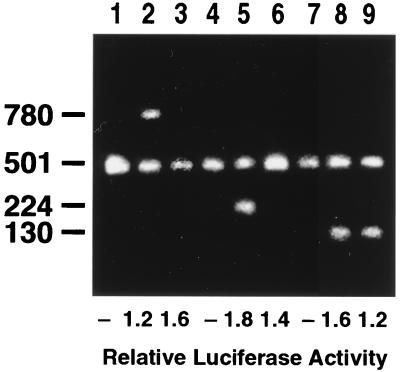
Glucocorticoids are potent anti-inflammatory agents widely used in the treatment of human disease. We have previously shown that the inflammatory cytokine monocyte chemoattractant protein 1 (MCP-1) is regulated posttranscriptionally by glucocorticoids in arterial smooth muscle cells (SMC). To elucidate the mechanism mediating this effect, in vitro-transcribed radiolabeled MCP-1 mRNA was incubated with cytoplasmic extracts from SMC and analyzed by gel electrophoresis. Extracts from SMC treated with platelet-derived growth factor (PDGF) did not degrade the transcripts for up to 3 h. In contrast, extracts from cells treated with 1 microM dexamethasone (Dex) alone or in combination with PDGF degraded the probe with a half-life of approximately 15 min. Dex had maximal effect at concentrations above 0.01 microM and was effective on both rat and human MCP-1 transcripts. By deletion analysis, the Dex-sensitive region of the MCP-1 mRNA was localized to the initial 224 nucleotides (nt) at the 5' end and did not involve an AU-rich sequence in the 3' untranslated end. The 224-nt region conferred Dex sensitivity to heterologous mRNA. These studies provide new insights into the molecular mechanisms underlying the effect of glucocorticoids on gene expression.


Figures

FIG. 1
FIG. 1
Dex mediated decay of MCP-1 mRNA in rat aortic SMC. (a) Cytoplasmic (S-100) extracts were isolated from SMC treated for 3 h with 1 μM Dex (Dex), 20 ng of PDGF BB (BB) per ml, or both (Dex/BB). 32P-labeled, in vitro-transcribed, full-length MCP-1 mRNA (Probe) was incubated with extracts for the times indicated and then analyzed by PAGE. (b) Cytoplasmic extracts were isolated from SMC treated for 3 h with various concentrations (from 10−13 to 10−6 M) of Dex. 32P-labeled, in vitro-transcribed, full-length MCP-1 mRNA (Probe) was incubated with extracts for 30 min and then analyzed by PAGE.
FIG. 2
FIG. 2
Rate of in vitro MCP-1 mRNA decay in nondenaturing gels. (a) SMC were incubated for 36 h with DMEM–10% serum and then treated for 3 h with 1 μM Dex (D) or left untreated (C). Cytoplasmic extracts were incubated with the radiolabeled MCP-1 probe or with a radiolabeled p-Bluescript (p-Bs) mRNA fragment for the times indicated. (b) MCP-1 mRNA decay curves generated with cytoplasmic extracts from untreated SMC (Con), SMC treated with Dex (Dex), and SMC treated with PDGF BB (BB). PAGE was performed as shown in panel a. Gels were analyzed by densitometry. Curves represent the average ± standard error of the mean of triplicate experiments.
FIG. 3
FIG. 3
In vitro MCP-1 mRNA decay in denaturing gels. Cytoplasmic extracts were isolated from untreated (Con) SMC (36 h after feeding with DMEM–10% serum) or from SMC treated for 3 h with Dex (Dex) and incubated with the radiolabeled MCP-1 probe for the times indicated (in minutes) and run on 4% denaturing polyacrylamide gels containing 6 M urea. The entire length of the gel is shown and is representative of three experiments.
FIG. 4
FIG. 4
Specificity of Dex-mediated effects on mRNA stability. (a) In vitro-transcribed, radiolabeled tissue factor (TF) and MCP-1 mRNAs (Probe) were incubated together for 30 min with cytoplasmic extracts from untreated SMC (Con) and SMC treated for 3 h with 1 μM Dex and analyzed by nondenaturing PAGE as described for Fig. 3. (b) Radiolabeled MCP-1 mRNA was incubated for 30 min with extracts from SMC treated for 3 h with 1 μM Dex, estrogen, progesterone, or fludrocortisone. (c) Cytoplasmic extracts were isolated from untreated normal human pulmonary fibroblasts (Con) or fibroblasts treated for 3 h with 1 μM Dex (Dex). 32P-labeled, in vitro-transcribed, full-length human MCP-1 (hMCP-1) and rat JE (rJE) mRNAs were incubated with extracts for 30 min and then analyzed by nondenaturing PAGE. (d) Cytoplasmic extracts were isolated from untreated rat SMC (Con) or SMC treated for 3 h with 1 μM Dex (Dex). 32P-labeled, in vitro-transcribed human MCP-1 mRNA was incubated with extracts for 30 min and then analyzed by PAGE. All gels are representative of duplicate experiments.
FIG. 5
FIG. 5
Dex-mediated effects on MCP-1 mRNA stability in SMC: cellular mechanisms. (a) Radiolabeled MCP-1 mRNA (Probe) was incubated with 1 μM Dex and cytoplasmic extract from PDGF-treated SMC. After 30 min, samples were analyzed by nondenaturing PAGE. (b) Radiolabeled MCP-1 mRNA was incubated for 3 h with cytoplasmic extracts from Dex-treated SMC (Dex). Aliquots of the same extract were treated for 30 min with 1 mg/ml proteinase K (Dex + Prot K) or heated at 95°C for 10 min (Dex + Heat) prior to incubation with the probe. (c) Radiolabeled MCP-1 or pBluescript (pBs) mRNA (Probe) was incubated with extracts from SMC treated for 3 h with Dex alone (Dex) or from SMC pretreated for 30 min with 10 μM actinomycin D (Act) or 10 μM cycloheximide (Cyclo) and then subsequently treated for 3 h with Dex in the presence of the same inhibitor. After 30 min, samples were analyzed by PAGE. All gels are representative of duplicate experiments.
FIG. 6
FIG. 6
In vitro decay analysis of truncated MCP-1 transcripts. MCP-1 mRNA was 32P-labeled by in vitro transcription from full-length or truncated rat MCP-1 cDNA constructs generated by PCR. In row 1, transcripts were also 5′ capped (m7GpppN), and polyadenylated (AAAAAA). Cytoplasmic extracts from untreated SMC (C) or SMC treated with Dex (D) for 3 h were incubated with the labeled transcripts for 30 min and then examined by nondenaturing PAGE. The location of the 3′ AUUUA sequences, the AUG initiation codon, and the UAG stop codon are noted. The broken line in construct 10 indicates a deletion between nt 80 and 145. Gels are representative of experiments performed in triplicate. The numbering of nucleotides is based on the full-length rat cDNA (accession no. AF058786); nt 1 corresponds to nt 1040 of the rat JE genomic DNA (accession no. X71053).
FIG. 7
FIG. 7
In vitro decay analysis of chimeric MCP-1 transcripts. Chimeric constructs were generated by ligating either full-length MCP-1 cDNA or MCP-1 cDNA fragments to a 290-bp fragment of the Dex-insensitive pBluescript DNA (pBS). In vitro-transcribed 32P-labeled chimeric transcripts were incubated for 30 min with cytoplasmic extracts from untreated SMC (C) or SMC treated for 3 h with Dex (D) and examined by nondenaturing PAGE. Gels are representative of triplicate experiments.
FIG. 8
FIG. 8
Analysis of MCP-1 transcripts in transfected SMC. DNA constructs containing nt 1 to 224 (lanes 1 to 3), nt 1 to 130 (lanes 4 to 6), or full-length rat MCP-1 (lanes 7 to 9) were ligated into the pIND vector and cotransfected with the pXP2 luciferase reporter plasmid into a retinoid X receptor-expressing SMC line. Cells were then incubated, as described in Materials and Methods, without pronesterone (lanes 1, 4, and 7), with pronesterone (lanes 2, 5, and 8), or with pronesterone plus 1 μM Dex for 3 h (lanes 3, 6, and 9). RNA was harvested and analyzed by RT-PCR using primers specific for each MCP-1 construct as well as primers corresponding to a 501-nt fragment of the neomycin resistance gene as an internal standard for transfection efficiency. For each construct, luciferase activity in pronesterone-treated cells (lanes 2, 3, 5, 6, 8, and 9) was normalized to that of cells not treated with pronesterone (lanes 1, 4, and 7). The gel is representative of duplicate experiments.

References

    1. Almawi W Y, Beyhum H N, Rahme A A, Rieder M J. Regulation of cytokine and cytokine receptor expression by glucocorticoids. J Leukoc Biol. 1996;60:563–572. - PubMed
    1. Amano Y, Lee S W, Allison A C. Inhibition by glucocorticoids of the formation of interleukin-1 alpha, interleukin-1 beta, and interleukin-6: mediation by decreased mRNA stability. Mol Pharmacol. 1993;43:176–182. - PubMed
    1. Bachurski C J, Theodorakis N G, Coulson R M, Cleveland D W. An amino-terminal tetrapeptide specifies cotranslational degradation of beta-tubulin but not alpha-tubulin mRNAs. Mol Cell Biol. 1994;14:4076–4086. - PMC - PubMed
    1. Barnes P J. Anti-inflammatory mechanisms of glucocorticoids. Biochem Soc Trans. 1995;23:940–945. - PubMed
    1. Beato M. Gene regulation by steroid hormones. Cell. 1989;56:335–344. - PubMed

Publication types