. 1999 Oct;19(10):6500-8.
doi: 10.1128/MCB.19.10.6500.

Mouse receptor interacting protein 3 does not contain a caspase-recruiting or a death domain but induces apoptosis and activates NF-kappaB

Affiliations

Mouse receptor interacting protein 3 does not contain a caspase-recruiting or a death domain but induces apoptosis and activates NF-kappaB

N J Pazdernik et al. Mol Cell Biol. 1999 Oct.

Abstract

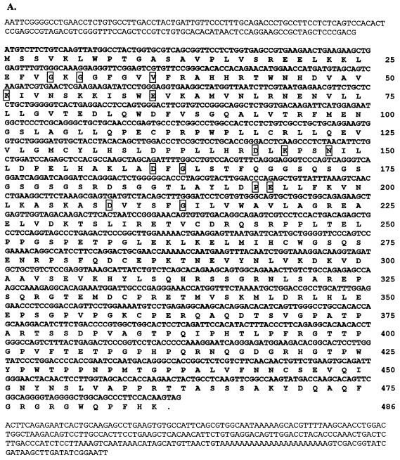
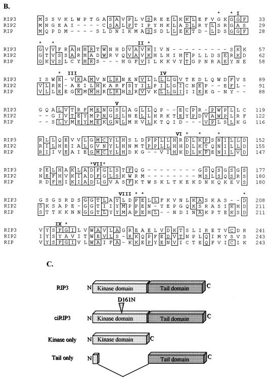
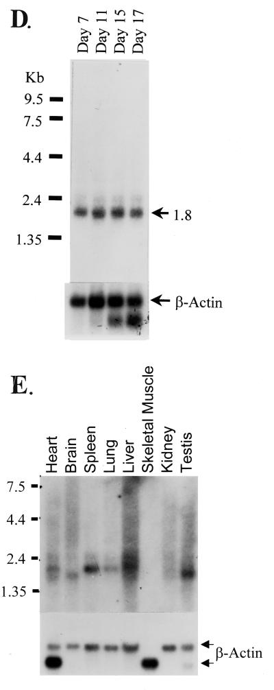
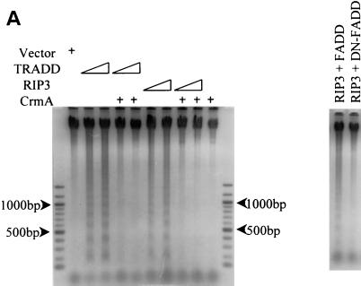
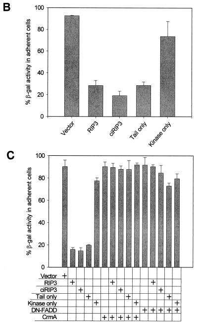
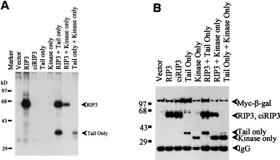
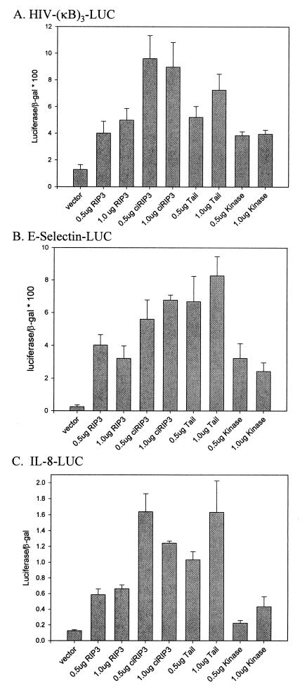
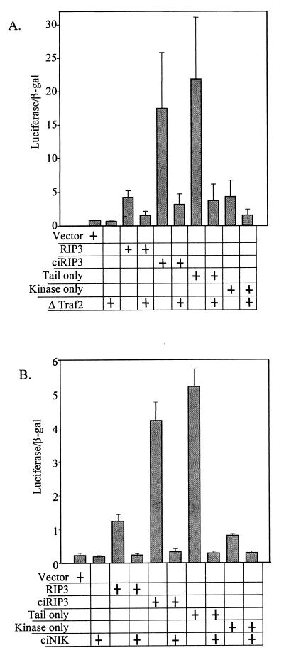
The death domain-containing receptor superfamily and their respective downstream mediators control whether or not cells initiate apoptosis or activate NF-kappaB, events critical for proper immune system function. A screen for upstream activators of NF-kappaB identified a novel serine-threonine kinase capable of activating NF-kappaB and inducing apoptosis. Based upon domain organization and sequence similarity, this novel kinase, named mRIP3 (mouse receptor interacting protein 3), appears to be a new RIP family member. RIP, RIP2, and mRIP3 contain an N-terminal kinase domain that share 30 to 40% homology. In contrast to the C-terminal death domain found in RIP or the C-terminal caspase-recruiting domain found in RIP2, the C-terminal tail of mRIP3 contains neither motif and is unique. Despite this feature, overexpression of the mRIP3 C terminus is sufficient to induce apoptosis, suggesting that mRIP3 uses a novel mechanism to induce death. mRIP3 also induced NF-kappaB activity which was inhibited by overexpression of either dominant-negative NIK or dominant-negative TRAF2. In vitro kinase assays demonstrate that mRIP3 is catalytically active and has autophosphorylation site(s) in the C-terminal domain, but the mRIP3 catalytic activity is not required for mRIP3 induced apoptosis and NF-kappaB activation. Unlike RIP and RIP2, mRIP3 mRNA is expressed in a subset of adult tissues and is thus likely to be a tissue-specific regulator of apoptosis and NF-kappaB activity. While the lack of a dominant-negative mutant precludes linking mRIP3 to a known upstream regulator, characterizing the expression pattern and the in vitro functions of mRIP3 provides insight into the mechanism(s) by which cells modulate the balance between survival and death in a cell-type-specific manner.


Figures

FIG. 1
FIG. 1
mRIP3 is a novel serine/threonine kinase related to the RIP family of kinases. (A) Nucleotide sequence of the mRIP3 cDNA and putative open reading frame with the number of amino acid residues listed at the right. Boxed residues are the conserved amino acids found in serine/threonine kinases. (B) Amino acid sequence alignments of RIP, RIP2, and mRIP3 N-terminal kinase domains. Roman numerals indicate the location of the kinase subdomains. Asterisks indicate the invariant kinase residues boxed in panel A. The analysis was performed by using the MegAlign program (DNAStar, Inc., Madison, Wis.). (C) Schematic of mRIP3 constructs. mRIP3 (wild-type protein), ciRIP3 (catalytically inactive mRIP3 in which the Mg2+-ATP binding site aspartic acid [amino acid 161] has been changed to asparagine), and tail-only and kinase-only (two deletion mutants in which each domain is expressed separately) are depicted. Kinase-only and tail-only proteins include amino acids 1 to 248 and amino acids 1 to 13 fused to 245 to 486, respectively. (D) mRIP3 is expressed in embryonic mouse tissues. A Clontech Mouse Multiple Tissue Embryo blot was probed with a radioactively labeled cDNA probe (see Materials and Methods for details). (E) mRIP3 mRNA is present in the heart, brain, spleen, lung, liver, kidney, and testis. The Clontech Adult Mouse Multiple Tissue Northern blot was probed with a radioactively labeled mRIP3 cDNA probe (see Materials and Methods for details). Size markers (in kilobases) are indicated on the left side of the panel.
FIG. 1
FIG. 1
mRIP3 is a novel serine/threonine kinase related to the RIP family of kinases. (A) Nucleotide sequence of the mRIP3 cDNA and putative open reading frame with the number of amino acid residues listed at the right. Boxed residues are the conserved amino acids found in serine/threonine kinases. (B) Amino acid sequence alignments of RIP, RIP2, and mRIP3 N-terminal kinase domains. Roman numerals indicate the location of the kinase subdomains. Asterisks indicate the invariant kinase residues boxed in panel A. The analysis was performed by using the MegAlign program (DNAStar, Inc., Madison, Wis.). (C) Schematic of mRIP3 constructs. mRIP3 (wild-type protein), ciRIP3 (catalytically inactive mRIP3 in which the Mg2+-ATP binding site aspartic acid [amino acid 161] has been changed to asparagine), and tail-only and kinase-only (two deletion mutants in which each domain is expressed separately) are depicted. Kinase-only and tail-only proteins include amino acids 1 to 248 and amino acids 1 to 13 fused to 245 to 486, respectively. (D) mRIP3 is expressed in embryonic mouse tissues. A Clontech Mouse Multiple Tissue Embryo blot was probed with a radioactively labeled cDNA probe (see Materials and Methods for details). (E) mRIP3 mRNA is present in the heart, brain, spleen, lung, liver, kidney, and testis. The Clontech Adult Mouse Multiple Tissue Northern blot was probed with a radioactively labeled mRIP3 cDNA probe (see Materials and Methods for details). Size markers (in kilobases) are indicated on the left side of the panel.
FIG. 2
FIG. 2
mRIP3 induces apoptosis. (A) mRIP3 overexpression induces DNA fragmentation. HEK 293 cells were transfected with 0.5 or 1.0 μg of TRADD with or without a fourfold excess of CrmA (lanes 2 to 5, respectively). In lanes 6 to 9, HEK 293 cells were transfected with 0.5 or 1.0 μg of mRIP3 with or without a fourfold excess of CrmA. In the right panel, 2 μg of mRIP3 was cotransfected with 2 μg of FADD or 10 μg of DN-FADD. At 24 h after transfection, genomic DNA was isolated and processed as described in Materials and Methods. (B) mRIP3 kinase domain does not induce apoptosis. HEK 293 cells were transfected with mRIP3, ciRIP3, and tail-only or kinase-only constructs plus 0.25 μg of pCMV-β-Gal. β-Gal activity was measured for adherent cells and floating apoptotic cells as described in Materials and Methods. The mean percentage of β-Gal in the adherent-cell fraction was calculated for three experiments and plotted with the standard deviation. (C) DN-FADD and CrmA inhibit mRIP3, ciRIP3, or mRIP3–tail-only-induced apoptosis. HEK 293 cells were transfected with the indicated mRIP3 constructs. Cell lysates were assayed as in panel B.
FIG. 2
FIG. 2
mRIP3 induces apoptosis. (A) mRIP3 overexpression induces DNA fragmentation. HEK 293 cells were transfected with 0.5 or 1.0 μg of TRADD with or without a fourfold excess of CrmA (lanes 2 to 5, respectively). In lanes 6 to 9, HEK 293 cells were transfected with 0.5 or 1.0 μg of mRIP3 with or without a fourfold excess of CrmA. In the right panel, 2 μg of mRIP3 was cotransfected with 2 μg of FADD or 10 μg of DN-FADD. At 24 h after transfection, genomic DNA was isolated and processed as described in Materials and Methods. (B) mRIP3 kinase domain does not induce apoptosis. HEK 293 cells were transfected with mRIP3, ciRIP3, and tail-only or kinase-only constructs plus 0.25 μg of pCMV-β-Gal. β-Gal activity was measured for adherent cells and floating apoptotic cells as described in Materials and Methods. The mean percentage of β-Gal in the adherent-cell fraction was calculated for three experiments and plotted with the standard deviation. (C) DN-FADD and CrmA inhibit mRIP3, ciRIP3, or mRIP3–tail-only-induced apoptosis. HEK 293 cells were transfected with the indicated mRIP3 constructs. Cell lysates were assayed as in panel B.
FIG. 3
FIG. 3
mRIP3 autophosphorylates site(s) in the C-terminal domain. HEK 293 cells were transfected with the indicated constructs plus CrmA and Myc-His-LacZ (Invitrogen). Cell lysates were harvested, and fusion proteins were immunoprecipitated as described in Materials and Methods. Immunoprecipitates were divided in half and were subjected to in vitro kinase assays (A) or Western blot analysis (B) to determine the protein expression level.
FIG. 4
FIG. 4
mRIP3 induces NF-κB activity. HEK 293 cells were transfected the indicated expression constructs plus HIV-(κB)3–LUC (A), E-selectin–LUC (B), or IL-8–LUC (C). Cell lysates were assayed for luciferase and β-Gal activity as described in Materials and Methods. The mean of four experiments with the standard error is shown.
FIG. 5
FIG. 5
mRIP3-induced NF-κB activity is blocked by ciNIK and ΔTRAF2. HEK 293 cells were transfected with the indicated constructs. Cell lysates were harvested and assayed as described in the legend to Fig. 4.

References

    1. Ashkenazi A, Dixit V M. Death receptors: signaling and modulation. Science. 1998;281:1305–1308. - PubMed
    1. Belvin M P, Anderson K V. A conserved signaling pathway: the Drosophila toll-dorsal pathway. Ann Rev Cell Dev Biol. 1996;12:393–416. - PubMed
    1. Bodmer J L, Burns K, Schneider P, Hofmann K, Steiner V, Thome M, Bornand T, Hahne M, Schroter M, Becker K, Wilson A, French L E, Browning J L, MacDonald H R, Tschopp J. TRAMP, a novel apoptosis-mediating receptor with sequence homology to tumor necrosis factor receptor 1 and Fas(Apo-1/CD95) Immunity. 1997;6:79–88. - PubMed
    1. Chaudhary P M, Eby M, Jasmin A, Bookwalter A, Murray J, Hood L. Death receptor 5, a new member of the TNFR family, and DR4 induce FADD-dependent apoptosis and activate the NF-κB pathway. Immunity. 1997;7:821–830. - PubMed
    1. Chinnaiyan A M, O’Rourke K, Tewari M, Dixit V M. FADD, a novel death domain-containing protein, interacts with the death domain of Fas and initiates apoptosis. Cell. 1995;81:505–512. - PubMed

Publication types

MeSH terms