. 1999 Oct;19(10):6690-8.
doi: 10.1128/MCB.19.10.6690.

Roles of cell division and gene transcription in the methylation of CpG islands

Affiliations

Roles of cell division and gene transcription in the methylation of CpG islands

C M Bender et al. Mol Cell Biol. 1999 Oct.

Abstract

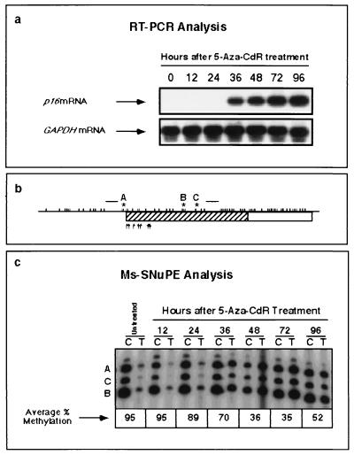
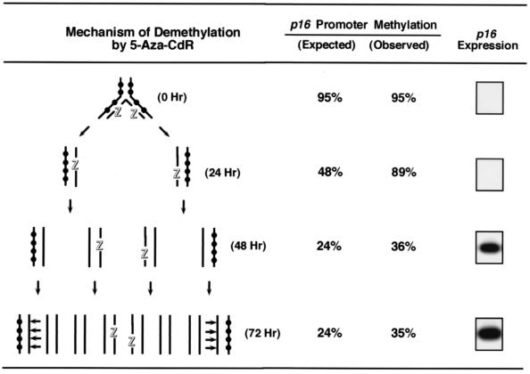
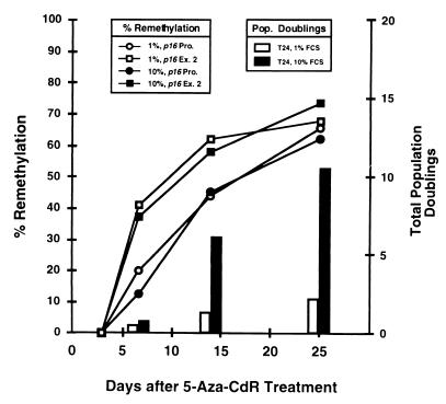
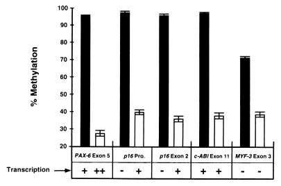
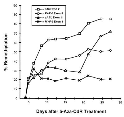
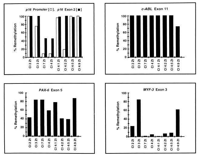
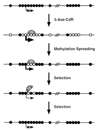
De novo methylation of CpG islands within the promoters of eukaryotic genes is often associated with their transcriptional repression, yet the methylation of CpG islands located downstream of promoters does not block transcription. We investigated the kinetics of mRNA induction, demethylation, and remethylation of the p16 promoter and second-exon CpG islands in T24 cells after 5-aza-2'-deoxycytidine (5-Aza-CdR) treatment to explore the relationship between CpG island methylation and gene transcription. The rates of remethylation of both CpG islands were associated with time but not with the rate of cell division, and remethylation of the p16 exon 2 CpG island occurred at a higher rate than that of the p16 promoter. We also examined the relationship between the remethylation of coding sequence CpG islands and gene transcription. The kinetics of remethylation of the p16 exon 2, PAX-6 exon 5, c-ABL exon 11, and MYF-3 exon 3 loci were examined following 5-Aza-CdR treatment because these genes contain exonic CpG islands which are hypermethylated in T24 cells. Remethylation occurred most rapidly in the p16, PAX-6, and c-ABL genes, shown to be transcribed prior to drug treatment. These regions also exhibited higher levels of remethylation in single-cell clones and subclones derived from 5-Aza-CdR-treated T24 cells. Our data suggest that de novo methylation is not restricted to the S phase of the cell cycle and that transcription through CpG islands does not inhibit their remethylation.


Figures

FIG. 1
FIG. 1
Kinetics of p16 mRNA induction and promoter demethylation by 5-Aza-CdR. T24 cells were exposed to 5-Aza-CdR (5 × 10⁻⁷ M) for 24 h. DNA and RNA were isolated at 12-h intervals after drug addition. (a) Levels of p16 mRNA expression at each time point were determined by RT-PCR analysis. Relative levels of GAPDH mRNA expression were measured to control for relative cDNA input. PCR products were resolved on 2% agarose, transferred to a nylon membrane, and hybridized with an internal oligonucleotide probe specific for the cDNA sequence of either p16 or GAPDH. (b) Schematic map of the p16 5′ CpG island. A 149-bp region (hatching) was amplified by PCR with primers specific for bisulfite-converted DNA (raised horizontal bars), and three CpG sites were analyzed by the Ms-SNuPE technique (sites A, B, and C). Each tick mark represents an individual CpG dinucleotide, and the arrows show putative transcription initiation sites. (c) Demethylation of the p16 promoter by 5-Aza-CdR at each time point, quantitated by Ms-SNuPE. Ms-SNuPE reaction mixtures were resolved on a 16% denaturing polyacrylamide gel and subsequently exposed by autoradiography, using purified bisulfite PCR products as the templates for primer extension. The presence of a band indicates primer extension at a given CpG site. A band in the “C” lane indicates the detection of DNA molecules which are methylated, and a band in the “T” lane indicates the detection of unmethylated molecules.
FIG. 2
FIG. 2
Model for the mechanism of DNA demethylation by 5-Aza-CdR. Unsynchronized T24 cells in the log phase of growth have a doubling time of 21 h (3) and were treated with 5-Aza-CdR (5 × 10⁻⁷ M) for 24 h. DNA and RNA were isolated at 24-h intervals, p16 promoter methylation levels were quantitated by Ms-SNuPE, and the presence of p16 mRNA was determined by RT-PCR. Cell numbers were assessed at each time point to determine the cell population doubling time. Strand breakage of 5-Aza-CdR-containing DNA after bisulfite treatment is likely because of the lability of the drug under alkaline conditions, which explains why the observed and expected values for methylation differ. Methylated CpG sites are indicated by black circles, DNA strands containing incorporated 5-Aza-CdR are each indicated by an outlined letter “Z,” and horizontal arrows show sites of hemimethylated DNA where maintenance methylation is expected to occur.
FIG. 3
FIG. 3
Investigation of the association between the rate of cell division and the rate of remethylation. T24 cells treated with 5-Aza-CdR (5 × 10⁻⁷ M) were maintained in medium supplemented with either 1 or 10% FCS. DNA was isolated at specific times after 5-Aza-CdR treatment and subsequently treated with sodium bisulfite (8, 11). Methylation of the p16 promoter (p16 Pro.) and exon 2 (p16 Ex. 2) CpG islands was determined by Ms-SNuPE (15) before and after 5-Aza-CdR treatment to ascertain whether remethylation of these regions was associated with the rate of cell division. Remethylation was determined as the degree of recovery (compared to original levels) following maximal demethylation at 72 h. T24 cells grown in medium supplemented with 10% FCS were analyzed as controls. If de novo methylation of p16 is linked to the rate of cell division, then cells maintained in 10% FCS would be expected to remethylate p16 more rapidly than cells dividing more slowly in 1% FCS.
FIG. 4
FIG. 4
Effects of 5-Aza-CdR on the demethylation and transcription of the p16 promoter and exon 2, PAX-6 exon 5, c-ABL exon 11, and MYF-3 exon 3 in T24 cells. Average methylation values for specific sites within the p16, PAX-6, c-ABL, and MYF-3 exonic CpG islands were measured in T24 cells prior to drug treatment (black bars) and 72 h after treatment with 5 × 10⁻⁷ M 5-Aza-CdR (white bars). Error bars indicate the ranges of values obtained. Relative transcription levels of each gene were also estimated by comparison of band intensities (−, +, or ++) before and after drug treatment by RT-PCR analysis (data not shown). The CpG and GC contents of these regions were also analyzed to determine if these exonic sequences fulfilled the criteria of CpG islands in which a DNA sequence of ≥200 bp must have a GC content of ≥0.50 and an observed/expected CpG ratio of ≥0.60 (12). Fragments of 800 bp from each gene, all of which fulfilled the established criteria for CpG islands, were analyzed. In untreated T24 cells, transcription through the p16 promoter and exon 2 CpG islands is not initiated from the p16 promoter but is initiated from the upstream p14 promoter (38). Pro., promoter.
FIG. 5
FIG. 5
Remethylation kinetics of CpG islands in T24 cells after 5-Aza-CdR treatment. T24 cells were treated with 5-Aza-CdR (5 × 10⁻⁷ M), and DNA was harvested every 1 to 3 days for up to 27 days. The population doubling time increased approximately 1.8-fold after 5-Aza-CdR treatment, as previously described (3). Only eight cell population doublings were attained between days 3 and 27 because cells transiently entered lag phase each time they were split and reseeded. Methylation of p16 exon 2, PAX-6 exon 5, c-ABL exon 11, and MYF-3 exon 3 was quantitated at each time point by the Ms-SNuPE technique, and the degree of remethylation at each locus was determined as the degree of methylation compared to the original level in untreated cells at 72 h. Methylation averages from three independent experiments are shown.
FIG. 6
FIG. 6
Remethylation of CpG islands after 5-Aza-CdR treatment in T24 clones and subclones. Clones 2, 3, 4, and 7 were isolated following treatment of parent T24 cultures with 5-Aza-CdR (3 × 10⁻⁷ M). This lower dose was utilized (instead of 5 × 10⁻⁷ M) to increase the cell survival rate and to facilitate the isolation of the clones. Clone 4 was used for the repeated isolation of single-cell subclones. DNA was isolated, and methylation of the p16 promoter, p16 exon 2, PAX-6 exon 5, c-ABL exon 11, and MYF-3 exon 3 CpG islands was quantitated by Ms-SNuPE. Clones 2, 3, 4, and 7 completed approximately 20 cell population doublings at the time DNA was harvested, whereas subclones derived from clone 4 (4:1, 4:2, 4:5, and 4:9) completed approximately 40 population doublings (data not shown). The degree of remethylation at each locus was determined as the degree of methylation recovery (compared to original levels) following maximal demethylation at 72 h. Methylation averages from three independent experiments are shown.
FIG. 7
FIG. 7
Model for the remethylation of promoter and coding sequence CpG islands of the p16 gene after 5-Aza-CdR treatment. Promoter and coding sequence CpG islands of growth-regulatory genes such as p16 become remethylated at different rates after 5-Aza-CdR treatment. Following 5-Aza-CdR-mediated demethylation and transcriptional activation, remethylation may first appear in a CpG island downstream of the promoter, whereas transcription factors (TF) associated with a demethylated promoter CpG island protect it from remethylation. Patches of demethylation have been observed by bisulfite genomic sequencing of the p16 promoter in single-cell clones after 5-Aza-CdR treatment, providing support for this interpretation (14). Cells which acquire promoter methylation in one or more growth-regulatory genes may subsequently exhibit selective growth advantages due to the obstruction of transcription factor binding, leading to gene silencing. The protection of demethylated promoter sequences by transcription factors may explain how exonic CpG islands can become remethylated more rapidly than promoter islands after 5-Aza-CdR treatment, as DNA-binding proteins may protect promoter sequences from methylation. Methylated CpG sites are depicted as filled circles; unmethylated sites are shown as open circles.

References

    1. Antequera F, Boyes J, Bird A. High levels of de novo methylation and altered chromatin structure at CpG islands in cell lines. Cell. 1990;62:503–514. - PubMed
    1. Baylin S B, Herman J G, Graff J R, Vertino P M, Issa J-P. Alterations in DNA methylation: a fundamental aspect of neoplasia. Adv Cancer Res. 1998;72:141–146. - PubMed
    1. Bender C M, Pao M M, Jones P A. Inhibition of DNA methylation by 5-aza-2′-deoxycytidine suppresses the growth of human tumor cell lines. Cancer Res. 1998;58:95–101. - PubMed
    1. Bender C M, Zingg J M, Jones P A. DNA methylation as a target for drug design. Pharmacol Res. 1998;15:175–187. - PubMed
    1. Bird A P. CpG islands as gene markers in the vertebrate nucleus. Trends Genet. 1987;3:342–347.

Publication types

MeSH terms

LinkOut - more resources