. 1999 Oct;19(10):6729-41.
doi: 10.1128/MCB.19.10.6729.

Regulation of cell cycle transcription factor Swi4 through auto-inhibition of DNA binding

Affiliations

Regulation of cell cycle transcription factor Swi4 through auto-inhibition of DNA binding

K Baetz et al. Mol Cell Biol. 1999 Oct.

Abstract

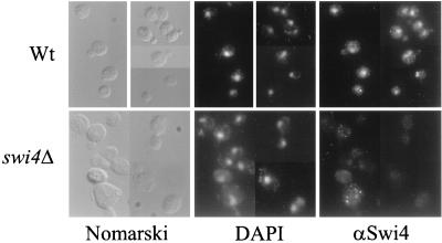
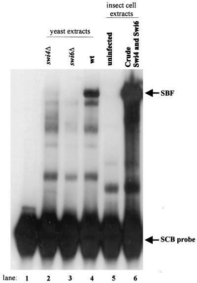
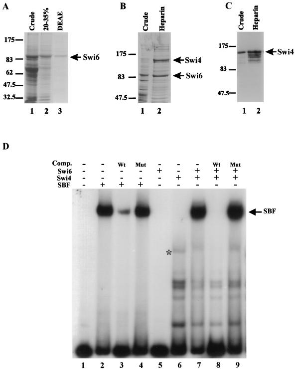
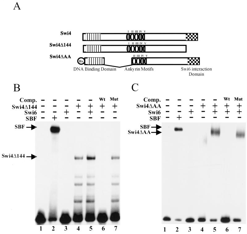
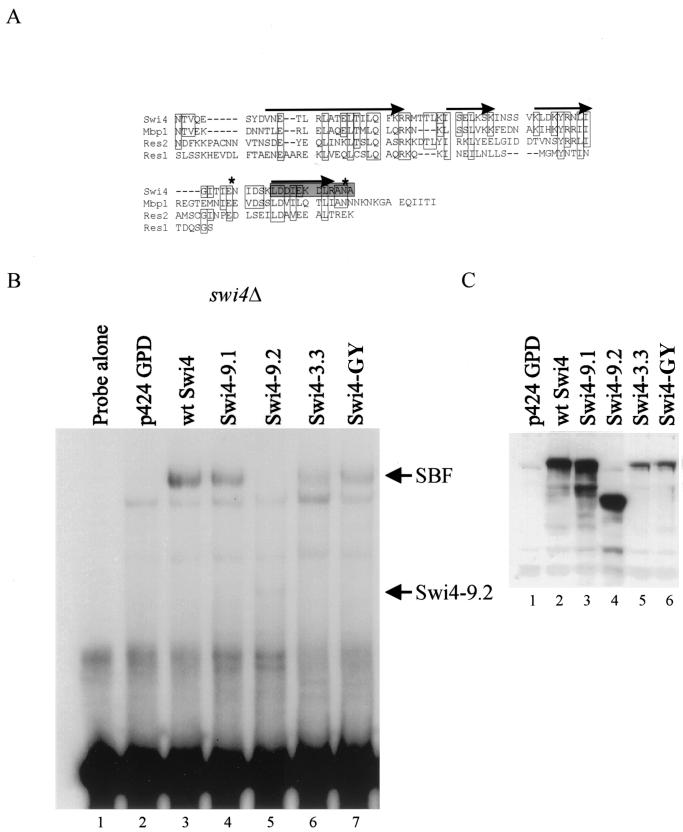
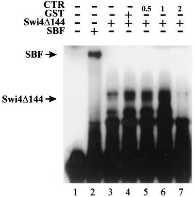
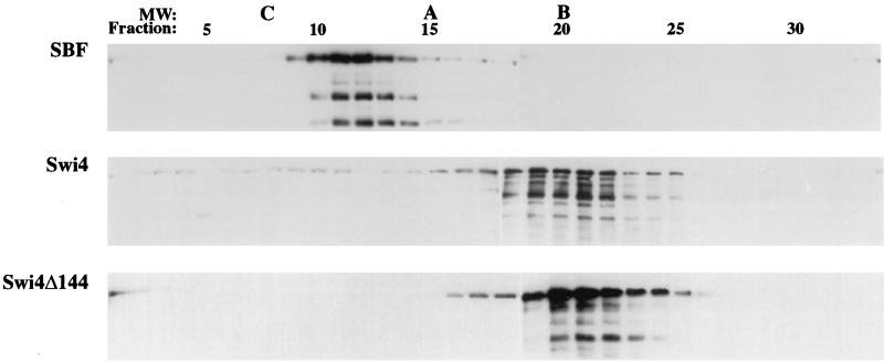
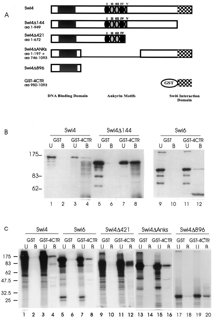
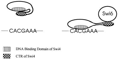
In Saccharomyces cerevisiae, two transcription factors, SBF (SCB binding factor) and MBF (MCB binding factor), promote the induction of gene expression at the G(1)/S-phase transition of the mitotic cell cycle. Swi4 and Mbp1 are the DNA binding components of SBF and MBF, respectively. The Swi6 protein is a common subunit of both transcription factors and is presumed to play a regulatory role. SBF binding to its target sequences, the SCBs, is a highly regulated event and requires the association of Swi4 with Swi6 through their C-terminal domains. Swi4 binding to SCBs is restricted to the late M and G(1) phases, when Swi6 is localized to the nucleus. We show that in contrast to Swi6, Swi4 remains nuclear throughout the cell cycle. This finding suggests that the DNA binding domain of Swi4 is inaccessible in the full-length protein when not complexed with Swi6. To explore this hypothesis, we expressed Swi4 and Swi6 in insect cells by using the baculovirus system. We determined that partially purified Swi4 cannot bind SCBs in the absence of Swi6. However, Swi4 derivatives carrying point mutations or alterations in the extreme C terminus were able to bind DNA or activate transcription in the absence of Swi6, and the C terminus of Swi4 inhibited Swi4 derivatives from binding DNA in trans. Full-length Swi4 was determined to be monomeric in solution, suggesting an intramolecular mechanism for auto-inhibition of binding to DNA by Swi4. We detected a direct in vitro interaction between a C-terminal fragment of Swi4 and the N-terminal 197 amino acids of Swi4, which contain the DNA binding domain. Together, our data suggest that intramolecular interactions involving the C-terminal region of Swi4 physically prevent the DNA binding domain from binding SCBs. The interaction of the carboxy-terminal region of Swi4 with Swi6 alleviates this inhibition, allowing Swi4 to bind DNA.


Figures

FIG. 1
FIG. 1
Subcellular localization of Swi4. Swi4 localization was assayed by indirect immunofluorescence with Swi4 antiserum and a fluorescein isothiocyanate-conjugated secondary antibody. Wild-type (Wt) and swi4Δ cells were photographed at a magnification of ×630 with an imaging system (see Materials and Methods). Photographs of the same fields of cells viewed with Nomarski optics and stained with DAPI to visualize cell nuclei are also shown.
FIG. 2
FIG. 2
Reconstitution of SBF in insect cells. A gel retardation assay with an SCB-containing probe and either crude yeast or insect cell lysates is shown. The labeled probe contained three SCB sequences from the upstream region of the HO gene (see reference 2). The following extracts were used in the binding assays: lane 1, probe alone; lanes 2 to 4, 10 μg of crude yeast extract from swi4Δ, swi6Δ, and wild-type strains, respectively; lane 5, 10 μg of crude cell lysate from uninfected insect cells; and lane 6, 10 μg of crude cell lysate from insect cells coinfected with Swi4- and Swi6-expressing baculovirus vectors. The migration positions of the SBF complex and the unbound probe are indicated to the right.
FIG. 3
FIG. 3
SCB binding activity of partially purified Swi4, Swi6, and SBF. (A) Purification of Swi6 protein expressed in insect cells. Swi6-containing fractions obtained during purification were analyzed by SDS–6% PAGE followed by Coomassie blue staining. Lane 1, crude lysate from insect cells expressing Swi6 protein; lane 2, 20 to 35% ammonium sulfate precipitation; lane 3, DEAE-Sepharose fraction. A 10-μl aliquot of each fraction was loaded per lane. (B) Purification of SBF expressed in insect cells. SBF-containing fractions obtained during purification were analyzed as described in panel A for Swi6. Lane 1, crude lysate from insect cells infected with both SWI4- and SWI6-expressing baculoviruses; lane 2, heparin-agarose fraction. A 10-μl aliquot each of the crude and partially purified fractions was loaded. (C) Enrichment of Swi4 expressed in insect cells. Swi4-containing fractions obtained during purification were separated by SDS–6% PAGE and analyzed by Western blotting with affinity-purified Swi4 antiserum. Lane 1, crude lysate from insect cells infected with a SWI4-expressing baculovirus vector (10 μg); lane 2, heparin-agarose fraction (10 μg). For panels A through C, the migration positions of molecular weight markers are indicated to the left (in thousands). (D) Gel retardation assay with partially purified SBF, Swi4, and Swi6. A labeled SCB-containing probe (see the legend to Fig. 2) was incubated with the following protein preparations: lane 1, no extract; lanes 2 to 4, SBF heparin-agarose fraction (1 μg); lane 5, Swi6 DEAE-Sepharose fraction (3 μg); lane 6, Swi4 heparin-agarose fraction (5 μg); and lanes 7 to 9, both partially purified Swi4 and Swi6 fractions. Where indicated above the lanes, a 100-fold molar excess of either wild-type SCB competitor (Comp.) DNA (Wt) or mutated SCB competitor DNA (Mut) was added. The migration position of SBF is shown to the right. The asterisk in lane 6 marks the migration position of a complex composed of the SCB-containing probe and either full-length Swi4 or a small C-terminal truncation of Swi4.
FIG. 4
FIG. 4
Analysis of SCB binding by deletion derivatives of Swi4. (A) Schematic of the Swi4 derivatives expressed in insect cells. The relative positions of the N-terminal DNA binding domain, the multiple ankyrin repeats, and the C-terminal Swi6 interaction domain are indicated. His indicates the presence of an N-terminal histidine tag. (B) Gel retardation assay with partially purified SBF or C-terminally truncated Swi4 (Swi4Δ144). A labeled SCB-containing probe (see the legend to Fig. 2) was incubated with the following protein preparations: lane 1, no extract; lane 2, SBF heparin-agarose fraction (1 μg); lane 3, 3 μg of purified Swi6; and lanes 4 to 7, 5 μg of partially purified Swi4Δ144. Lane 5 also contains 3 μg of a Swi6 DEAE-Sepharose fraction. In lanes 6 and 7, a 100-fold molar excess of either wild-type SCB competitor (Comp.) DNA (Wt) or mutated SCB competitor DNA (Mut) was added. (C) Gel retardation assay with partially purified SBF or a Swi4 internal deletion derivative (Swi4ΔAA). The labeled SCB-containing probe was incubated with the following protein preparations: lane 1, no extract; lane 2, SBF heparin-agarose fraction (1 μg); lane 3, 3 μg of purified Swi6; lane 4, 5 μg of partially purified Swi4ΔAA; and lanes 5 to 7, 5 μg of partially purified Swi4ΔAA and 3 μg of purified Swi6. In lanes 6 and 7, a 100-fold molar excess of either wild-type SCB competitor DNA (Wt) or mutated SCB competitor DNA (Mut) was added.
FIG. 5
FIG. 5
Gel retardation analysis of wild-type SWI4 and mutant swi4 alleles with yeast cell extracts. (A) Alignment of the extreme CTRs of Swi4 family members. Residues identical to those in Swi4 or conservative substitutions are boxed. Putative alpha helices are indicated by arrows, as predicted by the PHD protein structure algorithm. The shaded box shows the amino acids that were deleted in Swi4-3.3. The asterisks indicate the positions of the point mutations E1076G and N1092Y in mutant Swi4-GY. (B) A labeled SCB-containing probe was incubated with 10 μg of crude extract from a swi4Δ yeast strain (BY184) transformed with the following plasmids: lane 1, no extract; lane 2, empty vector, p424 GPD; lane 3, p424 GPD-Swi4 (wild-type [wt] SWI4); lane 4, p424 GPD-Swi4-9.1; lane 5, p424 GPD-Swi4-9.2; lane 6, p424 GPD-Swi4-3.3; and lane 7, p424 GPD-Swi4-GYr. The SWI4 mutations in the various plasmids are described in Table 2 and in the text. The migration positions of SBF and a complex of Swi4-9.2 and the SCB-containing probe are indicated to the right. (C) Western blot analysis of extracts used in the binding assay shown in panel B. Fifty micrograms of the crude yeast lysates used in the gel retardation analysis were separated by SDS–6% PAGE, and the Swi4 protein in the extracts was visualized with Swi4 antiserum. The Swi4 protein present in each extract is indicated above the lanes (see panel B).
FIG. 6
FIG. 6
Inhibition of Swi4Δ144-SCB complex formation by the CTR of Swi4. A gel retardation assay with an SCB-containing probe is shown (see the legend to Fig. 2). The probe was incubated with the following protein preparations: lane 1, no extract; lane 2, SBF heparin-agarose fraction (1 μg); and lanes 3 to 7, 2 μg of partially purified Swi4Δ144. Lane 4 also contained 1 μg of GST, and lanes 5 to 7 also contained increasing amounts (in micrograms) of purified Swi4 CTR (C-terminal 144 amino acids of Swi4). The migration positions of the SBF-SCB and Swi4Δ144-SCB complexes are indicated on the left.
FIG. 7
FIG. 7
Glycerol gradient sedimentation of Swi4, Swi4Δ144, and SBF. Fifty micrograms of partially purified Swi4, Swi4Δ144, and SBF was analyzed by glycerol gradient sedimentation. Glycerol gradients (4 ml of 10 to 40% [vol/vol] glycerol) were centrifuged in an SW60.1 rotor for 13 h at 55,000 rpm. Fractions from the gradients were analyzed on Western blots with anti-Swi4 antibody. The peak fractions of molecular weight (MW) standards run in parallel gradients are indicated by C (catalase, 232,000), A (aldolase, 158,000), and B (bovine albumin, 67,000). Fraction numbers are shown at the top of the figure, with the bottom of the gradients on the left.
FIG. 8
FIG. 8
Binding of the C-terminal 144 amino acids of Swi4 to Swi6 and N-terminal regions of Swi4 in vitro. (A) Schematic of the Swi4 proteins used in the assay. The relative positions of the DNA binding domain, ankyrin motifs, and C-terminal domain (Swi6 interaction domain) are depicted. aa, amino acids. (B) Ten micrograms of partially purified Swi4, Swi4Δ144, or Swi6 derived from insect cell extracts (see Fig. 2) was incubated with either GST or GST-4CTR immobilized on glutathione beads. The unbound (U) and bound (B) fractions were separated by SDS–6% PAGE. The gels were then blotted and incubated with Swi4 antiserum (lanes 1 to 8) or Swi6 antiserum (lanes 9 to 12) to identify the Swi4 or Swi6 proteins. The migration positions of molecular weight markers are indicated to the left (in thousands). (C) Seven microliters of in vitro-translated Swi4, Swi6, Swi4Δ421, Swi4ΔAnks, and Swi4Δ896 was incubated with either GST or GST-4CTR immobilized on glutathione beads. The unbound (U) and bound (B) fractions were separated by SDS–10% PAGE. The migration positions of molecular weight markers are indicated to the left (in thousands).
FIG. 9
FIG. 9
Model of the auto-inhibition of Swi4 binding to DNA. (Left) An intramolecular interaction involving the extreme C terminus of Swi4 (CTR) and a more N-terminal region of the protein is depicted. Our data suggest that when Swi4 is not bound to Swi6, the CTR of Swi4 is free to form an intramolecular interaction with the DNA binding domain of Swi4, preventing Swi4 binding to SCBs (CACGAAA). (Right) The binding of Swi6 to the CTR of Swi4 disrupts the intramolecular interaction of Swi4, allowing Swi4 to bind to SCBs.

Similar articles

Cited by

References

    1. Amon A, Tyers M, Futcher B, Nasmyth K. Mechanisms that help the yeast cell cycle clock tick: G2 cyclins transcriptionally activate G2 cyclins and repress G1 cyclins. Cell. 1993;74:993–1007. - PubMed
    1. Andrews B J, Herskowitz I. Identification of a DNA binding factor involved in cell-cycle control of the yeast HO gene. Cell. 1989;57:21–29. - PubMed
    1. Andrews B J, Herskowitz I. The yeast SWI4 protein contains a motif present in developmental regulators and is part of a complex involved in cell-cycle-dependent transcription. Nature. 1989;342:830–833. - PubMed
    1. Andrews B J, Moore L A. Interaction of the yeast Swi4 and Swi6 cell cycle regulatory proteins in vitro. Proc Natl Acad Sci USA. 1992;89:11852–11856. - PMC - PubMed
    1. Bassuk A G, Leiden J M. The role of Ets transcription factors in the development and function of the mammalian immune system. Adv Immunol. 1997;64:65–104. - PubMed

Publication types

MeSH terms

LinkOut - more resources