. 1999 Oct;19(10):6765-74.
doi: 10.1128/MCB.19.10.6765.

G alpha-q/11 protein plays a key role in insulin-induced glucose transport in 3T3-L1 adipocytes

Affiliations

G alpha-q/11 protein plays a key role in insulin-induced glucose transport in 3T3-L1 adipocytes

T Imamura et al. Mol Cell Biol. 1999 Oct.

Abstract

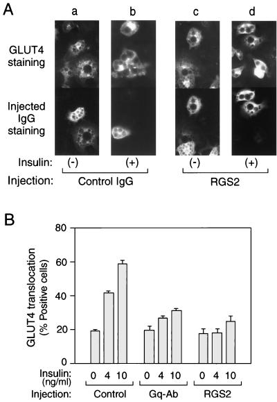
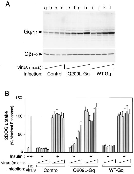
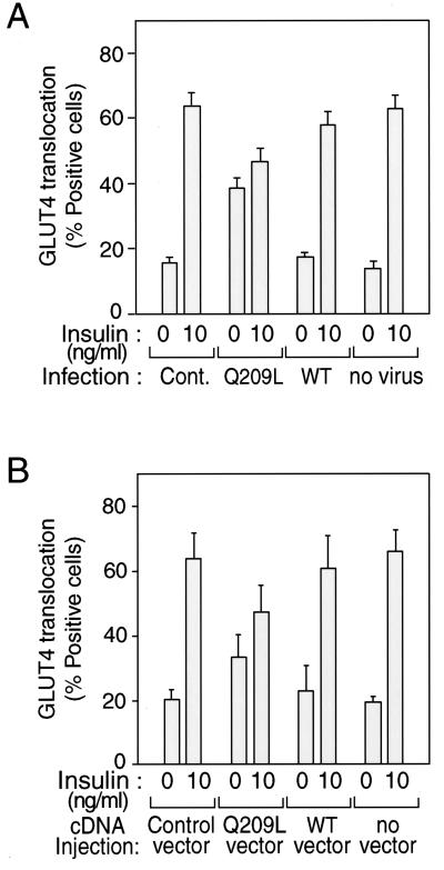
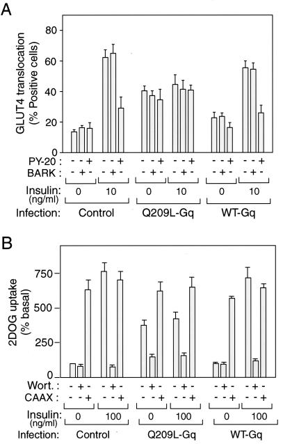
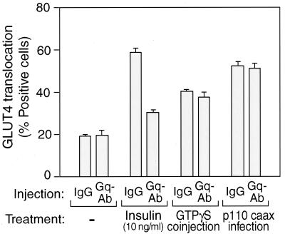
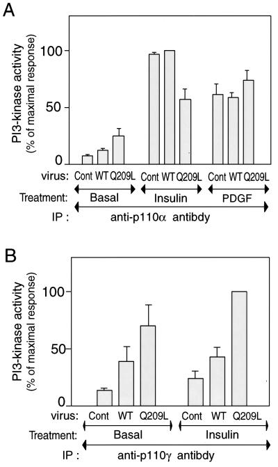
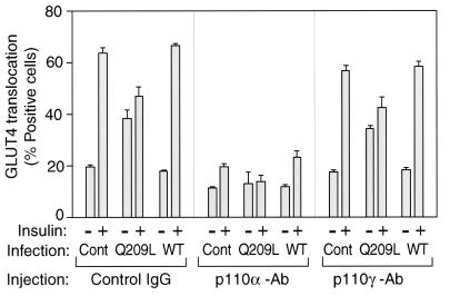
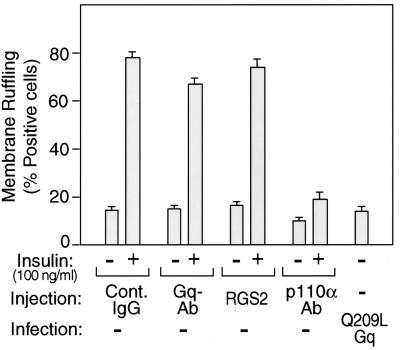
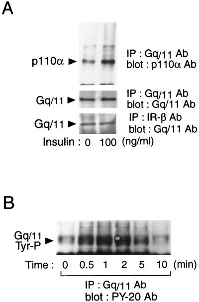
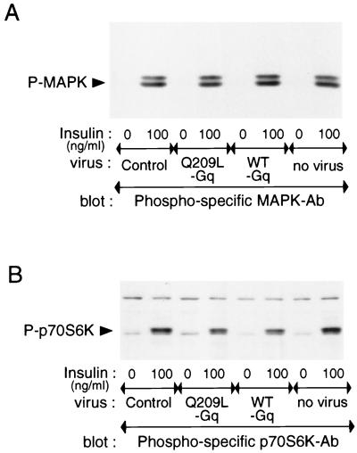
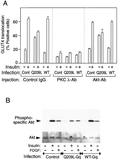
We evaluated the role of the G alpha-q (Galphaq) subunit of heterotrimeric G proteins in the insulin signaling pathway leading to GLUT4 translocation. We inhibited endogenous Galphaq function by single cell microinjection of anti-Galphaq/11 antibody or RGS2 protein (a GAP protein for Galphaq), followed by immunostaining to assess GLUT4 translocation in 3T3-L1 adipocytes. Galphaq/11 antibody and RGS2 inhibited insulin-induced GLUT4 translocation by 60 or 75%, respectively, indicating that activated Galphaq is important for insulin-induced glucose transport. We then assessed the effect of overexpressing wild-type Galphaq (WT-Galphaq) or a constitutively active Galphaq mutant (Q209L-Galphaq) by using an adenovirus expression vector. In the basal state, Q209L-Galphaq expression stimulated 2-deoxy-D-glucose uptake and GLUT4 translocation to 70% of the maximal insulin effect. This effect of Q209L-Galphaq was inhibited by wortmannin, suggesting that it is phosphatidylinositol 3-kinase (PI3-kinase) dependent. We further show that Q209L-Galphaq stimulates PI3-kinase activity in p110alpha and p110gamma immunoprecipitates by 3- and 8-fold, respectively, whereas insulin stimulates this activity mostly in p110alpha by 10-fold. Nevertheless, only microinjection of anti-p110alpha (and not p110gamma) antibody inhibited both insulin- and Q209L-Galphaq-induced GLUT4 translocation, suggesting that the metabolic effects induced by Q209L-Galphaq are dependent on the p110alpha subunit of PI3-kinase. In summary, (i) Galphaq appears to play a necessary role in insulin-stimulated glucose transport, (ii) Galphaq action in the insulin signaling pathway is upstream of and dependent upon PI3-kinase, and (iii) Galphaq can transmit signals from the insulin receptor to the p110alpha subunit of PI3-kinase, which leads to GLUT4 translocation.


Figures

FIG. 1
FIG. 1
Effects of anti-Gαq antibody or RGS2 protein microinjection on insulin-induced GLUT4 translocation in 3T3-L1 adipocytes. Serum-starved 3T3-L1 adipocytes on coverslips were incubated with or without insulin (4 and 10 ng/ml) for 20 min after the microinjection of sheep IgG, anti-Gαq/11 antibody, or RGS2 protein, mixed with sheep IgG as a marker, as described in Materials and Methods. (A) Representative cells of GLUT4 staining are shown in the top panels (a to d), and staining of injected RGS2/sheep IgG (c and d) or sheep IgG alone (a and b) is shown in the bottom panels. (B) Percentage of cells positive for GLUT4 translocation, calculated by counting at least 100 cells at each point. Injected materials are sheep IgG for Control, anti-Gαq/11 C-terminal antibody for Gq-Ab, and RGS2 protein mixed with sheep IgG for RGS2. The data are means and standard errors from four independent experiments.
FIG. 2
FIG. 2
Expression and effects of Gαq on glucose uptake into the 3T3-L1 adipocytes. (A) Differentiated 3T3-L1 adipocytes were infected with various concentrations (MOI = 1, 5, 10, and 20) of adenovirus expressing WT Gαq, Q209L-Gαq, or mock-infected control. After 60 h of infection, these cells were lysed, and total-cell lysates were analyzed by Western blotting with anti-Gαq/11 C-terminal antibody (top) or anti-Gβ1–5 antibody (bottom), as described in Materials and Methods. These experiments were repeated twice. (B) 3T3-L1 adipocytes were infected with various concentrations (MOI = 1, 5, 10, 20, 30, and 40) of adenovirus expressing WT Gαq, Q209L-Gαq, or mock control. After 60 h of infection, these cells were stimulated with 100 ng of insulin per ml for 1 h and 2-[3H]deoxyglucose uptake was measured as described in Materials and Methods. The data are the means and standard errors from four independent experiments.
FIG. 3
FIG. 3
Effects of Gαq expression on insulin-induced GLUT4 translocation in 3T3-L1 adipocytes. Serum-starved 3T3-L1 adipocytes on coverslips were incubated with or without insulin (10 ng/ml) for 20 min after 60 h of Gαq-expressing adenovirus infection (MOI = 10) (A) or after 24 h of nuclear microinjection with Gαq expression vector and with GFP expression vector as a marker (B). Fixed cells were stained with rabbit anti-GLUT4 antibody and incubated with TRITC-conjugated anti-rabbit IgG antibody, as described in Materials and Methods. The percentage of cells positive for GLUT4 translocation was calculated by counting at least 100 cells at each point. The data are means and standard errors from three independent experiments. (A) Cont., mock control adenovirus; Q209L, Q209L-Gαq-expressing adenovirus; WT, wild-type-Gαq-expressing adenovirus. (B) Control vector, GFP-expressing vector only.
FIG. 4
FIG. 4
Microinjection and treatments of inhibitors and p110caax expression on Gαq expressed 3T3-L1 adipocytes. (A) After 60 h of Gαq-expressing adenovirus infection (MOI = 10), 3T3-L1 cells were injected with antiphosphotyrosine antibody (PY-20), GST-BARK, or sheep IgG as a control, prior to 3 h of insulin stimulation. Fixed cells were stained with rabbit anti-GLUT4 antibody and incubated with TRITC-conjugated anti-rabbit IgG antibody and AMCA-conjugated anti-sheep IgG antibody, as described in Materials and Methods. (B) After 60 h of Gαq-expressing adenovirus infection (MOI = 10) with or without p110caax-expressing adenovirus coinfection (CAAX), 3T3-L1 adipocytes were incubated with 300 nM wortmannin (Wort) or 0.1% DMSO vehicle for 60 min and with insulin (100 ng/ml) for 50 min or left untreated. 2-[3H]deoxyglucose uptake was measured as described in Materials and Methods. The data are means and standard errors from three independent experiments.
FIG. 5
FIG. 5
Effect of Gαq antibody microinjection on GTPγS and p110caax signaling to GLUT4 translocation. 3T3-L1 cells infected with or without p110caax-expressing adenovirus were injected with anti-Gαq/11 antibody or sheep IgG with or without GTPγS. After 1 h, the cells were stimulated with insulin or left unstimulated, and then fixed. Fixed cells were stained with rabbit anti-GLUT4 antibody and incubated with TRITC-conjugated anti-rabbit IgG antibody and AMCA-conjugated anti-sheep IgG antibody as described in Materials and Methods.
FIG. 6
FIG. 6
PI3-kinase activities of p110α and p110γ subunit in 3T3-L1 adipocytes after Gαq expression. Serum-starved 3T3-L1 adipocytes were incubated in the absence or presence of insulin (100 ng/ml) for 10 min or PDGF (50 nM) for 2 min, after 60 h of Gαq-expressing adenovirus infection. Cell lysates were immunoprecipitated (IP) with anti-p110α or anti-p110γ antibody, and immune complexes were assayed for their ability to phosphorylate phosphatidylinositol. Reaction products (phosphatidylinositol-3 phosphate) were analyzed by thin-layer chromatography, and signals were quantitated on a PhosphorImager, as described in Materials and Methods. The data are means and standard errors from three independent experiments. (A) p110α PI3-kinase activity; (B) p110γ PI3-kinase activity. Cont., control.
FIG. 7
FIG. 7
Role of PI3-kinase in insulin-induced GLUT4 translocation in Gαq expressed 3T3-L1 adipocytes. After 60 h of Gαq-expressing adenovirus infection (MOI = 10), 3T3-L1 cells were injected with anti-p110α, anti-p110γ, or sheep IgG as a control (Cont). After insulin stimulation, fixed cells were stained with rabbit anti-GLUT4 antibody and incubated with TRITC-conjugated anti-rabbit IgG antibody and FITC-conjugated anti-goat or anti-sheep IgG antibody, as described in Materials and Methods. The data are means and standard errors from three independent experiments.
FIG. 8
FIG. 8
Effects of Gαq on insulin-induced membrane ruffling in 3T3-L1 adipocytes. 3T3-L1 cells were microinjected with anti-Gαq/11, anti-p110α antibody, RGS2 protein with sheep IgG, or sheep IgG as a control. After insulin stimulation, fixed cells were stained with rhodamine-phalloidin and FITC-conjugated anti-goat, anti-sheep, or anti-rabbit IgG antibody, as described in Materials and Methods. The data are means and standard errors from three independent experiments.
FIG. 9
FIG. 9
Gαq activity and association of Gαq with insulin receptor β-subunit and p110α subunit of PI3-kinase. 3T3-L1 adipocytes were lysed and immunoprecipitated (IP) with anti-Gαq/11 or IR-β antibodies. Immunoprecipitates were analyzed by Western blotting with anti-p110α antibody (A, top), anti-Gαq/11 antibody (A, middle and bottom panel), or PY-20 antibody (B). These experiments were repeated twice.
FIG. 10
FIG. 10
Effects of Q209L-Gq on MAPK or p70S6-kinase activities in 3T3-L1 adipocytes. Serum-starved 3T3-L1 adipocytes were incubated in the absence or presence of insulin (100 ng/ml) for 10 min, after 60 h of Gαq-expressing adenovirus infection. Whole-cell lysates were subjected to SDS-PAGE and immunoblotted with phosphospecific MAPK antibody (A) or phosphospecific p70S6K antibody (B), as described in Materials and Methods. These experiments were repeated twice.
FIG. 11
FIG. 11
Effects of anti-Akt or PKC-λ antibody injection on GLUT4 translocation in 3T3-L1 adipocytes. (A) After 60 h of Gαq-expressing adenovirus infection (MOI = 10), 3T3-L1 cells were injected with anti-Akt antibody (Akt-Ab), PKC-λ antibody (PKC λ-Ab), or sheep IgG as a control. After insulin stimulation, fixed cells were stained with rabbit anti-GLUT4 antibody and incubated with TRITC-conjugated anti-rabbit IgG antibody and FITC-conjugated anti-mouse or anti-sheep IgG antibody. The data are means and standard errors from three independent experiments. (B) Serum-starved 3T3-L1 adipocytes were incubated in the absence or presence of insulin (100 ng/ml) or PDGF (50 nM) for 10 min after 60 h of Gαq-expressing adenovirus infection. Whole-cell lysates were subjected to SDS-PAGE and immunoblotted with phosphospecific Akt antibody (top) or Akt antibody (bottom) for the gel shift assay, as described in Materials and Methods. These experiments were repeated twice.

Similar articles

Cited by

References

    1. Adams J W, Sakata Y, Davis M G, Sah V P, Wang Y, Liggett S B, Chien K R, Brown J H. Enhanced Galphaq signaling: a common pathway mediates cardiac hypertrophy and apoptotic heart failure. Proc Natl Acad Sci USA. 1998;95:10140–10145. - PMC - PubMed
    1. Araki E, Lipes M A, Patti M E, Brüning J C, Haag B, Johnson R S, Kahn C R. Alternative pathway of insulin signalling in mice with targeted disruption of the IRS-1 gene. Nature. 1994;372:186–190. - PubMed
    1. Chen D, Elmendorf J S, Olson A L, Li X, Earp H S. Osmotic shock stimulates GLUT4 translocation in 3T3L1 adipocytes by a novel tyrosine kinase pathway. J Biol Chem. 1999;274:27401–27410. - PubMed
    1. Clapham D E, Neer E J. G protein beta gamma subunits. Annu Rev Pharmacol Toxicol. 1997;37:167–203. - PubMed
    1. Daub H, Wallasch C, Ullrich A. Role of transactivation of the EGF receptor in signalling by G-protein-coupled receptors. Nature. 1996;379:557–560. - PubMed

Publication types

MeSH terms