. 1999 Oct;19(10):6872-90.
doi: 10.1128/MCB.19.10.6872.

Functional characterization of rpn3 uncovers a distinct 19S proteasomal subunit requirement for ubiquitin-dependent proteolysis of cell cycle regulatory proteins in budding yeast

Affiliations

Functional characterization of rpn3 uncovers a distinct 19S proteasomal subunit requirement for ubiquitin-dependent proteolysis of cell cycle regulatory proteins in budding yeast

E Bailly et al. Mol Cell Biol. 1999 Oct.

Abstract

By selectively eliminating ubiquitin-conjugated proteins, the 26S proteasome plays a pivotal role in a large variety of cellular regulatory processes, particularly in the control of cell cycle transitions. Access of ubiquitinated substrates to the inner catalytic chamber within the 20S core particle is mediated by the 19S regulatory particle (RP), whose subunit composition in budding yeast has been recently elucidated. In this study, we have investigated the cell cycle defects resulting from conditional inactivation of one of these RP components, the essential non-ATPase Rpn3/Sun2 subunit. Using temperature-sensitive mutant alleles, we show that rpn3 mutations do not prevent the G(1)/S transition but cause a metaphase arrest, indicating that the essential Rpn3 function is limiting for mitosis. rpn3 mutants appear severely compromised in the ubiquitin-dependent proteolysis of several physiologically important proteasome substrates. Thus, RPN3 function is required for the degradation of the G(1)-phase cyclin Cln2 targeted by SCF; the S-phase cyclin Clb5, whose ubiquitination is likely to involve a combination of E3 (ubiquitin protein ligase) enzymes; and anaphase-promoting complex targets, such as the B-type cyclin Clb2 and the anaphase inhibitor Pds1. Our results indicate that the Pds1 degradation defect of the rpn3 mutants most likely accounts for the metaphase arrest phenotype observed. Surprisingly, but consistent with the lack of a G(1) arrest phenotype in thermosensitive rpn3 strains, the Cdk inhibitor Sic1 exhibits a short half-life regardless of the RPN3 genotype. In striking contrast, Sic1 turnover is severely impaired by a temperature-sensitive mutation in RPN12/NIN1, encoding another essential RP subunit. While other interpretations are possible, these data strongly argue for the requirement of distinct RP subunits for efficient proteolysis of specific cell cycle regulators. The potential implications of these data are discussed in the context of possible Rpn3 function in multiubiquitin-protein conjugate recognition by the 19S proteasomal regulatory particle.


Figures

FIG. 1
FIG. 1
Sequence comparison of Rpn3 homologs. The following protein sequences (sizes and accession numbers) were aligned as indicated in Materials and Methods: S. cerevisiae Rpn3/Sun2 (523 amino acids [aa]; 603613), Homo sapiens p58/S3 (534 aa; D67025), Mus musculus P91A (529 aa; G387100), Drosophila melanogaster DoxA2 (510 aa; G157285), Caenorhabditis elegans open reading frame C30C11.2 product (504 aa; G156222), and S. pombe partial open reading frame SPBC 119.01 product (437 aa; g2959362). Identical amino acids are shown in black boxes, and similar residues are shaded in gray. Dashes indicate conserved residues. The bracket above the sequences indicates the PCI domain, as defined by Hofmann and Bucher (26), which is common to all Rpn3/Sun2 homologs shown here. D.m., H.s., and S.c. arrows mark the beginnings of the Drosophila, human, and budding yeast proteins, respectively, with complementing activity of a nullizygous rpn3 mutant (32, 35).
FIG. 2
FIG. 2
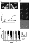
Thermosensitive phenotype of rpn3-4 and rpn3-7 mutants. (A) YE100 (rpn3-4) and YE101 (rpn3-7) mutant strains transformed with either an RPN3 gene-containing plasmid (YCp50::RPN3) or an empty vector (YCp50) were streaked on synthetic medium and incubated at 37°C for 2 days. (B) Wild-type YE46 (RPN3) cells and YE100 mutant (rpn3-4) cells were grown at 25°C to the early log phase and shifted at time zero (arrow) to 37°C. At hourly intervals, cell density was determined for both strains and plotted against time. (C) Wild-type YE46 (RPN3) cells and YE100 mutant (rpn3-4) cells were incubated as described for panel B and scored for bud morphology. Bars represent the percentages scored for each category. (D) Microtubule and nuclear phenotypes of rpn3-4 mutant cells at the restrictive temperature. YE100 (rpn3-4) cells were grown to the early log phase in YEPD medium at 25°C, shifted to 37°C, and incubated for 5 h prior to fixation and staining for mitotic spindles (tubulin) and nuclei (DAPI).
FIG. 3
FIG. 3
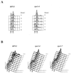
Cell cycle arrest phenotype of rpn3 mutants. (A) Wild-type YE46 (RPN3) cells and YE100 mutant (rpn3-4) cells were grown at 25°C to the early log phase and shifted to 37°C. Cell samples were withdrawn before (0 h) and every hour after the temperature upshift and analyzed for nuclear DNA content by flow cytometry. 1N and 2N indicate cells with unreplicated and fully replicated nuclear DNA, respectively. (B) Nuclear DNA profile of wild-type and ts rpn3 cells presynchronized in G1 with α-factor (αF) and released to 38.5°C. YE46 (RPN3), YE100 (rpn3-4), and YE101 (rpn3-7) strains were grown in YEPD medium to the early log phase, incubated with α-factor for 3.5 h at 25°C [αF 210 (25°C)], shifted to 38.5°C for an additional hour [αF 60 (38.5°C)], and finally resuspended in medium preequilibrated to 38.5°C and without the pheromone (0 min). Upon release from the G1 arrest, cell aliquots were taken at 20-min intervals and processed for flow cytometric analysis of nuclear DNA content. 1N and 2N indicate cells with unreplicated and fully replicated nuclear DNA, respectively.
FIG. 4
FIG. 4
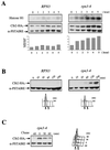
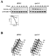
rpn3-4 mutants arrest with high levels of Clb2 and Clb2-associated histone H1 kinase activity and are defective in APC-dependent degradation of Clb2. (A) Wild-type YE44 (RPN3) cells and YE102 mutant (rpn3-4) cells carrying an HA-tagged CLB2 gene were grown and arrested as described in the legend to Fig. 3A. At hourly intervals, cell samples were recovered and Clb2-associated histone H1 kinase activity was determined by immunoprecipitation with an anti-HA antibody (upper panel; Histone H1). The same extracts were also analyzed by Western blotting with an anti-HA antibody to determine their Clb2 levels (lower panel; Clb2-HA3). Cdc28 immunodetected with an anti-PSTAIRE monoclonal antibody was used as a loading control (lower panel; α-PSTAIRE). Histograms represents quantification of the histone H1 kinase activity (percentage of Clb2-associated H1 kinase activity). (B) Accumulation of Clb2 in G1-arrested rpn3 mutant cells. YE106 (RPN3) cells and YE107 mutant (rpn3-4) cells harboring an HA-tagged CLB2 gene under the control of the inducible GAL1 promoter were grown to the early log phase in YEPR medium, arrested in G1 with α-factor for 2.5 h, and shifted to 37°C for an additional hour to inactivate the rpn3-4 gene product. At time zero, HA-tagged Clb2 expression was induced by adding galactose, and cell samples were collected at the indicated times for immunoblot analysis with an anti-HA antibody (Clb2-HA3). As in panel A, an immunoblot with an anti-PSTAIRE monoclonal antibody was used as a loading control (α-PSTAIRE). The effectiveness of the α-factor-induced G1 arrest in both experiments was assessed by FACScan analysis. 1N and 2N indicate cells with unreplicated and fully replicated nuclear DNA, respectively. (C) Clb2 is strongly stabilized in the rpn3 mutant. In an experiment similar to that shown in panel B, Clb2 expression was transiently induced with galactose for 60 min in G1-arrested YE107 (rpn3-4) cells and then repressed by transfer to prewarmed glucose-containing medium still in the presence of the pheromone. Cdc28 (α-PSTAIRE) is shown as a loading control.
FIG. 5
FIG. 5
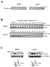
Role of Rpn3 in the proteolysis of Pds1. (A) Early-log-phase cultures of wild-type YE103 (RPN3) cells and YE104 mutant (rpn3-4) cells carrying an HA-tagged PDS1 gene were shifted from 25 to 37°C as described in the legend to Fig. 3A and analyzed by immunoblotting with an anti-HA antibody (Pds1-HA3) to monitor Pds1 levels at the indicated times. (B) Cell cycle regulation of Pds1 in the rpn3 mutant. Early-log-phase cultures of wild-type YE103 (RPN3) cells and YE104 mutant (rpn3-4) cells carrying an HA-tagged PDS1 gene were arrested in G1 with α-factor and then released into fresh medium lacking α-factor at 37°C. Samples for immunoblot analysis of HA-tagged Pds1 were withdrawn at the indicated times. Extracts from the wild-type strain with untagged PDS1 (no tag) were used as a control. A sample was also taken before the addition of α-factor as a source of asynchronous cells (Asyn). The 19S regulatory subunit Rpt1/Cim5 was immunodetected with specific antibodies as a loading control. (C) Reduced Pds1 turnover in the rpn3 mutant. Control YE108 (RPN3) cells and rpn3 mutant YE109 (rpn3-4) cells, both carrying an HA-tagged PDS1 gene under the control of the inducible GAL1 promoter, were grown in YEPR medium, arrested in G1, shifted to 37°C, and induced to express Pds1 for 60 min. Samples were collected before (lane −60) and after (lane 0) Pds1 induction and every 15 min after transfer of the cells to prewarmed α-factor-containing YEPD medium. A Cdc28 immunoblot is shown as a loading control (α-PSTAIRE). FACScan analysis of the first and last samples from this experiment is shown for each strain. (D) PDS1 deletion bypasses the metaphase arrest terminal phenotype of Rpn3-depleted cells. rpn3-54 (YE231) and rpn3-54 ΔPDS1 (YE416) cells, whose sole source of Rpn3 is expression from the GAL1 promoter, were inoculated into galactose-containing medium at 25°C. Rpn3 depletion was then induced at time zero by transferring both cultures to glucose-containing medium. At the indicated times, samples were collected for determination of their cellular DNA contents by flow cytometry. Cell samples were also recovered from the last (23-h) time point for phenotypic analysis of the arrested cells. DIC, differential interference contrast; DAPI, nuclear staining as observed with the DNA dye DAPI.
FIG. 5
FIG. 5
Role of Rpn3 in the proteolysis of Pds1. (A) Early-log-phase cultures of wild-type YE103 (RPN3) cells and YE104 mutant (rpn3-4) cells carrying an HA-tagged PDS1 gene were shifted from 25 to 37°C as described in the legend to Fig. 3A and analyzed by immunoblotting with an anti-HA antibody (Pds1-HA3) to monitor Pds1 levels at the indicated times. (B) Cell cycle regulation of Pds1 in the rpn3 mutant. Early-log-phase cultures of wild-type YE103 (RPN3) cells and YE104 mutant (rpn3-4) cells carrying an HA-tagged PDS1 gene were arrested in G1 with α-factor and then released into fresh medium lacking α-factor at 37°C. Samples for immunoblot analysis of HA-tagged Pds1 were withdrawn at the indicated times. Extracts from the wild-type strain with untagged PDS1 (no tag) were used as a control. A sample was also taken before the addition of α-factor as a source of asynchronous cells (Asyn). The 19S regulatory subunit Rpt1/Cim5 was immunodetected with specific antibodies as a loading control. (C) Reduced Pds1 turnover in the rpn3 mutant. Control YE108 (RPN3) cells and rpn3 mutant YE109 (rpn3-4) cells, both carrying an HA-tagged PDS1 gene under the control of the inducible GAL1 promoter, were grown in YEPR medium, arrested in G1, shifted to 37°C, and induced to express Pds1 for 60 min. Samples were collected before (lane −60) and after (lane 0) Pds1 induction and every 15 min after transfer of the cells to prewarmed α-factor-containing YEPD medium. A Cdc28 immunoblot is shown as a loading control (α-PSTAIRE). FACScan analysis of the first and last samples from this experiment is shown for each strain. (D) PDS1 deletion bypasses the metaphase arrest terminal phenotype of Rpn3-depleted cells. rpn3-54 (YE231) and rpn3-54 ΔPDS1 (YE416) cells, whose sole source of Rpn3 is expression from the GAL1 promoter, were inoculated into galactose-containing medium at 25°C. Rpn3 depletion was then induced at time zero by transferring both cultures to glucose-containing medium. At the indicated times, samples were collected for determination of their cellular DNA contents by flow cytometry. Cell samples were also recovered from the last (23-h) time point for phenotypic analysis of the arrested cells. DIC, differential interference contrast; DAPI, nuclear staining as observed with the DNA dye DAPI.
FIG. 6
FIG. 6
Cln2 stabilization and ubiquitination in an rpn3 mutant. (A) Control YE110 (RPN3) cells and rpn3 mutant YE111 (rpn3-4) cells, both with an HA-tagged CLN2 gene fused to the GAL1 promoter, were grown in YEPR medium and arrested in G1 with α-factor. The cultures were then shifted to 37°C for an additional hour and induced to express Cln2. After 45 min, Cln2 induction was terminated by returning the cells to prewarmed pheromone-free glucose-containing medium. Aliquots were taken before (lane −45) and after (lane 0) Cln2 induction and every 20 min after glucose repression. HA-tagged Cln2 and Rpt1 (used as a loading control) were immunodetected with anti-HA and anti-Rpt1 antibodies, respectively (top panel; Cln2-HA3 and Rpt1). CLN2-HA3 mRNAs were detected by Northern blot analysis (middle panel). The bottom panel shows ethidium bromide staining of the corresponding gel as a control for equivalent loading of total RNA in the different lanes. Quantification of HA-tagged Cln2 immunoreactivity at different times as measured by densitometry is shown in the graph. (B) Cln2-ubiquitin conjugates accumulate in the rpn3 mutant. Wild-type (strain YE110), rpn3-4 (strain YE111), and cdc34-2 (strain YE471) cells expressing HA-tagged Cln2 under the control of the GAL1 promoter were arrested in G1 with α-factor and then shifted to 37°C to inactivate Rpn3 and Cdc34. Cln2 expression was induced for 30 min by the addition of galactose. As a control for untagged Cln2, we used rpn3-54 strain YE231 (no tag). HA-tagged Cln2 was immunoprecipitated (IP) with a rabbit serum directed against the HA epitope; half of the immunoprecipitate was probed for ubiquitin conjugates by immunoblotting with a monoclonal antibody to polyubiquitin (top right panel), while the other half was checked for HA-tagged Cln2 by Western blotting (WB) with the anti-HA epitope monoclonal antibody 12CA5 (bottom right panel). Cell lysates used for the immunoprecipitation were separated by SDS-PAGE and analyzed with the antipolyubiquitin [anti-(Ub)n] monoclonal antibody to detect total cellular polyubiquitinated proteins (left panel).
FIG. 7
FIG. 7
Impaired degradation of Clb5 in a ts rpn3 mutant. (A) Wild-type YE46 (RPN3) cells and rpn3 mutant YE100 (rpn3-4) cells were transformed with a centromeric plasmid carrying an HA-tagged CLB5 gene fused to the GAL1 promoter (pGAL-CLB5HA). Cells were grown in selective medium with 2% raffinose to the early log phase, synchronized in G1 with a mating pheromone, and shifted to 37°C. After transient expression of Clb5 for 60 min by the addition of galactose, cells were transferred to prewarmed glucose-containing medium without the pheromone. Samples taken before (lane −60) or after (lane 0) Clb5 induction and at the indicated times following the termination of Clb5 expression were analyzed by SDS-PAGE and immunoblotting with an anti-HA antibody (Clb5-HA3). A Cdc28 immunoblot is shown as a loading control (α-PSTAIRE). Quantification of HA-tagged Clb5 immunoreactivity at the different times by densitometry is graphically represented. (B) G1 arrest failure of the rpn3 mutant upon ectopic expression of Clb5. Cell samples from the experiment shown in panel A were subjected to flow cytometric analysis. 1N and 2N indicate cells with unreplicated and fully replicated nuclear DNA, respectively.
FIG. 8
FIG. 8
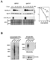
Sic1 degradation in an rpn3 mutant. (A) rpn3 mutants tolerate high levels of Sic1. Wild-type YE46 (RPN3) cells and rpn3 mutant YE100 and YE101 (rpn3-4 and rpn3-7) cells were transformed with a centromeric plasmid carrying an HA-tagged SIC1 gene under the control of the GAL1 promoter (YCpG-SIC1). Two independent transformants of each strain were streaked on selective medium containing either dextrose or galactose as a carbon source. Plates were photographed after 3 days of incubation at 30°C. (B) Sic1 levels in an arrested rpn3 mutant. Wild-type YE46 (RPN3) cells and rpn3 mutant YE100 (rpn3-4) cells were grown to the early log phase at 25°C and shifted to 37°C. Cells taken before (lane 0) and at hourly intervals after the shift were subjected to immunoblotting analysis with anti-Sic1 and anti-Rpt1 antisera. An extract from a SIC1-disrupted strain (ΔSIC1) was run in parallel as a control for the specificity of the anti-Sic1 antiserum. (C) Sic1 protein levels in synchronized rpn3 mutant cells. Control YE112 (RPN3) cells and rpn3 mutant YE113 (rpn3-4) cells, both carrying an HA-tagged CLN2 allele, were grown at 25°C to the early log phase, synchronized in G1 with α-factor, and shifted to 37°C. After release from the G1 arrest at the restrictive temperature, samples were withdrawn at the indicated times and probed by immunoblotting for HA-tagged Cln2, Sic1, and Rpt1 (as a loading control) with an anti-HA antibody (Cln2-HA3), Sic1-specific antiserum, and Rpt1/Cim5-specific antiserum, respectively. A graphic representation of the Sic1 and HA-tagged Cln2 (Cln2-HA3) immunoreactivities obtained by densitometry is also presented. (D) Sic1 turnover in an rpn3 mutant. Control YE114 (RPN3) cells and rpn3 mutant YE115 (rpn3-4) cells, both with an integrated GAL1:SIC1(HA)1 construct, were grown at 25°C in YEPR medium to the early log phase, arrested in G1 with α-factor, and shifted to 37°C. Galactose was added for 60 min to induce Sic1 expression, and the cells were returned to prewarmed glucose-containing medium to shut off the GAL1 promoter. Samples withdrawn before (lane −60) and after (lane 0) Sic1 induction and at the indicated times following glucose repression were subjected to Western blot analysis with anti-HA (Sic1-HA) and anti-PSTAIRE (α-PSTAIRE) antibodies to monitor HA-tagged Sic1 and Cdc28 (as a loading control), respectively. Flow cytometric analysis of the corresponding samples is also presented for each strain. A graphic representation of the Sic1 half-life, as estimated by immunoblotting, is also presented.
FIG. 8
FIG. 8
Sic1 degradation in an rpn3 mutant. (A) rpn3 mutants tolerate high levels of Sic1. Wild-type YE46 (RPN3) cells and rpn3 mutant YE100 and YE101 (rpn3-4 and rpn3-7) cells were transformed with a centromeric plasmid carrying an HA-tagged SIC1 gene under the control of the GAL1 promoter (YCpG-SIC1). Two independent transformants of each strain were streaked on selective medium containing either dextrose or galactose as a carbon source. Plates were photographed after 3 days of incubation at 30°C. (B) Sic1 levels in an arrested rpn3 mutant. Wild-type YE46 (RPN3) cells and rpn3 mutant YE100 (rpn3-4) cells were grown to the early log phase at 25°C and shifted to 37°C. Cells taken before (lane 0) and at hourly intervals after the shift were subjected to immunoblotting analysis with anti-Sic1 and anti-Rpt1 antisera. An extract from a SIC1-disrupted strain (ΔSIC1) was run in parallel as a control for the specificity of the anti-Sic1 antiserum. (C) Sic1 protein levels in synchronized rpn3 mutant cells. Control YE112 (RPN3) cells and rpn3 mutant YE113 (rpn3-4) cells, both carrying an HA-tagged CLN2 allele, were grown at 25°C to the early log phase, synchronized in G1 with α-factor, and shifted to 37°C. After release from the G1 arrest at the restrictive temperature, samples were withdrawn at the indicated times and probed by immunoblotting for HA-tagged Cln2, Sic1, and Rpt1 (as a loading control) with an anti-HA antibody (Cln2-HA3), Sic1-specific antiserum, and Rpt1/Cim5-specific antiserum, respectively. A graphic representation of the Sic1 and HA-tagged Cln2 (Cln2-HA3) immunoreactivities obtained by densitometry is also presented. (D) Sic1 turnover in an rpn3 mutant. Control YE114 (RPN3) cells and rpn3 mutant YE115 (rpn3-4) cells, both with an integrated GAL1:SIC1(HA)1 construct, were grown at 25°C in YEPR medium to the early log phase, arrested in G1 with α-factor, and shifted to 37°C. Galactose was added for 60 min to induce Sic1 expression, and the cells were returned to prewarmed glucose-containing medium to shut off the GAL1 promoter. Samples withdrawn before (lane −60) and after (lane 0) Sic1 induction and at the indicated times following glucose repression were subjected to Western blot analysis with anti-HA (Sic1-HA) and anti-PSTAIRE (α-PSTAIRE) antibodies to monitor HA-tagged Sic1 and Cdc28 (as a loading control), respectively. Flow cytometric analysis of the corresponding samples is also presented for each strain. A graphic representation of the Sic1 half-life, as estimated by immunoblotting, is also presented.
FIG. 9
FIG. 9
Sic1 regulation in an rpn12-1 mutant. (A) Hypersensitivity of the rpn12/nin1 mutant to elevated levels of Sic1. Control YK109-NIN1 (RPN12) cells and mutant YK109-nin1-1 (rpn12-1) cells were transformed with a centromeric plasmid carrying a SIC1 gene under the control of the GAL1 promoter. Two independent transformants of each strain were streaked on selective medium containing either dextrose or galactose as a carbon source. Plates were photographed after 3 days of incubation at 25°C. (B) Sic1 accumulation in the rpn12-1 mutant. Control YK109-NIN1 (RPN12) cells and mutant YK109-nin1-1 (rpn12-1) cells were grown to the early log phase at 25°C and shifted to 37°C. Samples taken before (lane 0) and at the indicated times after the shift to the restrictive temperature were probed by immunoblotting for Sic1 and Rpt1 (as an internal loading control) with antisera specific for these proteins. (C) Cln2 phosphoisoforms in the rpn12-1 mutant. Control YE112 (wild-type) cells, rpn12 mutant YE417 (rpn12-1) cells, and cdc34 mutant YE473 (cdc34-2) cells, all carrying an HA-tagged CLN2 allele, were grown at 25°C to the early log phase and shifted to 37°C. At the indicated times, samples were withdrawn for immunoblot analysis of Cln2 contents with an anti-HA-antibody (Cln2-HA3). Rpt1 was used as a loading control. (D) Asynchronous cultures of control YE112 (RPN12) cells and mutant YE417 (rpn12-1) cells, both carrying an HA-tagged CLN2 allele, were extracted and used for Cln2 immunoprecipitation with antibody 12CA5 directed against the HA epitope tag (anti-HA). As a control for the specificity of the HA epitope tag, the same cell lysates were immunoprecipitated with an irrelevant anti–glutathione S-transferase monoclonal antibody (control). Both immunoprecipitates were assayed for kinase activity with recombinant histidine-tagged Sic1 protein as a substrate. The incorporation of 32P-labeled phosphate into the Sic1 protein substrate (32PO4-Sic1) was monitored by autoradiography. (E) Control RPN12 (YE46) cells and rpn12-1 (YE413) cells harboring an HA-tagged CLN2 gene under the control of the tetracycline-repressible tetO2 promoter (pCM250) were grown to the early log phase at 25°C and shifted at time zero to 37°C. Samples were taken at hourly intervals for Cln2 immunoprecipitation with an anti-HA-antibody and for flow cytometric analysis of cellular DNA. Sic1 kinase activity present in Cln2 immunoprecipitates was assessed by the same in vitro assay as that used in panel D.
FIG. 9
FIG. 9
Sic1 regulation in an rpn12-1 mutant. (A) Hypersensitivity of the rpn12/nin1 mutant to elevated levels of Sic1. Control YK109-NIN1 (RPN12) cells and mutant YK109-nin1-1 (rpn12-1) cells were transformed with a centromeric plasmid carrying a SIC1 gene under the control of the GAL1 promoter. Two independent transformants of each strain were streaked on selective medium containing either dextrose or galactose as a carbon source. Plates were photographed after 3 days of incubation at 25°C. (B) Sic1 accumulation in the rpn12-1 mutant. Control YK109-NIN1 (RPN12) cells and mutant YK109-nin1-1 (rpn12-1) cells were grown to the early log phase at 25°C and shifted to 37°C. Samples taken before (lane 0) and at the indicated times after the shift to the restrictive temperature were probed by immunoblotting for Sic1 and Rpt1 (as an internal loading control) with antisera specific for these proteins. (C) Cln2 phosphoisoforms in the rpn12-1 mutant. Control YE112 (wild-type) cells, rpn12 mutant YE417 (rpn12-1) cells, and cdc34 mutant YE473 (cdc34-2) cells, all carrying an HA-tagged CLN2 allele, were grown at 25°C to the early log phase and shifted to 37°C. At the indicated times, samples were withdrawn for immunoblot analysis of Cln2 contents with an anti-HA-antibody (Cln2-HA3). Rpt1 was used as a loading control. (D) Asynchronous cultures of control YE112 (RPN12) cells and mutant YE417 (rpn12-1) cells, both carrying an HA-tagged CLN2 allele, were extracted and used for Cln2 immunoprecipitation with antibody 12CA5 directed against the HA epitope tag (anti-HA). As a control for the specificity of the HA epitope tag, the same cell lysates were immunoprecipitated with an irrelevant anti–glutathione S-transferase monoclonal antibody (control). Both immunoprecipitates were assayed for kinase activity with recombinant histidine-tagged Sic1 protein as a substrate. The incorporation of 32P-labeled phosphate into the Sic1 protein substrate (32PO4-Sic1) was monitored by autoradiography. (E) Control RPN12 (YE46) cells and rpn12-1 (YE413) cells harboring an HA-tagged CLN2 gene under the control of the tetracycline-repressible tetO2 promoter (pCM250) were grown to the early log phase at 25°C and shifted at time zero to 37°C. Samples were taken at hourly intervals for Cln2 immunoprecipitation with an anti-HA-antibody and for flow cytometric analysis of cellular DNA. Sic1 kinase activity present in Cln2 immunoprecipitates was assessed by the same in vitro assay as that used in panel D.

Similar articles

Cited by

References

    1. Amon A, Irniger S, Nasmyth K. Closing the cell cycle circle: G2 cyclin proteolysis initiated at mitosis persists until activation of G1 cyclins in the next cell cycle. Cell. 1994;77:1037–1050. - PubMed
    1. Bai C, Sen P, Hofmann K, Ma L, Goebl M, Harper J W, Elledge S J. SKP1 connects cell cycle regulators to the ubiquitin proteolysis machinery through a novel motif, the F-box. Cell. 1996;86:263–274. - PubMed
    1. Bailly, E. Unpublished results.
    1. Barral Y, Jentsch S, Mann C. G1 cyclin turnover and nutrient uptake are controlled by a common pathway in yeast. Genes Dev. 1995;9:399–409. - PubMed
    1. Baumeister W, Walz J, Zhul F, Seemuller E. The proteasome: paradigm of a self-compartmentalizing protease. Cell. 1998;92:367–380. - PubMed

Publication types

MeSH terms

Substances