. 1999 Oct;19(10):6891-7.
doi: 10.1128/MCB.19.10.6891.

The essential functions of human Rad51 are independent of ATP hydrolysis

Affiliations

The essential functions of human Rad51 are independent of ATP hydrolysis

C Morrison et al. Mol Cell Biol. 1999 Oct.

Abstract

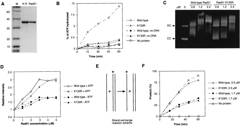
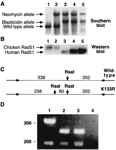
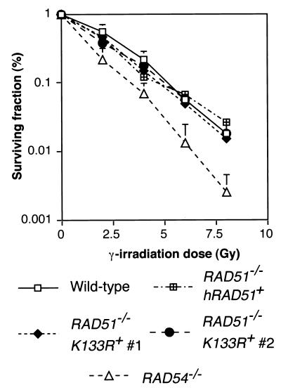
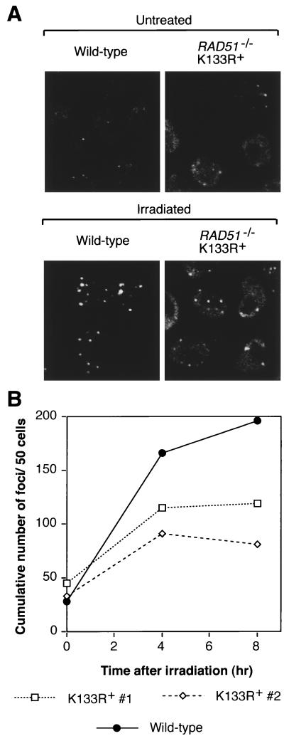
Genetic recombination and the repair of double-strand DNA breaks in Saccharomyces cerevisiae require Rad51, a homologue of the Escherichia coli RecA protein. In vitro, Rad51 binds DNA to form an extended nucleoprotein filament and catalyzes the ATP-dependent exchange of DNA between molecules with homologous sequences. Vertebrate Rad51 is essential for cell proliferation. Using site-directed mutagenesis of highly conserved residues of human Rad51 (hRad51) and gene targeting of the RAD51 locus in chicken DT40 cells, we examined the importance of Rad51's highly conserved ATP-binding domain. Mutant hRad51 incapable of ATP hydrolysis (hRad51K-133R) binds DNA less efficiently than the wild type but catalyzes strand exchange between homologous DNAs. hRad51 does not need to hydrolyze ATP to allow vertebrate cell proliferation, form nuclear foci, or repair radiation-induced DNA damage. However, cells expressing hRad51K-133R show greatly reduced targeted integration frequencies. These findings show that ATP hydrolysis is involved in DNA binding by hRad51 and suggest that the extent of DNA complexed with hRad51 in nucleoprotein influences the efficiency of recombination.


Figures

FIG. 1
FIG. 1
Biochemical characterization of hRad51. (A) Purification of Rad51 protein from E. coli. A total of 2 μg of hRad51K-133R (K-R) and 1.5 μg of the wild-type hRad (Rad51) protein were loaded in the lanes indicated. The sizes of the molecular mass markers (M) are shown at the left, in kilodaltons. (B) ATP hydrolysis mediated by hRad51 and hRad51K-133R proteins. hRad51 was used at 1 μM and ATP was used at 200 μM in the presence or absence of 20 μM poly(dT) as indicated. Results are the averages of five experiments. (C) Binding of φX174 DNA by wild-type hRad51 and hRad51K-133R in the presence of ATP. As shown in the left lane, no DNA is bound in the absence of protein. The arrows at the left indicate open circular (OC) and closed circular (CC) forms of the DNA. A total of 130 ng of DNA was incubated with various concentrations of the protein in the presence of ATP for 5 min and then complexes were analyzed by 0.9% agarose gel electrophoresis. Longer times (up to 30 min) of incubation of DNA with protein yielded the same results. (D) Binding of etheno-DNA by wild-type hRad51 and hRad51K-133R in the presence or absence of 1 mM ATP as indicated. Fluorescence was measured after 5 min of incubation, when the excitation values reached a plateau. (E) Diagram of the strand exchange reaction. The assay monitors the appearance of labelled ssDNA (indicated by asterisks), showing the exchange of the labelled and unlabelled strands. (F) DNA strand exchange mediated by wild-type hRad51 or hRad51K-133R. Different concentrations of protein were incubated first with unlabelled single-stranded oligonucleotide and then with labelled homologous dsDNA, and the exchange of labelled and unlabelled ssDNA over time was monitored by 12% polyacrylamide gel electrophoresis. Data are plotted as percentages of products over total input label as calculated with a phosphorimager.
FIG. 2
FIG. 2
Generation of RAD51−/− hRAD51K-133R+ DT40 cells. (A) Southern blot analysis of targeting of the chicken RAD51 locus (39). DNA from wild-type (lane 1), RAD51+/− (lane 2), RAD51−/− tet-hRAD51+ clone 110 (lane 3), RAD51−/− hRAD51K-133R+ clone 1 (lane 4), and RAD51−/− hRAD51K-133R+ clone 2 (lane 5) DT40 cells was used. (B) Western blot analysis. Total protein (10 μg/lane) was loaded from wild-type (lane 1), RAD51+/−(lane 2), RAD51−/− tet-hRAD51+ clone 110 (lane 3), RAD51−/− hRAD51K-133R+ clone 1 (lane 4), and RAD51−/− hRAD51K-133R+ clone 2 (lane 5) DT40 cells and immunoblotted with anti-hRad51 antiserum. (C) Scheme of diagnostic restriction digestion for the K-133R mutation. A second RsaI site in the PCR fragment shown was introduced as indicated into the mutant hRad51 gene. Expected sizes are indicated, in base pairs. (D) RT-PCR analysis of K-133R transgene expression. After reverse transcription and amplication of 1 μg of total RNA, 20 μl of each 100-μl reaction mixture was digested with RsaI and run on a 2.5% agarose gel. Shown are digests from RAD51−/− tet-hRAD51+ clone 110 (lane 1), RAD51−/− hRAD51K-133R+ clone 1 (lane 2), and RAD51−/− hRAD51K-133R+ clone 2 (lane 3) DT40 cells and a no reverse transcriptase control amplification (lane 4). Sizes are shown at the left, in base pairs.
FIG. 3
FIG. 3
Gamma ray sensitivity of RAD51−/− hRAD51K-133R+ DT40 cells. Survival 1 week after 137Cs irradiation with the indicated dose was quantified by colony formation in methylcellulose-containing medium relative to that of nonirradiated cells. Hypersensitive RAD54−/− cells (8) and a RAD51−/− cell line expressing comparably high levels of hRad51 (40) were included as controls. Plating efficiency was ≈100% for RAD51−/− hRAD51K-133R+ cells, and the data are the means ± the standard deviations of three experiments.
FIG. 4
FIG. 4
Formation of Rad51 foci by Rad51K-133R mutant proteins. (A) Immunofluorescent visualization of Rad51 foci in RAD51−/− hRAD51K-133R+ DT40 cells. Cells were fixed either directly or 8 h after gamma irradiation with 8 Gy of 137Cs. The high background levels in mutant cells are attributed to the very high hRad51 expression levels. This experiment was carried out at least twice for all genotypes shown. Images were processed with Adobe Photoshop, version 4.0J. (B) Kinetics of focus formation. Following microscopy and image processing with Adobe Photoshop, version 4.0J, color-inverted images were printed and distinct foci were counted. At least 80 cells from three randomly chosen frames were counted per time point.

Similar articles

Cited by

References

    1. Aboussekhra A, Chanet R, Adjiri A, Fabre F. Semidominant suppressors of Srs2 helicase mutations of Saccharomyces cerevisiae map in the RAD51 gene, whose sequence predicts a protein with similarities to procaryotic RecA proteins. Mol Cell Biol. 1992;12:3224–3234. - PMC - PubMed
    1. Basile G, Aker M, Mortimer R K. Nucleotide sequence and transcriptional regulation of the yeast recombinational repair gene RAD51. Mol Cell Biol. 1992;12:3235–3246. - PMC - PubMed
    1. Baumann P, Benson F E, Hajibagheri N, West S C. Purification of human Rad51 protein by selective spermidine precipitation. Mutat Res. 1997;384:65–72. - PubMed
    1. Baumann P, Benson F E, West S C. Human Rad51 protein promotes ATP-dependent homologous pairing and strand transfer reactions in vitro. Cell. 1996;87:757–766. - PubMed
    1. Baumann P, West S C. Role of the human Rad51 protein in homologous recombination and double-stranded break repair. Trends Biochem Sci. 1998;23:247–251. - PubMed

Publication types

MeSH terms

LinkOut - more resources