. 1999 Oct;19(10):6898-905.
doi: 10.1128/MCB.19.10.6898.

Delayed translational silencing of ceruloplasmin transcript in gamma interferon-activated U937 monocytic cells: role of the 3' untranslated region

Collaborators, Affiliations

Delayed translational silencing of ceruloplasmin transcript in gamma interferon-activated U937 monocytic cells: role of the 3' untranslated region

B Mazumder et al. Mol Cell Biol. 1999 Oct.

Abstract

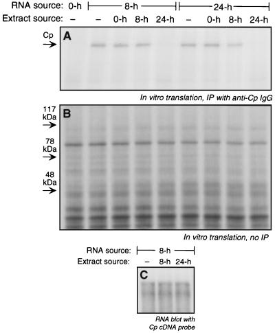
Ceruloplasmin (Cp) is an acute-phase protein with ferroxidase, amine oxidase, and pro- and antioxidant activities. The primary site of Cp synthesis in human adults is the liver, but it is also synthesized by cells of monocytic origin. We have shown that gamma interferon (IFN-gamma) induces the synthesis of Cp mRNA and protein in monocytic cells. We now report that the induced synthesis of Cp is terminated by a mechanism involving transcript-specific translational repression. Cp protein synthesis in U937 cells ceased after 16 h even in the presence of abundant Cp mRNA. RNA isolated from cells treated with IFN-gamma for 24 h exhibited a high in vitro translation rate, suggesting that the transcript was not defective. Ribosomal association of Cp mRNA was examined by sucrose centrifugation. When Cp synthesis was high, i.e., after 8 h of IFN-gamma treatment, Cp mRNA was primarily associated with polyribosomes. However, after 24 h, when Cp synthesis was low, Cp mRNA was primarily in the nonpolyribosomal fraction. Cytosolic extracts from cells treated with IFN-gamma for 24 h, but not for 8 h, contained a factor which blocked in vitro Cp translation. Inhibitor expression was cell type specific and present in extracts of human cells of myeloid origin, but not in several nonmyeloid cells. The inhibitory factor bound to the 3' untranslated region (3'-UTR) of Cp mRNA, as shown by restoration of in vitro translation by synthetic 3'-UTR added as a "decoy" and detection of a binding complex by RNA gel shift analysis. Deletion mapping of the Cp 3'-UTR indicated an internal 100-nucleotide region of the Cp 3'-UTR that was required for complex formation as well as for silencing of translation. Although transcript-specific translational control is common during development and differentiation and global translational control occurs during responses to cytokines and stress, to our knowledge, this is the first report of translational silencing of a specific transcript following cytokine activation.


Figures

FIG. 1
FIG. 1
Cessation of Cp synthesis in the presence of abundant Cp mRNA. (A) A steady-state amount of Cp mRNA was measured by mRNA blot analysis. U937 cells (108 cells in 50 ml) were treated with IFN-γ (500 U/ml) for up to 24 h. Poly(A)-selected mRNA was fractionated on 1% agarose–formaldehyde, transferred to Nytran membranes, and hybridized with a random primer-labeled 646-bp human Cp probe. The 18S and 28S rRNA bands are indicated by arrows. (B) The mRNA blot was stripped and rehybridized with a GAPDH cDNA probe. (C) The release of Cp into conditioned medium was measured by immunoblot analysis. U937 cells (2 × 10⁶ cells/ml) were treated with IFN-γ (500 U/ml) for up to 24 h. The conditioned medium was collected at the time indicated and replaced with fresh medium for the next collection. The conditioned medium was concentrated and subjected to SDS-PAGE and immunoblot analysis with rabbit anti-human Cp IgG. A purified human Cp standard (Std., 25 ng) is in the leftmost lane; the arrow indicates intact 132-kDa Cp. (D) Quantitation of Cp mRNA and protein synthesis. Cp protein made during each collection period in panel C was quantitated by densitometry, normalized by comparison to the Cp standard, and expressed as nanograms per hour (gray bars). Cp mRNA in panel A and GAPDH mRNA in panel B were quantitated by densitometry, and Cp mRNA was expressed as relative densitometric units after normalization with GAPDH mRNA (o). (E) The rate of Cp synthesis was measured by metabolic labeling. U937 cells (8 × 10⁶ cells in 4 ml) were treated with IFN-γ (500 U/ml) for 0, 8, or 24 h. At the end of each interval, cells were metabolically labeled by incubation with [35S]methionine in methionine-free medium for 2 h. The conditioned medium (CM) and lysates (Lys.) were immunoprecipitated (IP) with rabbit anti-human Cp IgG and resolved by SDS-PAGE, and radiolabeled bands were detected by fluorography. The arrow indicates the position of intact 132-kDa Cp.
FIG. 2
FIG. 2
In vitro translation of Cp mRNA from IFN-γ-treated U937 cells. U937 cells (108 cells) were incubated with IFN-γ (500 U/ml) for up to 24 h. Total RNA was isolated at the time shown, and an aliquot (100 μg) was subjected to in vitro translation for 1 h at 30°C with a rabbit reticulocyte lysate system in the presence of [35S]methionine. Translated Cp was immunoprecipitated (IP) with rabbit antihuman Cp IgG, resolved by SDS-PAGE (7% polyacrylamide), and detected by fluorography.
FIG. 3
FIG. 3
Release of Cp mRNA from polysomes by IFN-γ. U937 cells (5 × 10⁸ cells) were incubated with IFN-γ (500 U/ml) for 0, 8, and 24 h. The cells were homogenized in buffer containing cycloheximide (100 μg/ml) to prevent further elongation and centrifuged at low speed. The postmitochondrial supernatant was separated into polysomal (P) and nonpolysomal (NP) fractions by centrifugation through a sucrose (20% wt/vol) cushion. In one tube, cycloheximide was replaced by puromycin (100 μg/ml) to release mRNA from ribosomes. Total mRNA from both fractions was isolated by SDS-proteinase K digestion, Trizol reagent extraction, and poly(A) selection. (A) The blot was subjected to RNA blot analysis with a human Cp cDNA probe. The 18S and 28S rRNA bands are indicated by arrows. (B) The blot was stripped and rehybridized with a human γ-actin cDNA probe.
FIG. 4
FIG. 4
Inhibition of Cp translation by extracts from IFN-γ-treated U937 cells. U937 cells (5 × 10⁸ cells) were treated with IFN-γ (500 U/ml) for 0, 8, and 24 h. Total RNA (100 μg) was subjected to in vitro translation with the rabbit reticulocyte lysate system. Extracts (4 μg of protein) made from cells treated with IFN-γ for the same times were added to the translation reaction. (A) Translated, 35S-labeled Cp was immunoprecipitated (IP) with rabbit anti-human Cp IgG and resolved by SDS-PAGE, and radiolabeled bands were detected by fluorography. (B) Total in vitro protein synthesis was determined with an aliquot of the translated material described in panel A that was not subjected to immunoprecipitation. Translated, 35S-labeled protein was resolved by SDS-PAGE and detected by fluorography. (C) Analysis of Cp mRNA stability. U937 cells (5 × 10⁸ cells) were treated with IFN-γ (500 U/ml) for 8 and 24 h. Total RNA was isolated from the 8-h-treated cells, and 100 μg was incubated for 1 h at 30°C in a translation reaction mixture (without [35S]methionine) containing cell extract (4 μg of protein) from 8- and 24-h-treated cells. The RNA was reisolated with Trizol reagent and subjected to poly(A) selection and RNA blot analysis with a human Cp cDNA probe.
FIG. 5
FIG. 5
Cell specificity of the inhibitory activity. (A) The presence of translation inhibitory activity in HT1080, HeLa, HepG2, and human umbilical vein endothelial cells (HUVEC) was tested after incubating cells with IFN-γ for 24 h. Extracts (4 μg of protein) made from 108 cells were added to the in vitro translation reaction mixture in the presence of RNA prepared from U937 cells treated with IFN-γ for 8 h. Translated 35S-labeled Cp was immunoprecipitated (IP) with rabbit antihuman Cp IgG, resolved by SDS-PAGE (7% polyacrylamide), and detected by fluorography. (B) The presence of translation inhibitory activity in peripheral blood monocytes (PBM) was determined as in panel A.
FIG. 6
FIG. 6
RNA decoy experiment to evaluate the role of 3′-UTR in translational silencing of Cp mRNA. U937 cells (5 × 10⁸ cells) were incubated with IFN-γ (500 U/ml) for 0, 8, and 24 h. Total RNA was isolated from these cells, and 100 μg was subjected to in vitro translation using rabbit reticulocyte lysate in the presence of cytosolic extracts (4 μg of protein from 100,000 × g supernatant) made from IFN-γ-treated cells. Synthetic unlabeled transcripts of Cp 3′-UTR (247 nt), 15-lipoxygenase (LO) 3′-UTR (241 nt), and Cp exon 5 (255 nt) were tested for their ability to restore translation in the presence of the inhibitory extract. The unlabeled transcripts (500 ng) were preincubated for 10 min with extracts made from U937 cells treated with IFN-γ for 24 h and then added to the translation reaction mixture. Newly translated, 35S-labeled Cp was detected as in Fig. 2. Compet., competitor.
FIG. 7
FIG. 7
RNA decoy experiment using Cp 3′-UTR deletion fragments. (Top) Schematic representation of the Cp 3′-UTR deletion fragments and results from using these fragments. (Bottom) In vitro translation of U937 total RNA was done as described in the legend to Fig. 6, except that the cell extract was preincubated with unlabeled transcripts (500 ng) of each of the deletion fragments of the Cp 3′-UTR before addition to the translation reaction mixture. Compet., competitor.
FIG. 8
FIG. 8
RNA gel shift assay to detect protein or proteins binding to Cp 3′-UTR. α-32P-labeled Cp 3′-UTR transcript (10 fmol) was incubated for 30 min at room temperature with cytosolic extract (10 μg of protein) prepared from U937 cells treated with IFN-γ for 0, 8, and 24 h. RNA-protein complexes were resolved by electrophoresis on a nondenaturing 5% polyacrylamide gel and detected by autoradiography. In the lanes showing competition, a 25-fold molar excess of unlabeled Cp 3′-UTR transcript, 15-lipoxygenase 3′-UTR, and RNA corresponding to Cp exon 5 were preincubated for 10 min with the extract before addition of labeled probe.
FIG. 9
FIG. 9
RNA gel shift assay with Cp 3′-UTR deletion fragments. The RNA gel shift assay was done as in Fig. 8, except that binding of the extract to α-32P-labeled, full-length Cp 3′-UTR transcript (10 fmol) was competed for by a 25-fold molar excess of the full-length transcript or by a 25- or 100-fold molar excess of the deletion fragments illustrated in Fig. 7 (top).

References

    1. Al-Timimi D J, Dormandy T L. The inhibition of lipid autoxidation by human caeruloplasmin. Biochem J. 1977;168:283–288. - PMC - PubMed
    1. Askwith C, Eide D, Van Ho A, Bernard P S, Li L, Davis-Kaplan S, Sipe D M, Kaplan J. The FET3 gene of S. cerevisiae encodes a multicopper oxidase required for ferrous iron uptake. Cell. 1994;76:403–410. - PubMed
    1. Attieh Z K, Mukhopadhyay C K, Seshadri V, Tripoulas N A, Fox P L. Ceruloplasmin ferroxidase activity stimulates cellular iron uptake by a trivalent cation-specific transport mechanism. J Biol Chem. 1999;274:1116–1123. - PubMed
    1. Avni D, Shama S, Loreni F, Meyuhas O. Vertebrate mRNAs with a 5′-terminal pyrimidine tract are candidates for translational repression in quiescent cells: characterization of the translational cis-regulatory element. Mol Cell Biol. 1994;14:3822–3833. - PMC - PubMed
    1. Aziz N, Munro H N. Iron regulates ferritin mRNA translation through a segment of its 5′ untranslated region. Proc Natl Acad Sci USA. 1987;84:8478–8482. - PMC - PubMed

Publication types

LinkOut - more resources