. 1999 Oct;19(10):6940-52.
doi: 10.1128/MCB.19.10.6940.

Sperm chromatin decondensation by template activating factor I through direct interaction with basic proteins

Affiliations

Sperm chromatin decondensation by template activating factor I through direct interaction with basic proteins

K Matsumoto et al. Mol Cell Biol. 1999 Oct.

Abstract

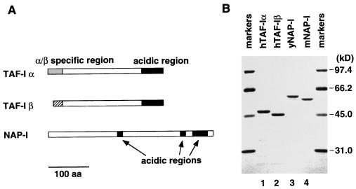
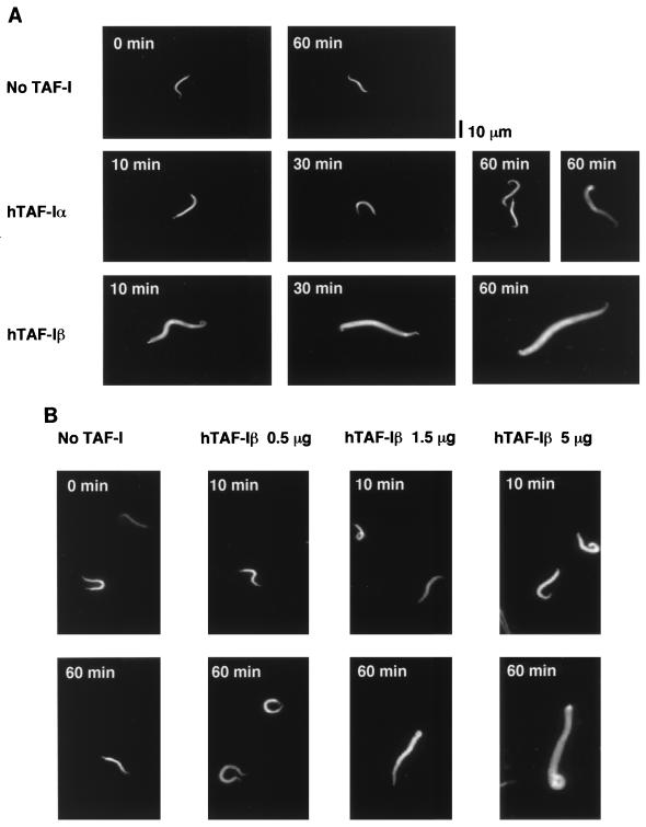
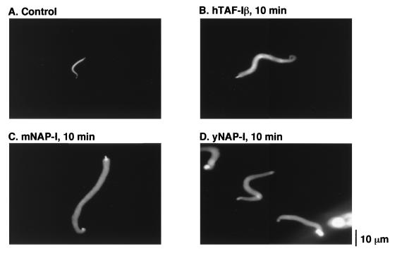
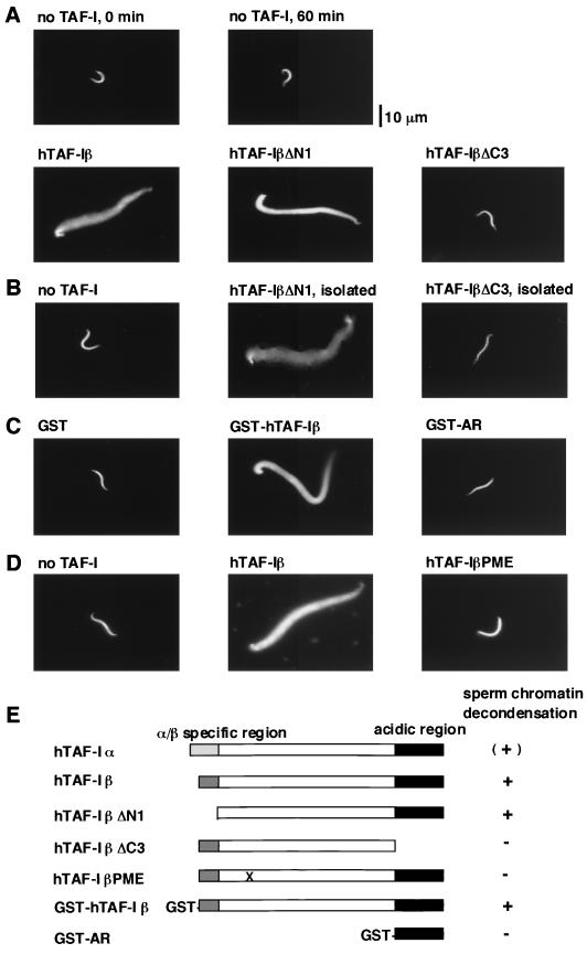
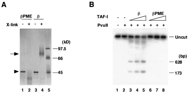
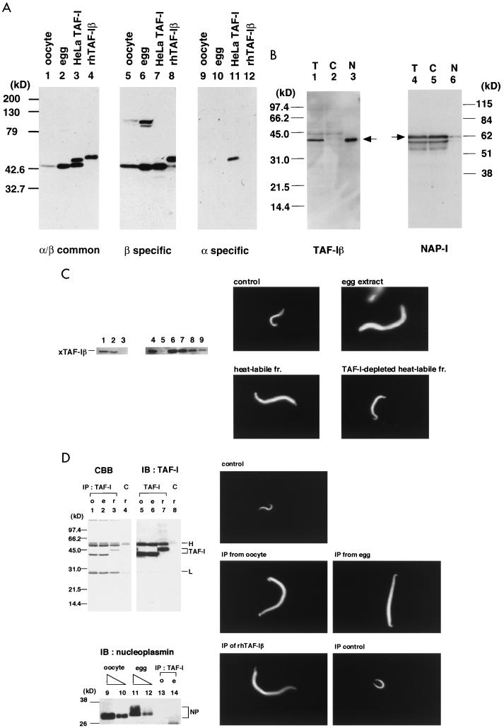
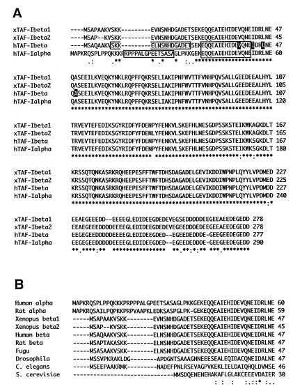
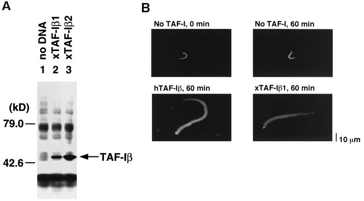
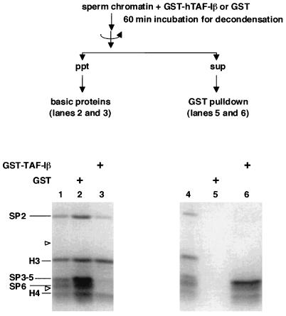
Template activating factor I (TAF-I) was originally identified as a host factor required for DNA replication and transcription of adenovirus genome complexed with viral basic proteins. Purified TAF-I was shown to bind to core histones and stimulate transcription from nucleosomal templates. Human TAF-I consists of two acidic proteins, TAF-Ialpha and TAF-Ibeta, which differ from each other only in their amino-terminal regions. Here, we report that TAF-I decondenses demembraned Xenopus sperm chromatin. Human TAF-Ibeta has a chromatin decondensation activity comparable to that of NAP-I, another histone binding protein, whereas TAF-Ialpha has only a weak activity. Analysis of molecular mechanisms underlying the chromatin decondensation by TAF-I revealed that TAF-I interacts directly with sperm basic proteins. Deletion of the TAF-I carboxyl-terminal acidic region abolishes the decondensation activity. Interestingly, the acidic region itself is not sufficient for decondensation, since an amino acid substitution mutant in the dimerization domain of TAF-I which has the intact acidic region does not support chromatin decondensation. We detected the beta form of TAF-I in Xenopus oocytes and eggs by immunoblotting, and the cloning of its cDNA led us to conclude that Xenopus TAF-Ibeta also decondenses sperm chromatin. These results suggest that TAF-I plays a role in remodeling higher-order chromatin structure as well as nucleosomal structure through direct interaction with chromatin basic proteins.


Figures

FIG. 1
FIG. 1
Structure of TAF-I and NAP-I. (A) Schematic diagrams of TAF-Iα, TAF-Iβ, and NAP-I. TAF-Iα- and TAF-Iβ-specific regions are indicated by gray and hatched boxes, respectively. The TAF-I acidic region and the NAP-I tripartite acidic regions are shown by black boxes. Bar, 100 amino acids. (B) Recombinant proteins used in this study. Purified recombinant human TAF-Iα and TAF-Iβ and yeast and mouse NAP-I proteins (yNAP-I and mNAP-I, respectively) were analyzed by SDS–10% PAGE and stained with Coomassie brilliant blue. The sizes of the molecular mass markers (Bio-Rad) are shown on the right.
FIG. 2
FIG. 2
Decondensation of Xenopus sperm chromatin by TAF-Iα and TAF-Iβ. (A) Time course of decondensation by TAF-Iα and TAF-Iβ. Sperm chromatin (5 × 10⁴ sperm) was incubated without (top panels) or with 5 μg of recombinant hTAF-Iα (middle panels) or hTAF-Iβ (lower panels) as described in Materials and Methods. At the indicated times, aliquots were mixed with fixation buffer containing Hoechst 33258 stain and the chromosomal DNA was visualized under a fluorescent microscope. For the sample from the 60-min incubation with hTAF-Iα, two panels are shown to represent a weak decondensation activity (see text for details). Bar, 10 μm. (B) Dose response of hTAF-Iβ in chromatin decondensation. Sperm chromatin (5 × 10⁴ sperm) was incubated with 0, 0.5, 1.5, and 5 μg of hTAF-Iβ for 10 and 60 min.
FIG. 3
FIG. 3
Chromatin decondensation by NAP-I. Sperm chromatin (5 × 10⁴ sperm) was incubated with 5 μg of hTAF-Iβ (B), mouse NAP-I (C), and yeast NAP-I (D) for 10 min. The chromosomal DNA was visualized as described in Materials and Methods. Sperm chromatin that was not incubated with any proteins is shown in panel A.
FIG. 4
FIG. 4
Domains required for chromatin decondensation. (A to D) Sperm chromatin was incubated with 5 μg of hTAF-Iβ and its derivatives for 60 min. The chromosomal DNA was stained with Hoechst 33258 stain and visualized under a fluorescent microscope. In panel B, the gel-isolated hTAF-IβΔN1 and hTAF-IβΔC3 were used (see Materials and Methods). (E) The results of domain analysis for sperm chromatin decondensation are summarized. The decondensation activity of each hTAF-I derivative is shown to the right of its schematic diagram. + and −, active and inactive in the decondensation assay, respectively. hTAF-Iα has a very weak activity, indicated by (+). Point mutations in hTAF-IβPME are shown by X in the schematic diagram.
FIG. 5
FIG. 5
Dimerization of TAF-I. (A) Chemical cross-linking of hTAF-I proteins. One hundred nanograms of hTAF-IβPME (lanes 1 and 2) or hTAF-Iβ (lanes 3 and 4) was cross-linked with (lanes 2 and 4) or without (lanes 1 and 3) 0.05% glutaraldehyde. Samples were analyzed by SDS–7.5% PAGE, and proteins were detected by silver staining. Lane 5 shows marker proteins. Monomer and dimer are indicated by an arrowhead and an arrow, respectively. The cross-linked product migrating faster than the monomer appears to be a compact form of TAF-I due to intramolecular cross-linking. (B) Restriction enzyme sensitivity assay. Adenovirus core (30 ng) was incubated at 30°C for 30 min without (lanes 1 and 2) or with 100 ng (lanes 3 and 6), 200 ng (lanes 4 and 7), or 400 ng (lanes 5 and 8) of hTAF-Iβ (lanes 3 to 5) and hTAF-IβPME (lanes 6 to 8) and then digested with PvuII. Lane 1 shows the undigested DNA. DNA was purified and separated by electrophoresis in a 1% agarose gel. DNA fragments around the E1A promoter region were detected by Southern blotting. One-hundred-seventy-three- and 628-bp-long fragments were shown by the probe after partial digestion with PvuII.
FIG. 6
FIG. 6
Existence of TAF-Iβ, but not TAF-Iα, in Xenopus oocytes and eggs. (A) Detection of TAF-Iβ in Xenopus oocytes and eggs. Xenopus oocyte lysates (30 μg of protein; lanes 1, 5, and 9), egg extracts (30 μg of protein; lanes 2, 6, and 10), purified HeLa TAF-I (lanes 3, 7, and 11), and recombinant human TAF-Iβ (40 ng; lanes 4, 8, and 12) were analyzed by SDS–10% PAGE and transferred to a polyvinylidene difluoride membrane. TAF-I was detected with monoclonal antibodies which recognize human TAF-Iα and TAF-Iβ common region (clone KM1725 in reference 40) (lanes 1 to 4), TAF-Iβ-specific region (clone KM1720) (lanes 5 to 8), or TAF-Iα-specific region (clone KM1712) (lanes 9 to 12) by immunoblotting (see Fig. 7 for the epitope of each antibody). It should be noted that recombinant hTAF-Iβ has a six-histidine tag at its amino terminus, which might have resulted in its lower mobility than that of the native hTAF-Iβ (compare a band in lane 4 and the lower band in lane 3). Molecular masses of marker proteins are shown on the left. (B) Localization of TAF-Iβ and NAP-I in oocytes. A Xenopus oocyte was separated into nucleus and cytoplasm. Lysates of total oocyte (T), nuclear (N), and cytoplasmic fractions (C) were analyzed by immunoblotting with anti-TAF-Iβ antibody (lanes 1 to 3). Immunoblotting with anti-NAP-I antibody of the same oocyte fractionation is also shown (lanes 4 to 6). The positions of TAF-I and NAP-I are indicated by arrows. (C) Chromatin decondensation activity of TAF-Iβ in heat-labile fraction of egg extracts. Xenopus egg extracts were heated at 80°C for 10 min and separated into heat-stable and heat-labile fractions. The heat-labile fraction was subjected to the denature-renature protocol, and TAF-I was depleted from the renatured heat-labile fraction. Left panels show immunoblotting analysis using anti-TAF-Iβ antibody of egg extracts (lane 1), heat-labile fraction solubilized in guanidine-containing buffer (lane 2), heat-stable fraction (lane 3), mock-depleted renatured heat-labile fraction (lane 4), TAF-I depleted renatured heat-labile fraction (lane 5), and sequential dilutions of renatured heat-labile fraction (lane 6, 100%; lane 7, 30%; lane 8, 10%; lane 9, 3%). Right panels show chromatin decondensation with these fractions. Sperm chromatin (104 sperm) was incubated for 10 min with egg extracts, mock-depleted renatured heat-labile fraction, and TAF-I-depleted renatured heat-labile fraction. The chromosomal DNA was stained with Hoechst 33258 stain and visualized under a fluorescent microscope. (D) Sperm chromatin decondensation by TAF-Iβ immunopurified from oocyte and egg extracts. The oocyte and egg extracts and recombinant hTAF-Iβ were subjected to immunoprecipitation with anti-TAF-Iβ antibody or control mouse IgG as described in Materials and Methods. The immunoprecipitates of oocyte extracts (labeled “o”) (lanes 1, 5, and 13), egg extracts (labeled “e”) (lanes 2, 6, and 14), and recombinant hTAF-Iβ (labeled “r”) (lanes 3 and 7) with anti-TAF-Iβ antibody or the immunoprecipitates of recombinant hTAF-Iβ with control mouse IgG (lanes 4 and 8) were analyzed by SDS-PAGE followed by staining with Coomassie brilliant blue (lanes 1 to 4) and by immunoblotting with anti-TAF-Iβ antibody (lanes 5 to 8) and with anti-nucleoplasmin monoclonal antibody (lanes 13 and 14). Two microliters (lanes 9 and 11) and 0.6 microliter (lanes 10 and 12) of oocyte (lanes 9 and 10) and egg (lanes 11 and 12) extracts were also analyzed by immunoblotting with antinucleoplasmin antibody. Positions of TAF-Iβ, IgG heavy chain (H) and light chain (L), and nucleoplasmin (NP) are indicated. It is important to note that the egg nucleoplasmin has lower mobility than the oocyte nucleoplasmin due to the hyperphosphorylation (32, 43, 48). The proteins released at pH 11 from the protein G beads were assayed for mediating the decondensation of sperm chromatin (2 × 10⁴) for 60 min. The panel labelled “control” shows the sperm chromatin incubated with no added protein. The panel labelled “IP control” shows the sperm chromatin incubated with the proteins released after the immunoprecipitation of recombinant hTAF-Iβ with control mouse IgG.
FIG. 7
FIG. 7
Sequence comparison of TAF-I. (A) The complete amino acid sequences of Xenopus TAF-Iβ1 and TAF-Iβ2, human TAF-Iβ (accession no. M93651), and TAF-Iα (D45198) aligned by use of a CLUSTALW program. Amino acids identical in all four sequences are indicated by asterisks, conserved substitutions are indicated by colons, and semiconserved substitutions are indicated by periods at the bottom of the alignment. The four amino acids mutated in hTAF-IβPME are highlighted with black shading. The peptide sequence to produce monoclonal antibodies against TAF-I (α and β common, EQQEAIEHIDEVQNE; α-specific, RPPPALGPEETSASA; β-specific, SKKELNSNHDGADET) are boxed. (B) Amino acid sequence alignment of amino-terminal regions of TAF-I/SET from different organisms. Sequences are aligned from human, rat (accession no. S68589 and S68987), Xenopus, puffer fish (fugu fish; AF007219), Drosophila (U30470), C. elegans (Z54236), and yeast (Z71522) TAF-I/SET. Only the amino-terminal portion of the alignment is shown.
FIG. 8
FIG. 8
Analysis of recombinant Xenopus TAF-Iβ. (A) Immunoblotting of cell-free translation products. Transcription-coupled translation in rabbit reticulocyte lysate was performed with no DNA (lane 1), expression plasmids of xTAF-Iβ1 (lane 2), and xTAF-Iβ2 (lane 3). Aliquots were subjected to immunoblotting with anti-TAF-Iβ antibody. (B) Decondensation activity of xTAF-I. Sperm chromatin (5 × 10⁴ sperm) was incubated with 5 μg of hTAF-Iβ or xTAF-Iβ1 for 60 min.
FIG. 9
FIG. 9
Interaction of TAF-I with chromatin basic proteins. Sperm chromatin was incubated with GST (lanes 2 and 5) or GST–hTAF-Iβ (lanes 3 and 6) under the conditions for chromatin decondensation for 60 min. The chromatin was precipitated by centrifugation, and chromatin-bound basic proteins were analyzed by SDS–15% PAGE (lanes 2 and 3). Proteins released during chromatin decondensation were subjected to a GST pulldown assay. Proteins eluted from the glutathione-Sepharose beads at 1 M NaCl were analyzed (lanes 5 and 6). Total proteins of sperm chromatin were electrophoresed in parallel (lane 1 and 4). The gel was stained with Coomassie brilliant blue. The position of sperm-specific basic proteins (SP2 to SP6) and core histones (H3 and H4) are indicated. Arrowheads on the left of the gel show the positions of marker proteins of 21.5 and 14.4 kDa.

Similar articles

Cited by

References

    1. Adachi Y, Pavlakis G N, Copeland T D. Identification and characterization of SET, a nuclear phosphoprotein encoded by the translocation break point in acute undifferentiated leukemia. J Biol Chem. 1994;269:2258–2262. - PubMed
    1. Adler H T, Nallaseth F S, Walter G, Tkachuk D C. HRX leukemic fusion proteins form a heterocomplex with the leukemia-associated protein SET and protein phosphatase 2A. J Biol Chem. 1997;272:28407–28414. - PubMed
    1. Almouzni G, Wolffe A P. Nuclear assembly, structure, and function: the use of Xenopus in vitro systems. Exp Cell Res. 1993;205:1–15. - PubMed
    1. Berger B, Wilson D B, Wolf E, Tonchev T, Milla M, Kim P S. Predicting coiled coils by use of pairwise residue correlations. Proc Natl Acad Sci USA. 1995;92:8259–8263. - PMC - PubMed
    1. Bradford M M. A rapid and sensitive method for the quantitation of microgram quantities of protein utilizing the principle of protein-dye binding. Anal Biochem. 1976;72:248–254. - PubMed

Publication types

MeSH terms

Associated data