. 1999 Oct;19(10):6972-9.
doi: 10.1128/MCB.19.10.6972.

Rsp5 ubiquitin-protein ligase mediates DNA damage-induced degradation of the large subunit of RNA polymerase II in Saccharomyces cerevisiae

Affiliations

Rsp5 ubiquitin-protein ligase mediates DNA damage-induced degradation of the large subunit of RNA polymerase II in Saccharomyces cerevisiae

S L Beaudenon et al. Mol Cell Biol. 1999 Oct.

Abstract

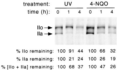
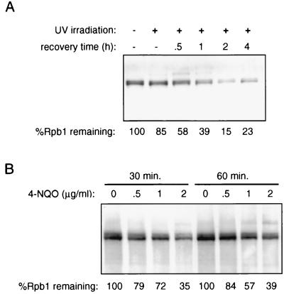
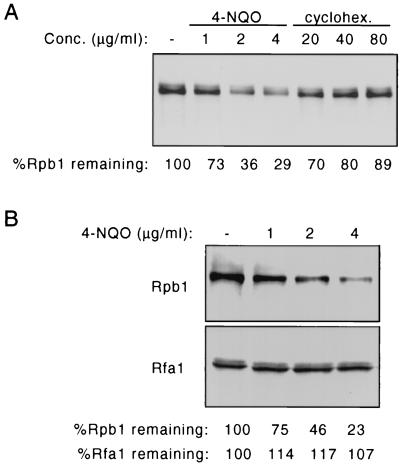
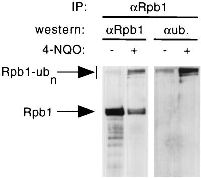
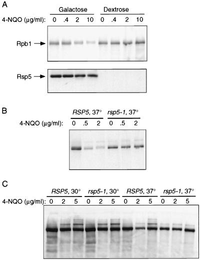
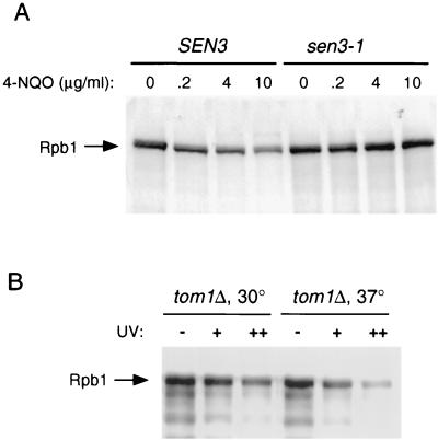
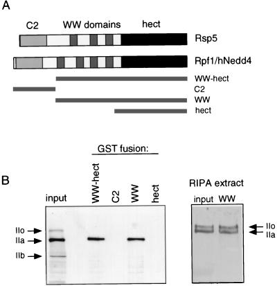
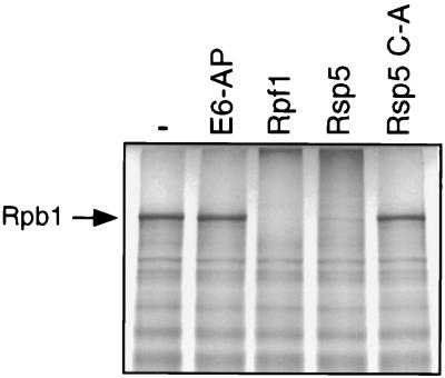
Rsp5 is an E3 ubiquitin-protein ligase of Saccharomyces cerevisiae that belongs to the hect domain family of E3 proteins. We have previously shown that Rsp5 binds and ubiquitinates the largest subunit of RNA polymerase II, Rpb1, in vitro. We show here that Rpb1 ubiquitination and degradation are induced in vivo by UV irradiation and by the UV-mimetic compound 4-nitroquinoline-1-oxide (4-NQO) and that a functional RSP5 gene product is required for this effect. The 26S proteasome is also required; a mutation of SEN3/RPN2 (sen3-1), which encodes an essential regulatory subunit of the 26S proteasome, partially blocks 4-NQO-induced degradation of Rpb1. These results suggest that Rsp5-mediated ubiquitination and degradation of Rpb1 are components of the response to DNA damage. A human WW domain-containing hect (WW-hect) E3 protein closely related to Rsp5, Rpf1/hNedd4, also binds and ubiquitinates both yeast and human Rpb1 in vitro, suggesting that Rpf1 and/or another WW-hect E3 protein mediates UV-induced degradation of the large subunit of polymerase II in human cells.


Figures

FIG. 1
FIG. 1
hRpb1 levels following UV irradiation and 4-NQO treatment of HeLa cells. HeLa cells were irradiated with 254-nm UV light at 22.5 J per m2 as described in Materials and Methods, and cell extracts were prepared immediately or 1 or 4 h postirradiation. For 4-NQO treatment, the chemical was added directly to the culture medium at a final concentration of 0.5 μg/ml, and cell extracts were prepared immediately or 1 or 4 h later. Relative hRpb1 levels were determined by SDS-PAGE and immunoblotting and quantitated by densitometry. Levels are expressed as the percentage of Rpb1 remaining relative to the level in untreated cells.
FIG. 2
FIG. 2
(A) Rpb1 levels following UV irradiation of yeast. Yeast cells (strain FY56) were irradiated at 22.5 J per m2 as described in Materials and Methods, and whole-cell extracts were made at the indicated times postirradiation. Rpb1 was detected by SDS-PAGE followed by immunoblotting with anti-CTD antibody. Rpb1 levels were quantitated by densitometry and are expressed as the percentage of Rpb1 remaining relative to the level in untreated cells. (B) Rpb1 levels following 4-NQO treatment. 4-NQO was added to liquid cultures of log-phase yeast at the indicated concentrations, and cells were collected at the indicated times following addition. Whole-cell extracts were prepared, and Rpb1 was detected by SDS-PAGE and immunoblotting.
FIG. 3
FIG. 3
(A) Yeast cells (FY56 [RSP5]) were treated with the indicated doses of 4-NQO or cycloheximide (cyclohex.) for 30 min, cell extracts were prepared, and Rpb1 levels were examined by SDS-PAGE and immunoblotting with anti-CTD antibody. (B) Yeast cells (FY56 [RSP5]) were treated with the indicated doses of 4-NQO for 30 min, cell extracts were prepared, and Rpb1 levels and Rfa1 levels were examined by SDS-PAGE and immunoblotting.
FIG. 4
FIG. 4
Antiubiquitin antibody recognizes high-molecular-weight forms of Rpb1 from 4-NQO-treated cells. Yeast cells were treated with 4-NQO at 4 μg/ml for 30 min, and whole-cell extracts were prepared. Rpb1 was immunoprecipitated (IP) in duplicate from each sample with anti-CTD antibody. The immunoprecipitates were then analyzed by SDS-PAGE followed by immunoblotting with either anti-CTD (αRpb1) or antiubiquitin (αub.) antibody.
FIG. 5
FIG. 5
(A) 4-NQO treatment of the Gal-RSP5 strain maintained in galactose or shifted to dextrose. The Gal-RSP5 strain was grown to early log phase in galactose-containing medium, and then the cells were either shifted to dextrose-containing medium for 48 h or maintained in galactose-containing medium. The cultures were then treated with 4-NQO at the indicated concentrations for 30 min, and whole-cell extracts were prepared and analyzed by SDS-PAGE and immunoblotting with either an anti-Rsp5 monoclonal antibody (bottom) or anti-CTD antibody (top). (B) 4-NQO treatment of the rsp5-1 temperature-sensitive mutant. Strains FY56 (RSP5) and FW1808 (rsp5-1) were grown to mid-log phase at 30°C and then shifted to 37°C for 1 h. 4-NQO was then added at the indicated concentrations for 30 min. Whole-cell extracts were prepared, and Rpb1 was detected by SDS-PAGE and immunoblotting. (C) Experiment similar to that in panel B, except that cells were treated with 4-NQO at both 30 and 37°C.
FIG. 6
FIG. 6
(A) 4-NQO treatment of SEN3 and sen3-1 strains at 37°C. 4-NQO was added at the indicated concentrations for 30 min. Whole-cell extracts were prepared, and Rpb1 was detected by SDS-PAGE and immunoblotting. (B) The tom1Δ mutant was grown at 30°C and then either maintained at 30°C or shifted for 4 h to 37°C. Cells were then irradiated at either 25 (+) or 50 (++) J/m2, followed by a 1-h recovery period at their respective temperatures. Whole-cell extracts were then prepared, and Rpb1 was detected by SDS-PAGE and immunoblotting.
FIG. 7
FIG. 7
(A) Schematic representation of yeast Rsp5 and human Rpf1/Nedd4. GST-Rpf1 fusions to the regions of Rpf1 indicated by the solid bars were made. (B) (Left) HeLa cell extract was prepared in NP-40 lysis buffer (see Materials and Methods). The binding of hRpb1 to GST-Rpf1 fusion proteins immobilized on glutathione-Sepharose was analyzed by SDS-PAGE and immunoblotting. The “input” shows hRpb1 in the extract with forms IIo, IIa, and IIb. (Right) Similar experiment, with HeLa cell extract prepared in radioimmunoprecipitation assay (RIPA) buffer. The input and binding to GST-WW are shown.
FIG. 8
FIG. 8
Ubiquitination of Rpb1 by Rpf1 in vitro. Rpb1 was translated in vitro in rabbit reticulocyte lysate in the presence of [35S]-methionine. Purified hect E3 proteins (human E6-AP, human Rpf1 [WW-hect; amino acids 193 to 901], yeast Rsp5, and the mutant of Rsp5 with a change of the active-site Cys to Ala [C-A]) were incubated as indicated with Rpb1 in the presence of ATP, ubiquitin, E1 enzyme, and E2 enzyme (Arabidopsis thaliana Ubc8) as previously described (18).

References

    1. Beaudenon, S. L., and J. M. Huibregtse. Unpublished results.
    1. Bedford M T, Chan D C, Leder P. FBP WW domains and the Abl SH3 domain bind to a specific class of proline-rich ligands. EMBO J. 1997;16:2376–2383. - PMC - PubMed
    1. Bedford M T, Reed R, Leder P. WW domain-mediated interactions reveal a spliceosome-associated protein that binds a third class of proline-rich motif: the proline glycine and methionine-rich motif. Proc Natl Acad Sci USA. 1998;95:10602–10607. - PMC - PubMed
    1. Bregman D B, Halaban R, van Gool A J, Henning K A, Friedberg E C, Warren S L. UV-induced ubiquitination of RNA polymerase II: a novel modification deficient in Cockayne syndrome cells. Proc Natl Acad Sci USA. 1996;93:11586–11590. - PMC - PubMed
    1. Chen H I, Sudol M. The WW domain of YES-associated protein binds a proline-rich ligand that differs from the consensus established for Src homology 3-binding modules. Proc Natl Acad Sci USA. 1995;92:7819–7823. - PMC - PubMed

Publication types

MeSH terms