. 1999 Oct;19(10):7011-9.
doi: 10.1128/MCB.19.10.7011.

Targeted disruption of CDK4 delays cell cycle entry with enhanced p27(Kip1) activity

Affiliations

Targeted disruption of CDK4 delays cell cycle entry with enhanced p27(Kip1) activity

T Tsutsui et al. Mol Cell Biol. 1999 Oct.

Abstract

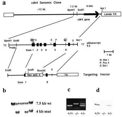
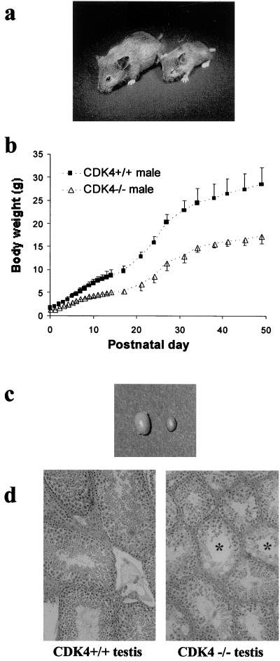
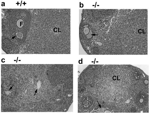
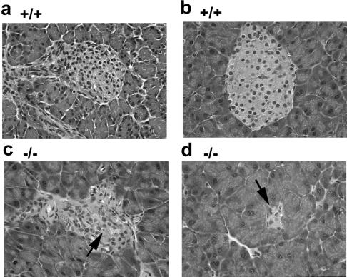
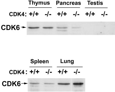
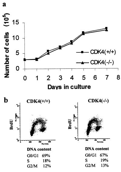
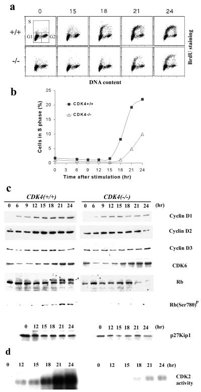
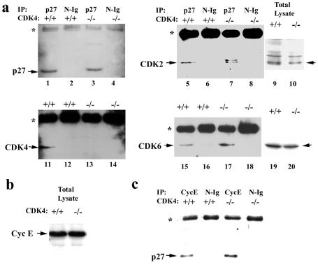
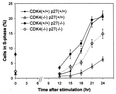
The mechanism by which cyclin-dependent kinase 4 (CDK4) regulates cell cycle progression is not entirely clear. Cyclin D/CDK4 appears to initiate phosphorylation of retinoblastoma protein (Rb) leading to inactivation of the S-phase-inhibitory action of Rb. However, cyclin D/CDK4 has been postulated to act in a noncatalytic manner to regulate the cyclin E/CDK2-inhibitory activity of p27(Kip1) by sequestration. In this study we investigated the roles of CDK4 in cell cycle regulation by targeted disruption of the mouse CDK4 gene. CDK4(-/-) mice survived embryogenesis and showed growth retardation and reproductive dysfunction associated with hypoplastic seminiferous tubules in the testis and perturbed corpus luteum formation in the ovary. These phenotypes appear to be opposite to those of p27-deficient mice such as gigantism and gonadal hyperplasia. A majority of CDK4(-/-) mice developed diabetes mellitus by 6 weeks, associated with degeneration of pancreatic islets. Fibroblasts from CDK4(-/-) mouse embryos proliferated similarly to wild-type embryonic fibroblasts under conditions that promote continuous growth. However, quiescent CDK4(-/-) fibroblasts exhibited a substantial ( approximately 6-h) delay in S-phase entry after serum stimulation. This cell cycle perturbation by CDK4 disruption was associated with increased binding of p27 to cyclin E/CDK2 and diminished activation of CDK2 accompanied by impaired Rb phosphorylation. Importantly, fibroblasts from CDK4(-/-) p27(-/-) embryos displayed partially restored kinetics of the G(0)-S transition, indicating the significance of the sequestration of p27 by CDK4. These results suggest that at least part of CDK4's participation in the rate-limiting mechanism for the G(0)-S transition consists of controlling p27 activity.


Figures

FIG. 1
FIG. 1
Gene targeting of the CDK4 gene in ES cells and generation of CDK4-null mice. (a) Targeting replacement vector. A genomic fragment containing the six exons encoding CDK4 was isolated from a 129/svj mouse genomic library, and a 6.0-kb EcoRI-NotI fragment was subcloned into the pBluescript plasmid vector. The homologous recombination was constructed by replacing a 2.2-kb MscI fragment of this plasmid with a neomycin-phosphotransferase gene under the control of the thymidine kinase promoter for the positive selection. An HSV thymidine kinase gene under the control of the polyoma enhancer was then inserted at the 3′ side of the 1.2-kb homologous flanking region for the negative selection. A 1.2-kb BamHI-EcoRI genomic fragment upstream of the 2.6-kb 5′ homologous region was used as a probe to distinguish the wild-type (wt) and mutant (mut) alleles. (b) Germ line transmission of the CDK4 mutation. The gene targeting event was identified by Southern blotting with BamHI-digested genomic DNAs and the probe shown in panel a. Germ line transmission was confirmed by the Southern blot with genomic DNAs of the F1 offspring from crossbreeding of chimeras with wild-type C57BL/6 mice. An analysis of a representative F1 litter is shown. (c) PCR genotyping of day 12.5 embryos obtained from intercross between CDK4-heterozyous mice. Wild-type (+/+), homozygous (−/−), and heterozygous (+/−) mice were identified by the presence of PCR products specific for either the wt or mut allele. (d) Immunoblotting for CDK4 with CDK4 monoclonal antibody (DCS-31) with extracts of fibroblasts obtained from (+/+), (−/−), and (+/−) embryos.
FIG. 2
FIG. 2
Growth retardation and testicular atrophy in CDK4−/− mice. (a) CDK4+/+ female (left) and the littermate CDK4−/− female (right) at 28 days after birth. (b) Growth curves of CDK4+/+ and CDK4−/− males. Body weights are indicated as means of mice with each genotype (n = 6), and bars represent standard deviations. (c) Testes from 12-week-old CDK4+/+ and CDK4−/− mice. Left, CDK4+/+; right, CDK4−/−. (d) Morphology of testes of 12-week-old mice. Sections (3 μm thick) were stained with hematoxylin-eosin. Asterisks indicate severely dysplastic seminiferous tubules. Magnification, ×200.
FIG. 3
FIG. 3
Perturbed differentiation of granulosa cells in CDK4−/− ovaries. (a) Ovary of 8-week-old CDK4+/+ wild-type mice. (b, c, d) Ovary of 8-week-old CDK4−/− mutant mice. Sections (3 μm thick) were stained with hematoxylin-eosin. Magnification, ×200. F, follicle; CL, corpus luteum. The arrows in panels a, b, and d indicate antral follicles, and those in panel c indicate oocytes trapped in structures showing abnormal luteinization.
FIG. 4
FIG. 4
Degeneration of endocrine islets in the pancrease of CDK4−/− mice. (a) Pancreas of wild-type mice at 3 weeks of age. A well-developed islet is surrounded by exocrine pancreatic tissue. (b) Pancreas of wild-type mice at 6 weeks. (c) Pancreas of CDK4−/− mice at 3 weeks. The arrow indicates a representative condensed nucleus in the islet. (d) Pancreas of CDK4−/− mice at 6 weeks. The arrow indicates a severely degenerated islet. Magnification, ×400.
FIG. 5
FIG. 5
Expression of CDK6 protein in tissues of 3-week-old CDK4+/+ and CDK4−/− mice. Extracts from the tissues indicated were analyzed by immunoblotting by using monoclonal CDK6 antibodies (K6.90 plus K6.83).
FIG. 6
FIG. 6
Continuous proliferation is not affected by targeted disruption of CDK4. (a) Growth curves of CDK4+/+ and CDK4−/− embryonic fibroblasts at passage 4. Embryonic fibroblasts were harvested from CDK4+/+ and CDK4−/− embryos (day 12.5 postcoitus) and cultured in the presence of 10% FBS. (b) Cell cycle analysis by flow cytometry after staining with BrdU for DNA replication and propidium iodide for DNA content. Cells at day 3 of passage 4 were pulse-labeled with 100 μM BrdU for 30 min, stained with propidium iodide and anti-BrdU antibody, and subjected to flow cytometry. These data represent three separate experiments with different clones of embryonic fibroblasts.
FIG. 7
FIG. 7
Delayed entry into the S phase of CDK4−/− fibroblasts after serum stimulation. (a) Cell cycle analysis by staining with BrdU and propidium iodide. Embryonic fibroblasts were harvested from CDK4+/+ and CDK4−/− embryos (day 12.5 postcoitus) and cultured in the presence of 10% FBS for three passages. Quiescence was then induced by culturing cells in 0.1% serum for 72 h (time zero), followed by stimulation of cells with medium containing 10% serum. Cells were pulse-labeled with 100 μM BrdU for 30 min and harvested at the times indicated and labeled hours after stimulation. Cells were stained with anti-BrdU antibody and propidium iodide and subjected to flow cytometry. The x axis represents the DNA content of each cell by the intensity of propidium iodide staining, and the y axis demonstrates the amount of BrdU incorporated into newly synthesized DNA in each cell by the intensity of BrdU staining. The upper left panel shows the gate settings for cells in G1, S, and G2/M. (b) Kinetics of S-phase entry. The percentage of cells in S phase from each time point was figured and plotted according to the flow cytometric analysis (a). (c) Expression of cell cycle-regulatory proteins and Rb phosphorylation. Cells were harvested at the times indicated, and lysates were prepared. Lysates with 50 μg of proteins were analyzed by SDS-PAGE, followed by immunoblotting for cell cycle-regulatory proteins as indicated. The asterisk in the lane for Rb indicates the hyperphosphorylated form of Rb. Rb(Ser780)P indicates immunoblotting with an antibody specific for Rb phosphorylated on the Ser-780 residue (New England Biolabs). (d) CDK2-associated kinase activity. CDK2-associated protein complexes were immunoprecipitated from cell lysates prepared at the times indicated, and histone H1-kinase activity was measured in vitro as described in Materials and Methods. These data represent three separate experiments with different clones of embryonic fibroblasts.
FIG. 8
FIG. 8
Increased binding of p27Kip1 to cyclin E-CDK2 in serum-stimulated fibroblasts with targeted disruption of CDK4. (a) CDK proteins in complex with p27. Fibroblasts from CDK4−/− and CDK4+/+ mouse embryos were serum starved for 72 h (time zero) and then restimulated with 10% FBS serum for 18 h, as described in Materials and Methods and the legend to Fig. 7. Cell lysates were immunoprecipitated with affinity-purified anti-p27 polyclonal antibody or normal IgG (N-Ig) as a control. The immune complexes were then analyzed by immunoblotting by using anti-p27 (lanes 1 to 4), -CDK2 (lanes 5 to 8), -CDK4 (lanes 11 to 14), and -CDK6 (lanes 15 to 18) monoclonal antibodies. Total lysates without immunoprecipitation were also analyzed by immunoblotting with anti-CDK2 and -CDK6 antibodies (lanes 9, 10, 19, and 20). The asterisks indicate heavy-chain IgG cross-reactive with secondary antibody used for immunoblotting. (b) Expression of cyclin E in CDK4−/− and CDK4+/+ fibroblasts at 18 h after serum stimulation measured by immunoblotting. (c) p27 protein in complex with cyclin E in in CDK4−/− and CDK4+/+ fibroblasts at 18 h after serum stimulation. These data represent three separate experiments with different clones of embryonic fibroblasts.
FIG. 9
FIG. 9
Restoration of cell cycle delay in CDK4-null fibroblasts by elimination of p27Kip1. Fibroblasts were harvested from embryos with the following genotypes: CDK4−/− p27+/+, CDK4+/+ p27−/−, CDK4−/− p27−/−, and CDK4+/+ p27+/+. Cells were serum starved for 72 h and then restimulated with 10% FBS. Cells were pulse-labeled with BrdU for 30 min before being harvested at the times indicated. Cells were stained with anti-BrdU antibody and subjected to flow cytometry, as described in Fig. 7. The x axis represents time (in hours) after the readdition of 10% FBS, and the y axis represents the percentages of cells in S phase determined by flow cytometry with propidium iodide and BrdU staining, as described in Materials and Methods and Fig. 7. Data are indicated as means ± standard deviations (n = 3), from experiments with different clones of embryonic fibroblasts.

Similar articles

Cited by

References

    1. Ando K, Ajchenbaum-Cymbalista F, Griffin J D. Regulation of G1/S transition by cyclins D2 and D3 in hematopoietic cells. Proc Natl Acad Sci USA. 1993;90:9571–9575. - PMC - PubMed
    1. Bates S, Bonetta L, MacAllan D, Parry D, Holder A, Dickson C, Peters G. CDK6 (PLSTIRE) and CDK4 (PSK-J3) are a distinct subset of the cyclin-dependent kinases that associate with cyclin D1. Oncogene. 1994;9:71–79. - PubMed
    1. Beijersbergen R L, Carlee L, Kerkhoven R M, Bernards R. Regulation of the retinoblastoma protein-related p107 by G1 cyclin complexes. Genes Dev. 1995;9:1340–1353. - PubMed
    1. Bignon Y J, Chen Y, Chang C Y, Riley D J, Windle J J, Mellon P L, Lee W H. Expression of a retinoblastoma transgene results in dwarf mice. Genes Dev. 1993;7:1654–1662. - PubMed
    1. Blain S W, Montalvo E, Massague J. Differential interaction of the cyclin-dependent kinase (Cdk) inhibitor p27Kip1 with cyclin A-Cdk2 and cyclin D2-Cdk4. J Biol Chem. 1997;272:25863–25872. - PubMed

Publication types

MeSH terms

LinkOut - more resources