. 1999 Oct;19(10):7061-75.
doi: 10.1128/MCB.19.10.7061.

Growth retardation, DNA repair defects, and lack of spermatogenesis in BRCA1-deficient mice

Affiliations

Growth retardation, DNA repair defects, and lack of spermatogenesis in BRCA1-deficient mice

V L Cressman et al. Mol Cell Biol. 1999 Oct.

Erratum in

Abstract

BRCA1 is a nuclear phosphoprotein expressed in a broad spectrum of tissues during cell division. The inheritance of a mutant BRCA1 allele dramatically increases a woman's lifetime risk for developing both breast and ovarian cancers. A number of mouse lines carrying mutations in the Brca1 gene have been generated, and mice homozygous for these mutations generally die before day 10 of embryonic development. We report here the survival of a small number of mice homozygous for mutations in both the p53 and Brca1 genes. The survival of these mice is likely due to additional unknown mutations or epigenetic effects. Analysis of the Brca1(-/-) p53(-/-) animals indicates that BRCA1 is not required for the development of most organ systems. However, these mice are growth retarded, males are infertile due to meiotic failure, and the mammary gland of the female mouse is underdeveloped. Growth deficiency due to loss of BRCA1 was more thoroughly examined in an analysis of primary fibroblast lines obtained from these animals. Like p53(-/-) fibroblasts, Brca1(-/-) p53(-/-) cells proliferate more rapidly than wild-type cells; however, a high level of cellular death in these cultures results in reduced overall growth rates in comparison to p53(-/-) fibroblasts. Brca1(-/-) p53(-/-) fibroblasts are also defective in transcription-coupled repair and display increased sensitivity to DNA-damaging agents. We show, however, that after continued culture, and perhaps accelerated by the loss of BRCA1 repair functions, populations of Brca1(-/-) p53(-/-) fibroblasts with increased growth rates can be isolated. The increased survival of BRCA1-deficient fibroblasts in the absence of p53, and with the subsequent accumulation of additional growth-promoting changes, may mimic the events that occur during malignant transformation of BRCA1-deficient epithelia.


Figures

FIG. 1
FIG. 1
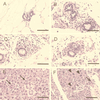
Abnormal end bud formation and decreased ductal branching in BRCA1-deficient mice. (A) Mammary epithelium of a 12-week-old p53−/− female mouse displays extensive branching and numerous end buds. (B) In contrast, very few branches from the primary ducts are seen in the mammary gland of the Brca1−/− p53−/− female mouse. At a higher magnification, many end buds can be visualized in the p53−/− female (C), while only a few underdeveloped end buds are seen in the Brca1−/− p53−/− mouse (D). Magnification bars: A and B, 500 μm; C and D, 200 μm.
FIG. 2
FIG. 2
Cellular abnormalities within the mammary and salivary glands of the Brca1−/− p53−/− mice. (A) Histological analysis of the mammary gland ducts from a p53−/− mouse shows a duct lined by one to two layers of low cuboidal epithelia. Mammary ducts of the Brca1−/− p53−/− female are dilated and lined by a single layer of flattened-to-low cuboidal cells. (B to D) In some regions, the ducts are surrounded by an array of individual cells. While acinar cells in the p53−/− salivary gland appear to be uniform (E), cytomegaly and karyomegaly (arrows) are seen sporadically throughout the acinar units of the salivary glands of the Brca1−/− p53−/− animals (F). Staining was done with hematoxylin and eosin. Magnification bar, 50 μm.
FIG. 3
FIG. 3
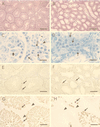
Absence of spermatids and spermiogenesis in a BRCA1-deficient male. The testes and seminiferous tubules of a Brca1−/− p53−/− male (B) are smaller than those of an age-matched p53−/− control male (A). While the seminiferous tubules of the p53−/− control male mouse (C) contained all of the cells involved in spermatogenesis (spermatogonia, thin arrows; spermatocyte, wide arrow; spermatid, large arrowhead; spermatozoa, small arrowhead), only spermatogonia (solid arrows) can be seen in the testes of the Brca1−/− p53−/− male (D). Sertoli cells (double arrow) were seen in abundance in the seminiferous tubules of the Brca1−/− p53−/− males and the p53−/− males (C and D). (E) Apoptosis as determined by the TUNEL assay (apoptotic cells, arrow) occurs rarely in spermatogenesis in p53−/− males. Apoptotic cells (arrows in panels E and F) appear to be more numerous in the testes of the Brca1−/− p53−/− male. However, to correct for the decreased size of the seminiferous tubules of the Brca1−/− p53−/− male in a given area, we counted the number of apoptotic cells present in 25 randomly chosen seminiferous tubules from the two animals. (F) TUNEL-positive cells were only increased approximately threefold in testes of the Brca1−/− p53−/− male. (G) Spermatocytes (arrows), spermatids, and spermatozoa can be detected in the testes of a p53−/− control male stained with HSP70-2 antibody. Incubation with the HSP70-2 antibody revealed the presence of spermatocytes (arrows) in the testes of a Brca1−/− p53−/− male. Staining: A and B, hematoxylin and eosin; C and D, toluidine blue; E and F, diaminobenzidine and methyl green; G and H, diaminobenzidine. Magnification bars: A and B, 200 μm; C and D, 20 μm; E and F, 100 μm; G and H, 67 μm.
FIG. 4
FIG. 4
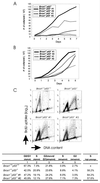
Growth rate of Brca1−/− p53−/− fibroblasts. (A) At early passages, Brca1−/− p53−/− and p53−/− control lines and a wild-type fibroblast line were plated at a low density and were counted daily. Brca1−/− p53−/− fibroblasts have a growth rate similar to that of the wild-type fibroblasts. In contrast, the Brca1+/− p53−/− control fibroblast lines have a higher growth rate, and contact inhibition occurs at a higher density. (B) However, at later passages, the growth rate of the Brca1−/− p53−/− fibroblasts has increased, and contact inhibition occurs at a higher cell density. (C) Brca1−/− p53−/− fibroblasts were incubated with BrdU and stained with anti-BrdU antibody and propidium iodide. Quantitation of the percentage of cells in each cell cycle stage was performed by flow cytometry. One Brca1+/− p53−/− fibroblast line and one wild-type fibroblast line were used as controls. Cells at passage numbers 3 and 4 were considered early-passage cells, while cells at passage numbers 10 through 14 were considered late-passage cells.
FIG. 5
FIG. 5
A higher rate of cellular death occurs in Brca1−/− p53−/− primary fibroblast cultures. (A) p21 protein levels are not elevated in the Brca1−/− p53−/− fibroblasts (lanes a and b) or the Brca1+/− p53−/− fibroblasts (lane c). In contrast, high levels of p21 were detected in wild-type fibroblasts (lanes d and e). (B) Nonadherent Brca1−/− p53−/− fibroblasts were collected and counted. This number was normalized to the number of adherent live cells in each fibroblast population. Nonadherent and adherent counts were obtained from six plates for each of the cell lines. The bars represent the standard errors.
FIG. 6
FIG. 6
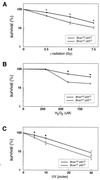
Brca1−/− p53−/− fibroblasts are sensitive to gamma radiation, hydrogen peroxide, and UV radiation. Fibroblasts from two Brca1−/− p53−/− cell lines and two Brca1+/− p53−/− cell lines were exposed to 0, 2.5, 5.0, or 7.5 Gy of gamma radiation (A); 0, 250, 500, or 750 μM hydrogen peroxide (B); or 0, 5, 10, or 30 J of UV radiation (C). Survival was determined in each cell line by normalizing surviving cells following DNA damage to the number of cells which were untreated. An asterisk indicates statistical significance (P < 0.05).
FIG. 7
FIG. 7
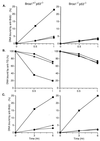
TCR is defective in the DHFR gene of Brca1−/− p53−/− fibroblasts after exposure to ionizing radiation and hydrogen peroxide but not after exposure to UV light. Cells were exposed to 10 Gy of gamma rays (A) or 10 J of UV radiation per m2 (C) and allowed to repair in the presence of 10 μM BrdU. Genomic DNA, digested with BamHI, was reacted with an antibody to BrdU. DNAs from the bound and free fractions were electrophoresed and transferred to a GeneScreen Plus membrane. The percentage of total DNA (▴) bound by the antibody was determined from the 3H prelabel. The percentage of the transcribed strand (■) and nontranscribed strand (●) of the DHFR gene was analyzed with a Bio-Rad phosphorimager. (B) Cells were exposed to 10 mM H2O2 for 15 min at 37°C. Purified DNA was digested with BamHI and incubated with an antibody against thymine glycols, and the extent of removal was determined as described above. The percentage of total DNA (▴) bound by the antibody was determined from the 3H prelabel. The percentage of the transcribed strand (■) and nontranscribed strand (●) of the DHFR gene was analyzed with a Bio-Rad phosphorimager.
FIG. 8
FIG. 8
Models for the role of mutations in BRCA1 in tumorigenesis. Experiments described in this study suggest that the loss of BRCA1 results in deficits in cellular growth. However, loss of BRCA1 is consistently seen in rapidly proliferating tumors from patients carrying a germ line mutation in BRCA1. Two models attempt to resolve these two conflicting concepts. In model 1, mutations in genes resulting in a growth advantage are necessary for the survival of cells which have lost BRCA1 function. The loss of BRCA1, as well as the growth-promoting mutations, results in an increased mutation rate, thus accelerating tumorigenesis. Therefore, in order for loss of BRCA1 to promote tumorigenesis, other growth promoting mutations must first occur. In model 2, cellular growth or death after the loss of BRCA1 function is dependent on the ability of the cell type to compensate by utilizing trophic factors. This model accounts for the specificity of tumor types resulting from the loss of BRCA1 in humans.

Comment in

  • Findings of scientific misconduct.
    [No authors listed] [No authors listed] NIH Guide Grants Contracts (Bethesda). 2006 Jun 9:NOT-OD-06-075. NIH Guide Grants Contracts (Bethesda). 2006. PMID: 16764107 Free PMC article. No abstract available.
  • Findings of Scientific Misconduct.
    [No authors listed] [No authors listed] Fed Regist. 2006 Jun 8;71(110):33308-33309. Fed Regist. 2006. PMID: 27737147 Free PMC article. No abstract available.

References

    1. Akashi M, Hachiya M, Osawa W, Spirin K, Suzuki G, Koeffler H. Irradiation induces WAF1 expression through a p53-independent pathway in KG-1 cells. J Biol Chem. 1995;270:19181–19187. - PubMed
    1. Blackshear P E, Goldsworthy S M, Foley J F, McAllister K A, Bennett L M, Collins N K, Bunch D O, Brown P, Wiseman R, Davis B J. Brca1 and Brca2 expression patterns in mitotic and meiotic cells of mice. Oncogene. 1998;16:61–68. - PubMed
    1. Callebaut I, Mornon J-P. From BRCA1 to RAP1: a widespread BRCT module closely associated with DNA repair. FEBS Lett. 1997;400:25–30. - PubMed
    1. Chapman M S, Verma I M. Transcriptional activation by BRCA1. Nature. 1996;382:678–679. - PubMed
    1. Chen C-F, Li S, Chen Y, Chen P-L, Sharp Z D, Lee W-H. The nuclear localization sequences of the BRCA1 protein interact with the importin-α subunit of the nuclear transport signal receptor. Proc Natl Acad Sci USA. 1996;271:32863–32868. - PubMed

Publication types

MeSH terms

LinkOut - more resources