. 1999 Oct;19(10):7076-87.
doi: 10.1128/MCB.19.10.7076.

The net repressor is regulated by nuclear export in response to anisomycin, UV, and heat shock

Affiliations

The net repressor is regulated by nuclear export in response to anisomycin, UV, and heat shock

C Ducret et al. Mol Cell Biol. 1999 Oct.

Abstract

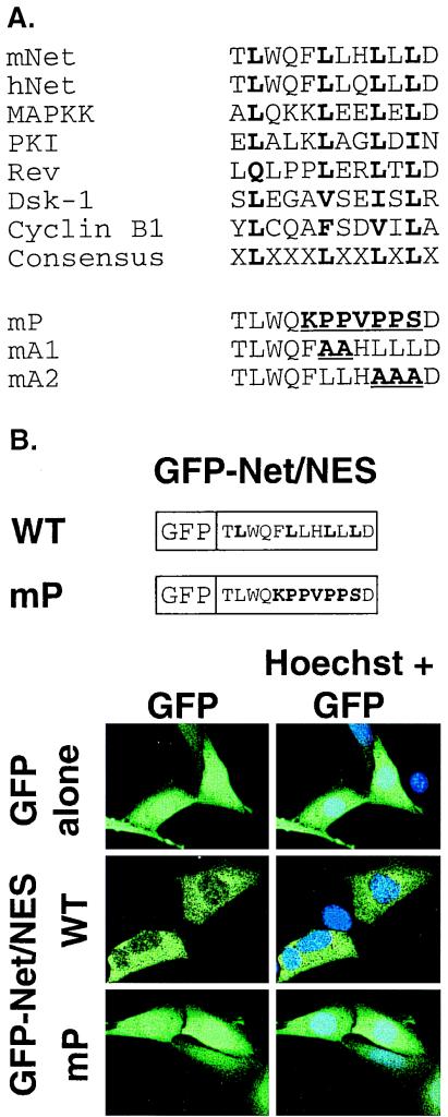
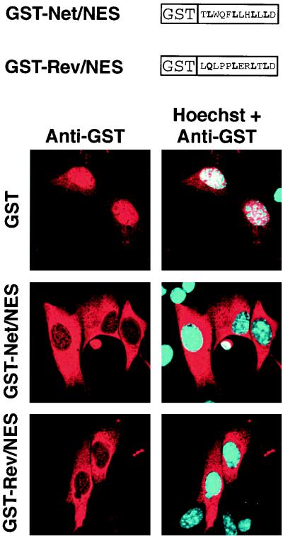
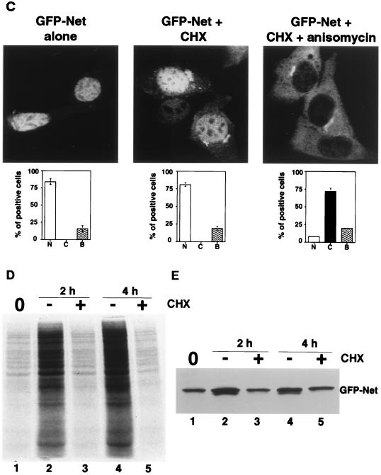
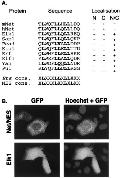
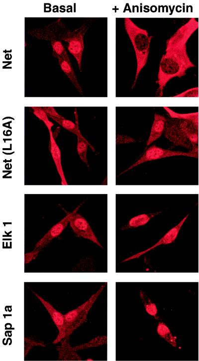
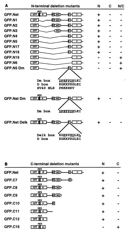
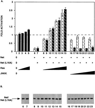
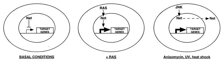
The ternary complex factors (TCFs) are targets for Ras/mitogen-activated protein kinase signalling pathways. They integrate the transcriptional response at the level of serum response elements in early-response genes, such as the c-fos proto-oncogene. An important aim is to understand the individual roles played by the three TCFs, Net, Elk1, and Sap1a. Net, in contrast to Elk1 and Sap1a, is a strong repressor of transcription. We now show that Net is regulated by nuclear-cytoplasmic shuttling in response to specific signalling pathways. Net is mainly nuclear under both normal and basal serum conditions. Net contains two nuclear localization signals (NLSs); one is located in the Ets domain, and the other corresponds to the D box. Net also has a nuclear export signal (NES) in the conserved Ets DNA binding domain. Net is apparently unique among Ets proteins in that a particular leucine in helix 1, a structural element, generates a NES. Anisomycin, UV, and heat shock induce active nuclear exclusion of Net through a pathway that involves c-Jun N-terminal kinase kinase and is inhibited by leptomycin B. Nuclear exclusion relieves transcriptional repression by Net. The specific induction of nuclear exclusion of Net by particular signalling pathways shows that nuclear-cytoplasmic transport of transcription factors can add to the specificity of the response to signalling cascades.


Figures

FIG. 1
FIG. 1
The NES of Net. (A) Alignment of NES sequences. The NES-like sequence in the ets domain of Net (residues 6 to 18 in mice and humans) is aligned with known NESes. The functionally important hydrophobic residues are shown in boldface. Sequences used for comparison: MAPKK (10), cAMP-dependent protein kinase inhibitor (PKI) (79), HIV-1 Rev (7), Dsk-1 (9), and cyclin B1 (73). The Net NES mutants mP (F10K, L11P, L12P, H13V, L14P, L15P, L16S), mA1 (L11A, L12A), and mA2 (L14A, L15A, L16A) have the amino acids changes shown in parentheses. (B) Cellular localization of GFP, GFP-Net/NES, and GFP-Net/NES mP. Drawings representing GFP fused to NES wild-type (WT) and mP sequences are shown. NIH 3T3 cells transiently expressing GFP, GFP-Net/Nes, and GFP-Net/NES mP, were analyzed by confocal microscopy. Hoechst was used to stain nuclei.
FIG. 2
FIG. 2
Net NES mediates nuclear export. GST and GST fused to NES sequences from either Net or HIV Rev (see diagrams) were purified from E. coli and injected into the nuclei of NIH 3T3 cells. Six hours later, injected cells were stained with an antibody against GST and a Texas red-coupled secondary antibody. Hoechst was used to stain nuclei.
FIG. 3
FIG. 3
Anisomycin, UV, and heat shock induce nuclear export of Net. (A) Cellular localization of GFP-Net and GFP-Net(L16A) exposed to stress conditions. NIH 3T3 cells transiently transfected with GFP-Net or GFP-Net(L16A) (see diagrams for structures) were treated with anisomycin (50 ng/ml), UV (40 J/m2), heat shock (42°C), sorbitol (600 mM), and TNF-α (20 ng/ml) and, as a control, were left untreated (Alone). After 30 min, cells were fixed and visualized by confocal microscopy. About 100 positive cells were analyzed to calculate the percentages with GFP-Net in the nucleus (N), cytoplasm (C), or both (B). (B) Nuclear exclusion of endogenous Net. NIH 3T3 cells were treated with anisomycin, UV, heat shock, TNF-α and sorbitol. The cells were fixed and stained with an antibody raised against Net (PAb 376), as shown for the anisomycin treatment. C, cytoplasmic; N, nuclear. Hoechst was used to stain nuclei. (C to E) Cellular localization of GFP-Net in the presence of cycloheximide and anisomycin. (C) Cells transfected with the GFP-Net expression vector were either left untreated (left) or pretreated with cycloheximide (CHX; 15 μg/ml) and then either mock treated (middle) or incubated with anisomycin (right). The middle panels show percentages of cells with GFP-Net in the nucleus (N), cytoplasm (C), and both (B). (D) Protein synthesis was measured by labelling with [35S]methionine followed by SDS-PAGE and autoradiography. The cells were preincubated with [35S]methionine for 15 min, and then at time zero (lane 1) incubation was continued in the absence (−) or presence (+) of cycloheximide for 2 or 4 h. (E) GFP-Net levels were determined by Western blotting of whole-cell extracts with anti-GFP antibodies before (lane 1) or after 2 h (lanes 2 and 3) or 4 h (lanes 4 and 5) of incubation in the absence (−) or presence (+) of cycloheximide. (F) Activation of endogenous p38 was monitored by Western blotting of whole-cell extracts with antibodies specific for dual-phosphorylated activated p38 (lanes 1 to 7) or ERK (lanes 8 to 11). The cells were either untreated (lanes 1 and 8), transfected with a vector expressing MKK3(Glu) (lane 3) or MKK6(Glu) (lane 5), treated with sorbitol (600 mM) after transfection with a vector expressing MKK3(Ala) (lane 4) or MKK6(Ala) (lane 6), treated with sorbitol (600 mM) without (lane 2) or with SB203580 (20 μM), transfected with a vector that expresses Ha-Ras (lane 9), and treated with EGF (0.1 μg/ml) in the absence (lane 10) or presence (lane 11) of PD98059 (20 μM).
FIG. 3
FIG. 3
Anisomycin, UV, and heat shock induce nuclear export of Net. (A) Cellular localization of GFP-Net and GFP-Net(L16A) exposed to stress conditions. NIH 3T3 cells transiently transfected with GFP-Net or GFP-Net(L16A) (see diagrams for structures) were treated with anisomycin (50 ng/ml), UV (40 J/m2), heat shock (42°C), sorbitol (600 mM), and TNF-α (20 ng/ml) and, as a control, were left untreated (Alone). After 30 min, cells were fixed and visualized by confocal microscopy. About 100 positive cells were analyzed to calculate the percentages with GFP-Net in the nucleus (N), cytoplasm (C), or both (B). (B) Nuclear exclusion of endogenous Net. NIH 3T3 cells were treated with anisomycin, UV, heat shock, TNF-α and sorbitol. The cells were fixed and stained with an antibody raised against Net (PAb 376), as shown for the anisomycin treatment. C, cytoplasmic; N, nuclear. Hoechst was used to stain nuclei. (C to E) Cellular localization of GFP-Net in the presence of cycloheximide and anisomycin. (C) Cells transfected with the GFP-Net expression vector were either left untreated (left) or pretreated with cycloheximide (CHX; 15 μg/ml) and then either mock treated (middle) or incubated with anisomycin (right). The middle panels show percentages of cells with GFP-Net in the nucleus (N), cytoplasm (C), and both (B). (D) Protein synthesis was measured by labelling with [35S]methionine followed by SDS-PAGE and autoradiography. The cells were preincubated with [35S]methionine for 15 min, and then at time zero (lane 1) incubation was continued in the absence (−) or presence (+) of cycloheximide for 2 or 4 h. (E) GFP-Net levels were determined by Western blotting of whole-cell extracts with anti-GFP antibodies before (lane 1) or after 2 h (lanes 2 and 3) or 4 h (lanes 4 and 5) of incubation in the absence (−) or presence (+) of cycloheximide. (F) Activation of endogenous p38 was monitored by Western blotting of whole-cell extracts with antibodies specific for dual-phosphorylated activated p38 (lanes 1 to 7) or ERK (lanes 8 to 11). The cells were either untreated (lanes 1 and 8), transfected with a vector expressing MKK3(Glu) (lane 3) or MKK6(Glu) (lane 5), treated with sorbitol (600 mM) after transfection with a vector expressing MKK3(Ala) (lane 4) or MKK6(Ala) (lane 6), treated with sorbitol (600 mM) without (lane 2) or with SB203580 (20 μM), transfected with a vector that expresses Ha-Ras (lane 9), and treated with EGF (0.1 μg/ml) in the absence (lane 10) or presence (lane 11) of PD98059 (20 μM).
FIG. 4
FIG. 4
NES-like sequences in other ets domains do not mediate nuclear export. (A) Comparison of mouse and human Net (mNet and hNet) NES with corresponding sequences in other Ets family members. The Ets proteins used for the alignment are human Elk1, human Sap1a, mouse Pea3, mouse Ets1, human Erf, human Elf1, Drosophila Yan (Pok), and human Pu1. Cons., consensus. Cellular localization of the corresponding GFP-NES fusion proteins is summarized on the right. N, nuclear; C, cytoplasmic; N/C, both nuclear and cytoplasmic. (B) Cellular localization of GFP fusion proteins. Proteins with GFP linked to the sequences listed in panel A were transiently expressed in NIH 3T3 cells and localized by confocal microscopy. Only GFP-Net/NES and GFP-Elk1 are shown. The localization of the other GFP-Ets proteins was similar to that of GFP-Elk1, as summarized in panel A.
FIG. 5
FIG. 5
Anisomycin induces export of Net but not Elk1 or Sap1a. Cells transiently transfected with pTL2-Net, pTL2-Net(L16A), pTL1-Elk1, or pKOZ1-Sap1a were left untreated (Basal) or exposed to anisomycin (50 ng/ml) for 30 min (+ Anisomycin), fixed, and stained with an antibody against Net (PAb 376), Elk1 (PAb 512), or Sap1a (PAb 643) followed by a Texas red-conjugated anti-mouse antibody.
FIG. 6
FIG. 6
The D box and the Ets domain of Net are required for nuclear import. N-terminal (A) and C-terminal (B) deletion mutants of Net fused to GFP were expressed in NIH 3T3 cells and analyzed for cellular location. The sequences of the D box, the D box mutants (Dm and Delk), and the SV40 NLS are shown. Localizations: N, nuclear; C, cytoplasmic; N/C, nuclear and cytoplasmic. A (ets domain), B, C, D, and NID represent domains of Net.
FIG. 7
FIG. 7
Net and Net(L16A) exhibit different transcriptional responses to JNKK and Ras. (A) NIH 3T3 cells were cotransfected with the reporter (1 μg of Palx8-TK-LUC; all lanes), the empty vector lacking Net coding sequences (lanes 2 to 5), or expression vectors for Net [250 ng of pTL2Net; lanes 6, 8, 10, 12, 14, 16, 18, 20, and 22], Net(L16A) [250 ng of pTL2Net(L16A); lanes 7, 9, 11, 13, 15, 17, 19, 21, and 23], Ha-Ras (pRasCTBx2; 100 ng [lanes 2, 8, and 9], 250 ng [lanes 3, 10, and 11], 500 ng [lanes 4, 12, and 13], 1,000 ng [lanes 5, 14, and 15]), or JNKK (pSRα-mJNKK; 100 ng [lanes 16 and 17], 250 ng [lanes 18 and 19], 500 ng [lanes 20 and 21], 1,000 ng [lanes 22 and 23]). Fold activation was calculated relative to the basal activity of the reporter in the presence of empty vector (pSG5). (B) Expression levels of Net and Net(L16A) were assessed by Western blotting using an antibody against Net (PAb 375). The specific band is indicated on the left.
FIG. 8
FIG. 8
A model for two signalling pathways regulating Net. Under basal conditions, Net is nuclear and inhibits the transcription of target genes. Ras-mediated signals switch Net from a negative to a positive factor in the nucleus, leading to activation of transcription by Net. Stress signal activation of the JNK signalling cascade induces nuclear export of Net, releasing genes from Net repression.

References

    1. Batchelor A H, Piper D E, de la Brousse F C, McKnight S L, Wolberger C. The structure of GABPα/β: an ETS domain-ankyrin repeat heterodimer bound to DNA. Science. 1998;279:1037–1041. - PubMed
    1. Cavigelli M, Dolfi F, Claret F X, Karin M. Induction of c-fos expression through JNK-mediated TCF/Elk-1 phosphorylation. EMBO J. 1995;14:5957–5964. - PMC - PubMed
    1. Chen C, Okayama H. High-efficiency transformation of mammalian cells by plasmid DNA. Mol Cell Biol. 1987;7:2745–2752. - PMC - PubMed
    1. Derijard B, Raingeaud J, Barrett T, Wu I H, Han J, Ulevitch R J, Davis R J. Independent human MAP-kinase signal transduction pathways defined by MEK and MKK isoforms. Science. 1995;267:682–685. . (Erratum, 269:17.) - PubMed
    1. Dittmer J, Nordheim A. Ets transcription factors and human disease. Biochim Biophys Acta. 1998;1377:F1–F11. - PubMed

Publication types

MeSH terms