. 1999 Oct;19(10):7168-80.
doi: 10.1128/MCB.19.10.7168.

Concerted regulation of wild-type p53 nuclear accumulation and activation by S100B and calcium-dependent protein kinase C

Affiliations

Concerted regulation of wild-type p53 nuclear accumulation and activation by S100B and calcium-dependent protein kinase C

C Scotto et al. Mol Cell Biol. 1999 Oct.

Abstract

The calcium ionophore ionomycin cooperates with the S100B protein to rescue a p53-dependent G(1) checkpoint control in S100B-expressing mouse embryo fibroblasts and rat embryo fibroblasts (REF cells) which express the temperature-sensitive p53Val135 mutant (C. Scotto, J. C. Deloulme, D. Rousseau, E. Chambaz, and J. Baudier, Mol. Cell. Biol. 18:4272-4281, 1998). We investigated in this study the contributions of S100B and calcium-dependent PKC (cPKC) signalling pathways to the activation of wild-type p53. We first confirmed that S100B expression in mouse embryo fibroblasts enhanced specific nuclear accumulation of wild-type p53. We next demonstrated that wild-type p53 nuclear translocation and accumulation is dependent on cPKC activity. Mutation of the five putative cPKC phosphorylation sites on murine p53 into alanine or aspartic residues had no significant effect on p53 nuclear localization, suggesting that the cPKC effect on p53 nuclear translocation is indirect. A concerted regulation by S100B and cPKC of wild-type p53 nuclear translocation and activation was confirmed with REF cells expressing S100B (S100B-REF cells) overexpressing the temperature-sensitive p53Val135 mutant. Stimulation of S100B-REF cells with the PKC activator phorbol ester phorbol myristate acetate (PMA) promoted specific nuclear translocation of the wild-type p53Val135 species in cells positioned in early G(1) phase of the cell cycle. PMA also substituted for ionomycin in the mediating of p53-dependent G(1) arrest at the nonpermissive temperature (37.5 degrees C). PMA-dependent growth arrest was linked to the cell apoptosis response to UV irradiation. In contrast, growth arrest mediated by a temperature shift to 32 degrees C protected S100B-REF cells from apoptosis. Our results suggest a model in which calcium signalling, linked with cPKC activation, cooperates with S100B to promote wild-type p53 nuclear translocation in early G(1) phase and activation of a p53-dependent G(1) checkpoint control.


Figures

FIG. 1
FIG. 1
S100B stabilizes wild-type p53. (A) The upper panel shows a comparison of S100B contents in MEF−/− cells and J-β subclones selected by limiting dilution. Cells were metabolically labeled with [35S]Met-Cys mix, and S100B was immunoprecipitate with rabbit polyclonal anti-S100B antibody as previously described (52). The asterisk indicates nonspecific binding. The lower panel shows levels of p53 in MEF−/− cells and J-β3, J-β6, and J-β8 subclones following transient transfection as determined by Western blotting. Transfection efficiencies were determined by analysis of GFP expression. (B) Levels of p53 in S100B-MEF (clone J-β8) cells and MEF−/− cells in transient transfection assays using different p53 plasmid concentrations. Upper panel, transfection efficiencies determined by Western blot analysis of GFP expression in parallel with that of p53; lower panel, transfection efficiencies determined by analyzing luciferase activities for each cell line, using a plasmid encoding luciferase (Luc.) under the control of the SV40 enhancer and promoter. Note that p53 expression in S100B-producing J-β8 cells resulted in cell growth arrest with a high incidence of cell death and decrease in luciferase activities. Hence, loading samples for Western blot analysis of p53 were adjusted to equal luciferase activities determined in the absence of p53 plasmids.
FIG. 2
FIG. 2
Ionomycin stimulation, but not permissive temperature, promotes specific activation of wild-type p53 in S100B-MEF cells. (A) Comparison of p53Val135 contents in clone J-β2p53 cells grown at 39°C (lane 1), kept at 32°C for 22 h (lane 2), or stimulated with ionomycin (Iono) for 22 h at 39°C (lane 3). (B) Ionomycin stimulation promotes nuclear accumulation of the wild-type p53Val135 species in S100B-MEF cells grown at 39°C. Clone J-β2p53 cells grown at 39°C were stimulated for 12 h with 1 μM ionomycin prior to labeling with [35S]Met-Cys mix (100 μCi/ml) for 5 h. Nuclear extracts were prepared, and 35S-labeled p53Val135 was immunoprecipitated with anti-MyoD immunoglobulin G used as a control (lane 1), the wild-type-specific PAb246 (lane 2), the mutant-specific PAb240 (lane 3), or the pan-specific PAb421 (lane 4). (C) Flow cytometry analysis of the DNA content of clone J-β2P53 cells grown at 39°C (39°C), shifted to 32°C for 24 h (32°C), or grown at 39°C and stimulated with ionomycin for 36 h (39°C-Iono). (D) Immunofluorescence analysis of PAb246 and PAb240 immunoreactivities in subconfluent clone J-β2p53 cells grown at 39°C not stimulated (control) or stimulated with 1 μM ionomycin (Iono.) for 20 h.
FIG. 2
FIG. 2
Ionomycin stimulation, but not permissive temperature, promotes specific activation of wild-type p53 in S100B-MEF cells. (A) Comparison of p53Val135 contents in clone J-β2p53 cells grown at 39°C (lane 1), kept at 32°C for 22 h (lane 2), or stimulated with ionomycin (Iono) for 22 h at 39°C (lane 3). (B) Ionomycin stimulation promotes nuclear accumulation of the wild-type p53Val135 species in S100B-MEF cells grown at 39°C. Clone J-β2p53 cells grown at 39°C were stimulated for 12 h with 1 μM ionomycin prior to labeling with [35S]Met-Cys mix (100 μCi/ml) for 5 h. Nuclear extracts were prepared, and 35S-labeled p53Val135 was immunoprecipitated with anti-MyoD immunoglobulin G used as a control (lane 1), the wild-type-specific PAb246 (lane 2), the mutant-specific PAb240 (lane 3), or the pan-specific PAb421 (lane 4). (C) Flow cytometry analysis of the DNA content of clone J-β2P53 cells grown at 39°C (39°C), shifted to 32°C for 24 h (32°C), or grown at 39°C and stimulated with ionomycin for 36 h (39°C-Iono). (D) Immunofluorescence analysis of PAb246 and PAb240 immunoreactivities in subconfluent clone J-β2p53 cells grown at 39°C not stimulated (control) or stimulated with 1 μM ionomycin (Iono.) for 20 h.
FIG. 3
FIG. 3
Nuclear translocation of p53 is down regulated by Gö6976, a specific cPKC inhibitor. S100B-MEF clone J-β8 cells were cotransfected with EGFP and plasmids encoding wild-type p53, mutant p53-Ala, or mutant p53-Asp as indicated. After 12 h, cells culture medium was changed to medium without or with 1 μM Gö6976 as indicated. After 20 h cells were fixed. Immunofluorescence of PAb246 immunoreactivity was analyzed in parallel with DNA staining with Hoechst 33258 and EGFP autofluorescence.
FIG. 4
FIG. 4
Down regulation of cPKC by Gö6976 counteracts nuclear accumulation of the wild-type p53Val135 conformational species in S100B-MEF cells. (A) Western blot analysis of p53Val135 protein accumulation in clone J-β2p53 cells stimulated with 1 μM ionomycin in the absence (S100B-MEF) or in the presence of 1 μM Gö6976 (S100B-MEF-Gö6976). p53 was detected by a mixture of monoclonal antibodies PAb421 and PAb240. The asterisk indicates a cross-reacting protein that serves as internal loading control. (B) Microscopic analysis of PAb246 immunoreactivities in S100B-MEF clone J-β2p53 cells grown at 37.5°C and stimulated for 18 h with 1 μM ionomycin in the absence (Iono.) or in the presence (Iono. Gö6976) of 1 μM Gö6976. Left panels show DNA staining with Hoechst 33258.
FIG. 4
FIG. 4
Down regulation of cPKC by Gö6976 counteracts nuclear accumulation of the wild-type p53Val135 conformational species in S100B-MEF cells. (A) Western blot analysis of p53Val135 protein accumulation in clone J-β2p53 cells stimulated with 1 μM ionomycin in the absence (S100B-MEF) or in the presence of 1 μM Gö6976 (S100B-MEF-Gö6976). p53 was detected by a mixture of monoclonal antibodies PAb421 and PAb240. The asterisk indicates a cross-reacting protein that serves as internal loading control. (B) Microscopic analysis of PAb246 immunoreactivities in S100B-MEF clone J-β2p53 cells grown at 37.5°C and stimulated for 18 h with 1 μM ionomycin in the absence (Iono.) or in the presence (Iono. Gö6976) of 1 μM Gö6976. Left panels show DNA staining with Hoechst 33258.
FIG. 5
FIG. 5
Effect of PMA on cellular localization of wild-type and mutant p53Val135 in S100B-REF cells synchronized in the early G1 phase of the cell cycle. Shown are the results of confocal microscope analysis of PAb246 and PAb240 immunoreactivities in clone 6β cells synchronized by mitotic detachment and grown for 1 h on polylysine-coated coverslips allowing cells to pass through mitosis. Cells were either untreated (control) or stimulated with 8 nM PMA for 5 min (PMA).
FIG. 6
FIG. 6
PMA stimulates nuclear translocation and DNA binding of wild-type p53Val135 in S100B-REF cells. (A) Interaction of wild-type p53 with biotinylated p53-CON target DNA. Clone 6β cell nuclear extracts were prepared, and 35S-labeled p53 was immunoprecipitated with anti-MyoD immunoglobulin G used as a control (lane 1) or wild-type-specific PAb246 (lane 2). Nuclear extracts were also incubated with streptavidin-agarose and biotinylated p53-CON DNA target in the absence (lane 3) or presence (lane 4) of 20 μg of nonbiotinylated p53-CON DNA used as a specific competitor. (B) PMA stimulates wild-type p53 (PAb246+) nuclear translocation. Clone 6β cells were not stimulated (−) or were stimulated with 15 nM PMA for 2, 5, 10, and 15 min as indicated. Wild-type p53 was immunoprecipitated with PAb246. (C) PMA stimulates wild-type p53 binding to biotinylated target DNA. Clone 6β cells were not stimulated (−) or stimulated with 15 nM PMA for 2 or 5 min as indicated. Lanes 1 to 3, nuclear extracts were incubated with biotinylated p53-CON DNA target and streptavidin (Strep.)-agarose. Lanes 4 and 5, nuclear extracts were first incubated with PAb246 and protein G-agarose; the remaining supernatants were then incubated with biotinylated p53-DNA target and streptavidin-agarose. Arrows indicate positions of 35S-labeled p53 which bound to PAb246 or to a biotinylated DNA probe that was visualized by gel electrophoresis and autoradiography.
FIG. 7
FIG. 7
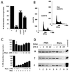
PMA induces G1 phase growth arrest of S100B-REF cells. (A) Effect of PMA concentration on the rate of DNA synthesis of clone 6 (⧫), clone 6β (○), and clone 9β (□) cells grown at 37.5°C. Subconfluent cells were treated for 20 h with a single dose of PMA as indicated, and [3H]thymidine uptake was measured during the last 2 h. (B) Flow cytometry analysis of the DNA content of clone 6 and clone 6β cells stimulated with 8 nM PMA for 8 and 20 h as indicated. (C) Effect of PMA on cell cycle regulatory proteins in S100B-REF cells. Shown is a time course of p21 protein induction and Rb dephosphorylation in clone 6β after PMA stimulation. Cells grown at 37.5°C were not stimulated (lane 1) or stimulated with 8 nM PMA for 1 h (lane 2), 2 h (lane 3), 3 h (lane 4), 4 h (lane 5), 8 h (lane 6), and 16 h (lane 7). Total cell extracts were analyzed by Western blotting using anti-β-tubulin, anti-p27, anti-p21, and anti-Rb antibodies as indicated. (D) Time course of B99 protein induction in clone 6β cells after PMA stimulation (lanes 1 to 7) and comparison with growth-arrested cells at 32°C (lane 8). The asterisk indicates a cross-reacting protein that serves as internal loading control.
FIG. 8
FIG. 8
cPKC inhibition suppresses the long-term cytostatic effect of PMA. (A) Subconfluent S100B-REF clone 6β cells were treated for a total of 20 h with 15 nM PMA in the absence or in the presence of the PKC inhibitor Gö6976 (1 μM) as indicated, and [3H]thymidine uptake was measured during the last 2 h. Results are the averages of values from three experiments performed in duplicate. C, control. (B) Effect of Gö6976 (1 μM) on the cell cycle parameters of clone 6β cells that were not stimulated or stimulated with 15 nM PMA for 20 h as indicated. (C) Upper panel, effect of bryostatin (Bryo.) concentration on the rate of DNA synthesis of clone 6β cells grown at 37.5°C; lower panel, effect of bryostatin concentration on PMA-mediated inhibition of DNA synthesis of clone 6β cells. Subconfluent cells were treated for 20 h with a single dose of bryostatin without (upper panel) or with (lower panel) 15 nM PMA as indicated, and [3H]thymidine uptake was measured during the last 2 h. (D) Comparison of the effects of PMA (PMA) and bryostatin (Bryo.) on down regulation of PKC isoenzymes in clone 6β cells. Cells grown at 37.5°C were not stimulated (lanes 1 and 5) or stimulated with PMA (15 nM) (lanes 2 to 4) or bryostatin (20 nM) (lanes 6 to 8) for 2 h (lanes 2 and 6), 8 h (lanes 3 and 7), and 22 h (lanes 4 and 8). Lane 9 is bovine brain extract, which was used as control. Total cell extracts (50 μg) were analyzed by Western blotting using anti-PKCα, anti-PKCγ, and anti-PKCɛ antibodies as indicated.
FIG. 9
FIG. 9
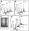
S100B cooperates with PMA in triggering apoptosis of clone 6 cells upon UV irradiation. (A and B) Time course of induction of apoptosis in clone 6 (A) and clone 6β (B) cells by UV irradiation. Clone 6 and clone 6β cells were stimulated for 24 h with 4 nM PMA (PMA/24h). Cells were then irradiated (10 J/m2), changed to fresh medium without PMA, and collected 8 h (UV/8h) or 24 h (UV/24h) after irradiation. (C) Agarose gel analysis of DNA fragmentation in apoptotic clone 6β cells. Lane 1, control cells. Lanes 2 and 3, cells were first stimulated with 4 nM PMA, UV irradiated (10J/m2), and analyzed after 8 h (lane 2) or 24 h (lane 3). Lane 4, cells were as in lane 3 but incubated with actinomycin D (2.5 mg ml−1) for 1 h prior to irradiation. After irradiation, actinomycin D was kept in the medium for another 24 h prior to cell analysis. Lanes 5 and 6 correspond to cells in panel D (32°C, UV and 32°C-PMA, UV). (D) PMA stimulation but not temperature shift restores full G1 checkpoint control. Clone 6β cells were growth arrested by shifting the temperature to 32°C. After 24 h at 32°C, cells were UV irradiated (32°C, UV). Cells were first growth arrested at 32°C; after 24 h, cells were stimulated with PMA for another 20 h and UV irradiated (32°C, PMA-UV). Cells were stimulated with PMA at the time of the temperature shift to 32°C. After 24 h, cells were UV irradiated (32°C-PMA, UV). In all experiments, PMA was used at an 8 nM final concentration, the irradiation dose was 10 J/m2, and the culture media were replaced by media without PMA after irradiation. Cells were analyzed 24 h postirradiation.

Similar articles

Cited by

References

    1. Aloni-Grinstein R, Schwartz D, Rotter V. Accumulation of wt p53 protein upon γ-irradiation induces a G2 arrest-dependent immunoglobulin κ light chain gene expression. EMBO J. 1995;14:1392–1401. - PMC - PubMed
    1. Bates S, Vousden K. p53 in signalling checkpoint arrest or apoptosis. Curr Opin Genet Dev. 1996;6:12–19. - PubMed
    1. Baudier J, Glasser N, Gérard D. Ions binding to S100 protein. Calcium and zinc-binding properties of bovine brain S100αα, S100a (αβ), and S100B (ββ) protein: Zn2+ regulates Ca2+ binding on S100B protein. J Biol Chem. 1986;261:8192–8203. - PubMed
    1. Baudier J, Delphin C, Grunwald D, Khoshbin S, Lawrence J J. Characterization of the tumor suppressor protein p53 as a protein kinase C substrate and a S100B-binding protein. Proc Natl Acad Sci USA. 1992;89:11627–11631. - PMC - PubMed
    1. Baudier J, Bergeret E, Bertacchi N, Weintraub H, Gagnon J, Garin J. Interactions of myogenic bHLH transcription factors with calcium-binding calmodulin and S100a (αα) proteins. Biochemistry. 1995;34:7834–7846. - PubMed

Publication types