. 1999 Oct;19(10):7203-15.
doi: 10.1128/MCB.19.10.7203.

Multiple signaling pathways of the insulin-like growth factor 1 receptor in protection from apoptosis

Affiliations

Multiple signaling pathways of the insulin-like growth factor 1 receptor in protection from apoptosis

F Peruzzi et al. Mol Cell Biol. 1999 Oct.

Abstract

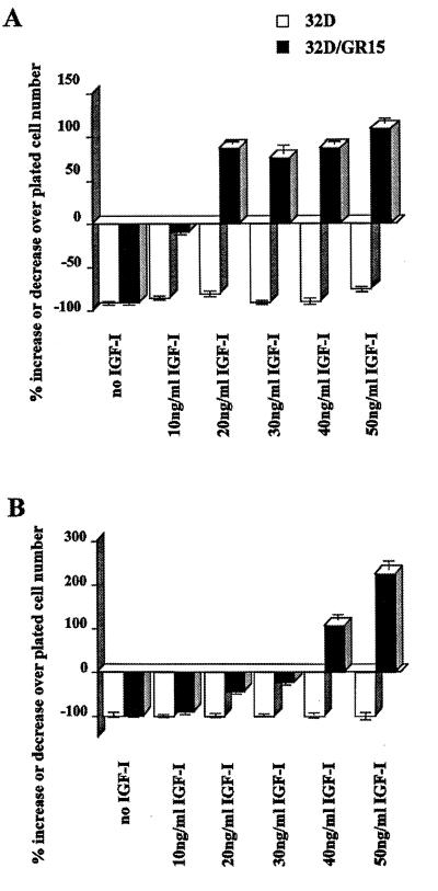
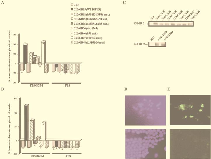
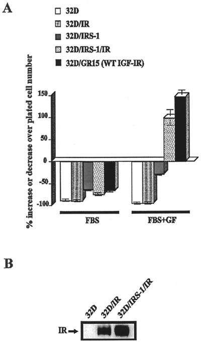
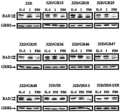
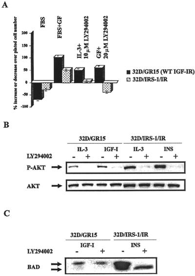
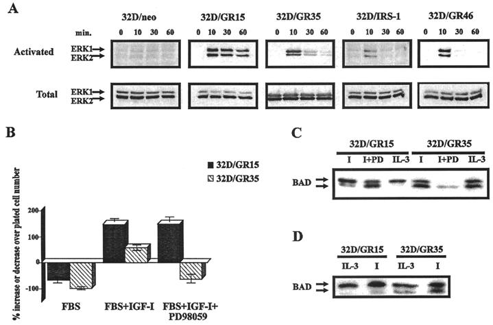
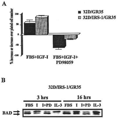
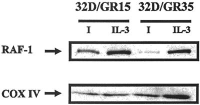
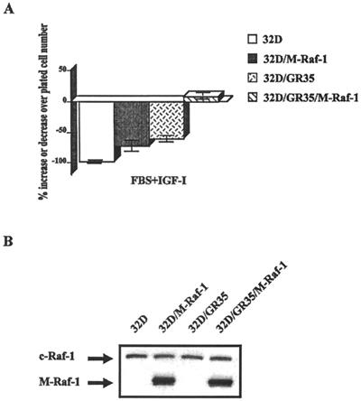
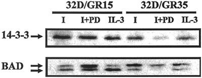
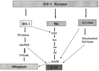
The type 1 insulin-like growth factor receptor (IGF-1R), activated by its ligands, protects several cell types from a variety of apoptotic injuries. The main signaling pathway for IGF-1R-mediated protection from apoptosis has been previously elucidated and rests on the activation of phosphatidylinositol 3-kinase, Akt/protein kinase B, and the phosphorylation and inactivation of BAD, a member of the Bcl-2 family of proteins. In 32D cells (a murine hemopoietic cell line devoid of insulin receptor substrate 1 [IRS-1]), the IGF-1R activates alternative pathways for protection from apoptosis induced by withdrawal of interleukin-3. One of these pathways leads to the activation of mitogen-activated protein kinase, while a third pathway results in the mitochondrial translocation of Raf and depends on the integrity of a group of serines in the C terminus of the receptor that are known to interact with 14.3.3 proteins. All three pathways, however, result in BAD phosphorylation. The presence of multiple antiapoptotic pathways may explain the remarkable efficacy of the IGF-1R in protecting cells from apoptosis.


Figures

FIG. 1
FIG. 1
Effect of IGF-1 concentrations on survival of 32D cells expressing the wild-type IGF-1R. Parental 32D cells (open bars) and 32D cells expressing the wild-type IGF-1R (closed bars) were cultured in medium containing 10% FBS and the indicated concentrations of IGF-1. The percentages of surviving cells were determined at 24 h (A) and 48 h (B) after removal of IL-3 (WEHI-conditioned medium).
FIG. 2
FIG. 2
Survival of 32D cells expressing wild-type (WT) and mutant IGF-1R. 32D cells were stably transduced with retroviral vectors expressing the indicated receptors (see also Table 1), and survival was determined at 24 h (A) and 48 h (B) after IL-3 withdrawal. FBS+IGF-1, 10% FBS plus IGF-1 (50 ng/ml), no IL-3; FBS, 10% fetal bovine serum, no IGF-1 or IL-3. The behavior in FBS supplemented with IL-3 was omitted since all cell lines grew vigorously in this medium. mut., mutation; del., deletion. (C) Western blots for the IGF-1R of the cell lines used in panels A and B. In all instances, we used an antibody to the C terminus of the IGF-1R (see Materials and Methods), except for GR36, that is truncated at residue 1245. For this receptor, we used an antibody to the α subunit. (D) Staining for DNA. (E) TUNEL staining. The upper figures are for the GR18 mutant; the lower figures are for the GR15 cells (wild-type receptor).
FIG. 3
FIG. 3
Survival of 32D cells expressing the IR and/or IRS-1. The cell lines used were 32D cells expressing either IRS-1 only (32D/IRS-1), the insulin receptor only (32D/IR), or both (32D/IRS-1/IR). (A) Survival was determined at 24 h after IL-3 withdrawal. 32D/GR15 cells and parental 32D cells are also shown for comparison. FBS+GF, FBS supplemented with insulin (50 ng/ml), with the exception of the 32D/GR15 cells, which were supplemented with IGF-1 (50 ng/ml). WT, wild type. (B) Levels of expression of the IR in the two cell lines overexpressing it.
FIG. 4
FIG. 4
BAD phosphorylation in 32D cells expressing the wild-type and mutant IGF-1 receptors. BAD phosphorylation was determined, as described in Materials and Methods, under three different sets of conditions, all in medium with 10% FBS: with IL-3, with IGF-1 (I), or with no additions. BAD phosphorylation was determined at 3 h after IL-3 withdrawal. The cell lines and the treatments are indicated above the BAD blots. In the case of the cell lines expressing IRS-1, the IR, or both, the FBS was supplemented with insulin (50 ng/ml). In all other cases, IGF-1 (50 ng/ml) was used. Under the BAD blots are Western blots of Grb2, used as indicators of protein amounts in each lane.
FIG. 5
FIG. 5
Effect of PI3-ki inhibitors on survival of 32D cells. (A) Cell survival. 32D/GR15 and 32D/IRS1/IR cells were incubated in medium with FBS only or with FBS supplemented with the respective growth factors (GF) (IGF-1 for 32D/GR15, insulin for 32D/IRS1/IR cells, or IL-3), plus or minus the indicated concentrations of LY294002, an inhibitor of PI3-ki. The numbers of surviving cells were determined at 24 h after IL-3 withdrawal and addition of the growth factors and the inhibitor. WT, wild type. (B) Phosphorylation (upper panel) and amounts (lower panel) of Akt/PKB in the same cell lines and with different treatments. The presence (+) or absence (−) of LY294002 (10 μM) is indicated. P-AKT, phosphorylated Akt. (C) BAD phosphorylation under the same conditions.
FIG. 6
FIG. 6
Role of MAPK in IGF-1R-mediated survival of 32D cells. (A) MAPK activation in selected cell lines. MAPK phosphorylation and protein amounts were determined as described in Materials and Methods. Stimulation was with IGF-1 (50 ng/ml), and lysates were prepared at the times (in minutes) indicated above the blots. The cell lines are also indicated above the blots. (B) Survival of 32D/GR15 and 32D/GR35 cells in the presence of PD98059, an inhibitor of MEK. The indicated cell lines were incubated in FBS (no IL-3) for 24 h, in the presence of only IGF-1 or both IGF-1 and PD98059. Survival is expressed as a percentage of cell number increase or decrease over the number of cells plated. (C) BAD phosphorylation in the same cell lines, with or without PD98059 (I, IGF-1; PD, the inhibitor). (D) Same procedures as in Fig. 4, except that 32D/GR15 and 32D/GR35 cells were examined at 16 h after IL-3 withdrawal. Lysates were prepared from cells in FBS supplemented with either IL-3 or IGF-1 (I).
FIG. 7
FIG. 7
IRS-1 protects GR35 cells from apoptosis. The two cell lines tested were 32D/GR35 (4-serine mutant) and 32D/IRS1/GR35 (expressing IRS-1). (A) Survival was determined after 24 h as in previous experiments. The cells were tested in medium containing serum supplemented with IGF-1, plus or minus the MEK inhibitor PD98059. (B) Lysates from 32D/IRS1/GR35 cells, under the same conditions, were examined for BAD phosphorylation. I, IGF-1; PD, PD98059.
FIG. 8
FIG. 8
Presence of mitRaf in 32D/GR15 and 32D/GR35 cells. Mitochondrial lysates were prepared as described in Materials and Methods, and the same amounts of protein were blotted for Raf-1 (upper panel). The amounts of mitochondrial proteins in each lane were monitored with an antibody to cytochrome oxidase (COX IV) (lower panel). I, IGF-1.
FIG. 9
FIG. 9
Effect of mitRaf on cell survival. (A) 32D and 32D/GR35 cells were stably transfected with constitutively activated mutant Raf that localizes to the mitochondria. Transduction and selection of cell lines are described in Materials and Methods. Survival was determined at 48 h after IL-3 withdrawal, under the usual conditions. (B) Detection of mitRaf in the various cell lines. Detection was carried out with an anti-Raf1 antibody (Santa Cruz). The size of the mutant Raf is different from that of wild-type Raf-1, which is visible above the mutant Raf.
FIG. 10
FIG. 10
Coprecipitation of 14.3.3 and BAD. BAD immunoprecipitation, detection of 14.3.3, and detection of BAD itself on the stripped blot were carried out as described in Materials and Methods. The cell lines used were 32D/GR15 and 32D/GR35. PD, MEK inhibitor PD98059 at the same concentration as that used for Fig. 7; I, IGF-1 (50 ng/ml).
FIG. 11
FIG. 11
Diagram of the multiple antiapoptotic pathways of the IGF-1R. The diagram is based on the results presented in this paper and previous papers from this and other laboratories.

Similar articles

Cited by

References

    1. Avruch J. Insulin signal transduction through protein kinase cascades. Mol Cell Biochem. 1998;182:31–48. - PubMed
    1. Backer J M, Myers M G, Jr, Sun X J, Chi D J, Shoelson S E, Miralpeix M, White M F. Association of IRS-1 with insulin receptor and the phosphatidylinositol 3′-kinase. J Biol Chem. 1993;268:8204–8212. - PubMed
    1. Baserga R, Hongo A, Rubini M, Prisco M, Valentinis B. The IGF-I receptor in cell growth, transformation and apoptosis. Biochim Biophys Acta. 1997;1332:105–126. - PubMed
    1. Baserga R. The price of independence. Exp Cell Res. 1997;236:1–3. - PubMed
    1. Brunet A, Bonni A, Zigmond M J, Lin M Z, Jun P, Hu L S, Anderson M J, Arden K C, Blenis J, Greenberg M E. Akt promotes cell survival by phosphorylating and inhibiting a forkhead transcription factor. Cell. 1999;96:857–868. - PubMed

Publication types

MeSH terms

Substances