. 1999 Oct;19(10):7255-63.
doi: 10.1128/MCB.19.10.7255.

zfh-1, the Drosophila homologue of ZEB, is a transcriptional repressor that regulates somatic myogenesis

Affiliations

zfh-1, the Drosophila homologue of ZEB, is a transcriptional repressor that regulates somatic myogenesis

A A Postigo et al. Mol Cell Biol. 1999 Oct.

Abstract

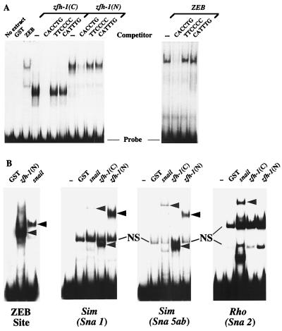
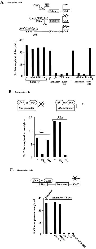
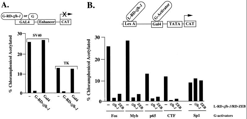
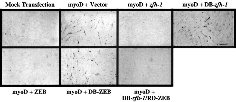
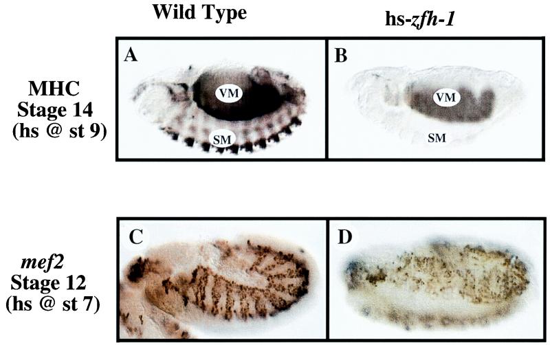
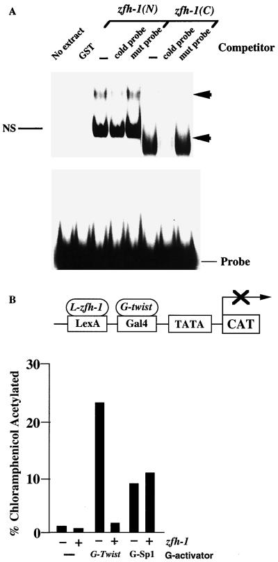
zfh-1 is a member of the zfh family of proteins, which all contain zinc finger and homeodomains. The roles and mechanisms of action of most family members are still unclear. However, we have shown previously that another member of the family, the vertebrate ZEB protein, is a transcriptional repressor that binds E box sequences and inhibits myotube formation in cell culture assays. zfh-1 is downregulated in Drosophila embryos prior to myogenesis. Embryos with zfh-1 loss-of-function mutation show alterations in the number and position of embryonic somatic muscles, suggesting that zfh-1 could have a regulatory role in myogenesis. However, nothing is known about the nature or mechanism of action of zfh-1. Here, we demonstrate that zfh-1 is a transcription factor that binds E box sequences and acts as an active transcriptional repressor. When zfh-1 expression was maintained in the embryo beyond its normal temporal pattern of downregulation, the differentiation of somatic but not visceral muscle was blocked. One potential target of zfh-1 in somatic myogenesis could be the myogenic factor mef2. mef2 is known to be regulated by the transcription factor twist, and we show here that zfh-1 binds to sites in the mef2 upstream regulatory region and inhibits twist transcriptional activation. Even though there is little sequence similarity in the repressor domains of ZEB and zfh-1, we present evidence that zfh-1 is the functional homologue of ZEB and that the role of these proteins in myogenesis is conserved from Drosophila to mammals.


Figures

FIG. 1
FIG. 1
zfh-1 binds E box sequences. (A) Gel retardation assays using a probe containing CACCTG sites (36). Recombinant proteins encoding the C- and N-terminal zinc fingers of zfh-1 [zfh-1(C) and zfh-1(N), respectively] and N-terminal zinc fingers of ZEB were obtained from overexpression in bacteria as described in Materials and Methods. zfh-1 and ZEB binding was competed with a 50-fold excess of unlabeled probe but not with the mutant non-E box probe (TTCCCC) or an unrelated E box sequence (CATTTG). (B) Snail and zfh-1 share DNA binding specificities. A probe containing a ZEB binding site (E361/E399 in the α4 integrin promoter [36]), the highest-affinity sites for snail from the single-minded (sim) promoter (Sna1 and Sna5b as in reference 23) and from the rhomboid promoter (Sna2 as in reference 22) were used in gel shift experiments to test the binding of GST-snail and GST–N- and C-terminal zfh-1 zinc fingers [zfh-1(N) and zfh-1(C), respectively]. Equal molar amounts of GST fusion proteins (as determined by Western blotting with anti-GST antibodies [data not shown]) were used. Note that zfh-1 binds to snail sites in the single-minded promoter (even to the highest-affinity site, Sna5ab [23]) with even higher affinity than snail. However, binding of zfh-1 to the snail sites in the rhomboid promoter were weak or neglible (this figure and data not shown). Arrowheads indicate specific complex; NS denotes a nonspecific band. Free probes are indicated at the bottom.
FIG. 2
FIG. 2
zfh-1 is an active transcriptional repressor. (A) Five micrograms of a reporter containing two copies of a zfh-1 site cloned either 30 or 300 bp upstream of an enhancer element (from the α4 integrin promoter [38]) was cotransfected in Drosophila Schneider cells with 8 μg of actin 5 promoter-driven constructs for zfh-1, ZEB, or snail (sna). A reporter lacking the zfh-1 site was used as a control. (B) zfh-1 represses the single-minded promoter by binding through snail binding sites. Ten micrograms of a CAT reporter construct driven by the single-minded promoter and 2 μg of a CAT reporter driven by the rhomboid promoter were cotransfected in Drosophila Schneider cells with 8 μg of actin 5 promoter-driven constructs for snail or zfh-1. (C) zfh-1 represses transcription in mammalian cells. DB-zfh-1 and DB-ZEB are able to displace endogenous ZEB and release transcriptional repression. Full-length zfh-1, full-length ZEB, and DB-zfh-1–RD-ZEB block transcription. Three micrograms of a reporter construct containing two copies of the CACCTG site upstream of the enhancer (38) was cotransfected in human C33a cells (and HT1080 cells, with identical results) with equal molar concentrations of zfh-1, DB-zfh-1, DB-ZEB, and DB-zfh-1–RD-ZEB. CAT results are averages of duplicate assays and are all representative of at least five separate experiments with standard deviations below 15%.
FIG. 3
FIG. 3
zfh-1 is an active and selective transcriptional repressor. (A) The region between the zinc finger domains of zfh-1 acts a repressor when fused to the DNA binding domain of the yeast Gal4 protein (GRD-zfh-1). Three micrograms of a reporter construct containing five Gal4 sites upstream of the SV40 enhancer/promoter and the TK promoter were cotransfected in human HT1080 cells (or C33a cells, with identical results) with 3 μg of Gal4–RD–zfh-1. No effect was observed when the reporter was cotransfected with the molar equivalent amount of the control Gal4 expression vector. (B) zfh-1 is a selective transcriptional repressor. The region of zfh-1 between the zinc finger regions was fused to the bacterial protein LexA and tested for its ability to block the activity of a set of Gal4 activators by cotransfection with the pLG reporter construct (45) containing six LexA binding sites upstream of two or five Gal4 binding sites (results were the same with either construct); 0.8 μg of the pLG construct was cotransfected with 0.1 to 0.8 μg of different Gal4 activators and 3 μg of L–RD–zfh-1 into HT1080 cells. As a control, LexA was also cotransfected instead of LexA–zfh-1. LexA had no effect on the activity of the different Gal4 activators (data not shown). CAT results are averages of duplicate assays with standard deviations below 15%.
FIG. 4
FIG. 4
zfh-1 blocks myogenesis in vertebrate cells by active transcriptional repression. 10T1/2 mouse fibroblasts were transfected with 0.5 μg of a myoD expression vector (or its parent vector; mock transfection) along with equal molar concentrations of expression vectors for full-length zfh-1 and ZEB, DB-zfh-1, DB-ZEB, DB-zfh-1–RD-ZEB, or the empty vector as a control. After 5 to 6 days, cells were immunostained for MHC and developed with HRP and DAB as previously described (36). The images are representative of at least five different myogenic differentiation assays. The expression levels of ZEB, zfh-1, DB-ZEB, DB-zfh-1, and DB-zfh-1–RD-ZEB proteins were similar by Western blot assay (results not shown).
FIG. 5
FIG. 5
Overexpression of zfh-1 inhibits somatic muscle differentiation in Drosophila embryos and disrupts mef2 expression. MHC expression was analyzed as a marker of myogenic differentiation in wild-type and zfh-1-overexpressing embryos (hs-zfh-1). Embryos were analyzed with antibodies against MHC and mef2 as described in Materials and Methods. The embryos are oriented with the anterior to the left in lateral views. (A) Wild-type embryos were heat shocked (hs) for 15 min at 37°C at stage (st) 9 as described in Materials and Methods and immunostained for MHC at stage 14. VM, visceral muscle; SM, somatic muscle. (B) hs-zfh-1 embryos were heat shocked at stage 9 and immunostained for MHC at stage 14. Visceral muscle (VM) remained relatively unaffected, whereas somatic muscle (SM) showed a complete absence of MHC-positive cells. (C) Staining for mef2 in wild-type embryos (stage 12, lateral view); embryos were heat shocked at stage 7 for 15 min at 37°C as described in Materials and Methods. mef2 expression is restricted to the visceral (internal) and somatic (external) mesodermal layers and the cephalic mesoderm (most anterior part of the embryo). (D) Staining for mef2 in hs-zfh-1 embryos (stage 12, in ventrolateral view); embryos were heat shocked at stage 7. Maintained expression of zfh-1 causes inhibition of mef2 expression and severe derangement of its pattern in the mesoderm.
FIG. 6
FIG. 6
zfh-1 binds sites from the mef2 gene promoter and blocks transcriptional activation by twist. (A) Gel retardation assays using a probe containing zfh-1 sites in the mef2 promoter sequence (35a). Sites at bp −2782 and −8564 were tested. Recombinant proteins encoding the C- and N-terminal zinc fingers of zfh-1 [zfh-1(C) and zfh-1(N), respectively] were obtained from expression in bacteria as described in Materials and Methods. zfh-1 binding was competed with a 50-fold excess of unlabeled probe (CACCTG or CACCTA) but not with the mutant non-E box probe (TTCCCC). Arrowheads indicate specific complex; NS denotes a nonspecific band. (B) zfh-1 represses twist activity. The ability of L–RD–zfh-1 (as in Fig. 3B) to block the activity of Gal4-twist was tested by cotransfection with the pLG reporter (as in Fig. 3B and reference 45); 0.8 μg of pLG reporter was cotransfected with 0.4 of Gal4-twist or 0.6 μg of Gal4-Sp1 activators and 3 μg of LexA–zfh-1. As a control, LexA was also cotransfected instead of L–RD–zfh-1. LexA had no effect on the activity of the different GAL4 activators (data not shown). L–RD–zfh-1 failed to repress the activity of Sp1 which was included as a control. CAT results are averages of duplicate assays with standard deviations below 15%.

Similar articles

Cited by

References

    1. Bate M. The mesoderm and its derivatives. In: Bate M, Martinez-Arias A, editors. The development of Drosophila melanoganster. Cold Spring Harbor, N.Y: Cold Spring Harbor Laboratory Press; 1993. pp. 1013–1090.
    1. Baylies M K, Bate M. Twist: a myogenic switch in Drosophila. Science. 1996;272:1481–1484. - PubMed
    1. Baylies M K, Bate M, Gomez M R. Myogenesis: a view from Drosophila. Cell. 1998;93:921–927. - PubMed
    1. Benezra R, Davis R L, Lockshon D, Turner D L, Weintraub H. The protein Id: a negative regulator of helix-loop-helix DNA binding proteins. Cell. 1990;61:49–59. - PubMed
    1. Bour B A, O’Brien M A, Lockwood W L, Goldstein E S, Bodmer R, Taghert P H, Abmayr S M, Nguyen H T. Drosophila Mef2, a transcription factor that is essential for myogenesis. Genes Dev. 1995;15:730–741. - PubMed

Publication types

LinkOut - more resources