Comparative Study
. 1999 Oct;19(10):7276-86.
doi: 10.1128/MCB.19.10.7276.

De novo synthesis of sphingolipids is required for cell survival by down-regulating c-Jun N-terminal kinase in Drosophila imaginal discs

Affiliations
Comparative Study

De novo synthesis of sphingolipids is required for cell survival by down-regulating c-Jun N-terminal kinase in Drosophila imaginal discs

T Adachi-Yamada et al. Mol Cell Biol. 1999 Oct.

Abstract

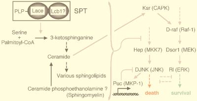
Mitogen-activated protein kinase (MAPK) is a conserved eukaryotic signaling factor that mediates various signals, cumulating in the activation of transcription factors. Extracellular signal-regulated kinase (ERK), a MAPK, is activated through phosphorylation by the kinase MAPK/ERK kinase (MEK). To elucidate the extent of the involvement of ERK in various aspects of animal development, we searched for a Drosophila mutant which responds to elevated MEK activity and herein identified a lace mutant. Mutants with mild lace alleles grow to become adults with multiple aberrant morphologies in the appendages, compound eye, and bristles. These aberrations were suppressed by elevated MEK activity. Structural and transgenic analyses of the lace cDNA have revealed that the lace gene product is a membrane protein similar to the yeast protein LCB2, a subunit of serine palmitoyltransferase (SPT), which catalyzes the first step of sphingolipid biosynthesis. In fact, SPT activity in the fly expressing epitope-tagged Lace was absorbed by epitope-specific antibody. The number of dead cells in various imaginal discs of a lace hypomorph was considerably increased, thereby ectopically activating c-Jun N-terminal kinase (JNK), another MAPK. These results account for the adult phenotypes of the lace mutant and suppression of the phenotypes by elevated MEK activity: we hypothesize that mutation of lace causes decreased de novo synthesis of sphingolipid metabolites, some of which are signaling molecules, and one or more of these changes activates JNK to elicit apoptosis. The ERK pathway may be antagonistic to the JNK pathway in the control of cell survival.


Figures

FIG. 1
FIG. 1
lace alleles and the adult phenotypes. (A) The lace alleles used in this study. Homozygotes of the P element-inserted allele l(2)k05305 and all of the heteroallelic combinations with l(2)k05305 partially grew to the adult stage with similar aberrant morphologies. The Df(2L)TE35D-GW1 allele lacks three loci (lace and the adjacent two loci, sna and cycE) (Fig. 3A). All flies with genotypes which had a strong lace allele either inter se or when heterozygous with deletion died just after hatching. EMS, ethyl methanesulfonate. (B to I) Adult lace phenotypes in a lace mutant and wild type. (B, D, F, and H) Wild-type Canton-S. (C, E, G, and I) Transheterozygote lacel(2)k05305/laceHG34. (B and C) Wing. Anterior area is to the left. In the lace mutant, incision of the wing margin and the ectopic crossvein are marked by the filled and open arrows, respectively. The laced vein pattern, a previously identified lace phenotype, is thought to be allele specific or caused by a mutation of a different gene that is linked with lace on the same chromosome. (D and E) Compound eye. Anterior area is to the left, and dorsal area is to the top. In the lace mutant, the hexagonal array of ommatidia is disordered along the equatorial plane (arrow). The deep pseudopupil pattern is normal (1). (F and G) Nota. Dorsal view. Anterior area is to the right. In the lace mutant, microchaetae (arrowheads) and macrochaetae (arrows) were frequently missing. (H and I) Aristae, distal portion of antennae. Anterior area is to the bottom, and dorsal area is to the right. The secondary projection of arista (arrow) was rarely present in the lace mutant.
FIG. 2
FIG. 2
Suppression of lacel(2)k05305/laceHG34 adult phenotypes by Dsor1Su1 and rlSem. The frequency of occurrence of each lace phenotype in the indicated number of individuals, N, is shown. The genotypes are indicated in the box. The frequency of occurrence of all of the lace phenotypes was decreased by single-copy introduction of Dsor1Su1. Since more evident restoration was found when Dsor1Su1 was introduced in the male hemizygously [Dsor1Su1/Y; lacel(2)k05305/laceHG34 [1]), suppression occurs in a manner dependent on Dsor1-Rl activity. The rlSem fly itself shows dominant phenotypes of rough eye and an increased number of veins (17). Thus, the lace mutant fly heterozygous for rlSem stably showed these phenotypes. However, the other lace phenotypes such as loss of microchaetae and incision of wing margin were markedly restored by introduction of rlSem.
FIG. 3
FIG. 3
Structural organization of the lace gene and alignment of amino acid sequences of Lace and its homologs in other organisms. (A) Schematic representation of the lace gene. The 35D region of the second chromosome is shown on the top, and the approximately 10-kb region of DNA containing the lace gene is shown on the bottom. The arrows below the chromosome denote the transcriptional direction of each gene. The positions of the coding exons and noncoding introns (filled and open boxes, respectively) of the lace gene are indicated in the lower diagram. The P-lacW insertion site in the lacel(2)k05305 allele is indicated by the inverted triangle. The HindIII restriction sites are also indicated. (B) The primary amino acid sequence of Lace in Drosophila was compared with its homologs in other organisms by the Higgins method (DNASIS program; Hitachi Software Engineering Co., Ltd., Yokohama, Japan). DmLACE indicates the amino acid sequence of the lace gene product. HsLCB2, MmLCB2, ScLCB2, KlLCB2, and SpLCB2 represent the amino acid sequences of the LCB2 homologs in humans (Homo sapiens), mice (Mus musculus), budding yeast (S. cerevisiae), another budding yeast (Kluyveromyces lactis), and fission yeast (Schizosaccharomyces pombe), respectively. Gaps were introduced into the sequences to optimize the alignment. Identical residues are indicated with periods. The underlined segment denotes the putative transmembrane helices predicted by Nagiec et al. (68). The N-terminal signal peptide present in many types of membrane proteins is absent in this family of proteins. The asterisk denotes the lysine residue conserved in many members of the aminolevulinate synthase (pyridoxal phosphate-containing acyltransferase) superfamily. The lysine residue forms a Schiff base with pyridoxal phosphate, thereby making up a part of the catalytic site (69). The Asp-86 residue in DmLACE is replaced with His in the sequence presented in the Berkeley Drosophila Genome Project database. It seems to be a polymorphism.
FIG. 4
FIG. 4
Expression of lace during development of normal Drosophila. In situ hybridization with the digoxigenin-labeled antisense RNA probe was performed as previously described (5). (A through D) Embryos. (A) Stage 3. (B) Stage 10. (C) Stage 13. (D) Stage 16. Anterior area is to the left. (A, B, and D) Lateral view; dorsal area is to the top. (C) Dorsal view. lace expression was observed in most of the examined cells. Stronger hybridization signals were detected in the embryonic midgut (arrows) and the head sensory organs (arrowhead). (E) Eye-antennal disc. (F) Wing disc. Expression of lace was observed ubiquitously in these tissues. Control hybridization with the sense lace RNA probe showed no apparent staining.
FIG. 5
FIG. 5
Rescue of lace mutant by feeding with sphingosine. Female lacel(2)k05305 heterozygotes [lacel(2)k05305/CyO] were crossed with male laceHG34 heterozygotes (laceHG34/CyO), and progeny were reared on diets containing various concentrations of sphingosine. The number of surviving adult fly progeny of each genotype was counted. The genotype CyO/CyO is lethal. In all cases, the number of lacel(2)k05305 heterozygote progeny was standardized as 1.0. In these experiments, a low-nutrient diet (2% dry yeast [Asahi Beer Co. Ltd., Tokyo, Japan], 2% standard agar, and 0 μM sphingosine) was used to reduce the supply of sphingolipids from the diet. This led to decreased viability of the hypomorphic lace mutant in comparison with the viability of mutants fed the standard diet (Fig. 2). When d-erythrosphingosine was added to the diet, the viability of the lace mutant was strikingly restored. Various adult phenotypes were also suppressed (1). Viability values over 1.0 were derived from the toxicity of a higher concentration of sphingosine in the lacel(2)k05305 heterozygote. The degree of sphingosine toxicity appears to vary among genotypes. The heterozygote of the mild lace allele, lacel(2)k05305, was the most sensitive to sphingosine toxicity; the heterozygote of a strong lace mutant allele (laceHG34), was less sensitive; and a hypomorphic lace mutant, lacel(2)k05305/laceHG34, was most resistant. The toxicity of sphingosine to wild-type cells has also been documented (70).
FIG. 6
FIG. 6
Rescue ability, expression, and subcellular localization of HA-tagged Lace protein (HAlace). (A) The wing phenotype of lace was rescued by expression of HAlace. (Upper panel) A wing from lacel(2)k05305/laceVT5 carrying 69B-GAL4, as a control. Incision of the wing margin was not rescued by 69B-GAL4 alone. (Lower panel) A wing from lacel(2)k05305/laceVT5 expressing UAS-HAlace1 by 69B-GAL4. In 90% of the adults, incision of the wing margin was completely restored. By using a stronger UAS line (UAS-HAlace2 [see also panel B]) and other GAL4 drivers, all of the lace phenotypes could be rescued. (B) Detection of HAlace protein expressed in the adult fly by Western blot analysis. The genotype is UAS-HAlace/+; hs (heat shock)-GAL4/+; flies were reared at 25°C without heat shock. The basal activity of the heat shock promoter was sufficient to achieve constitutive expression of GAL4; thus, HAlace was induced even in the absence of heat treatment. The UAS-HAlace1 and UAS-HAlace2 lines expressed the HAlace protein (expected size, 68 kDa, recognized by anti-HA antibody) at a low and a high level, respectively. The HAlace protein was absent in the control fly, which has only hs-GAL4 (control). The chemiluminescent image shown was overexposed to visualize the low expression of HAlace1. In a weaker exposed image, bands other than HAlace were not seen in the strain HAlace2 (1). (C) Subcellular localization of HAlace protein in the polyploid cells of the salivary gland of a HAlace producer, visualized by indirect immunofluorescent cytochemistry. The method was as described elsewhere (5). Left panels are photomicrographs of the salivary gland with visible light. The polyploid nuclei of the salivary gland cells can be seen as white spots. The dark areas along the edge of the salivary gland are the fat bodies. The image on the right is a fluorescent image of that on the left. (Upper panels) A salivary gland from wild-type Canton-S (negative control). Faint staining can be seen at the boundaries between polyploid cells and in the fat bodies. (Lower panels) A salivary gland from a HAlace producer (UAS-HAlace2/+; hs-GAL4/+). Flies were reared as described for panel B. Cells of the salivary gland expressed various levels of HAlace. In cells which expressed lower levels of HAlace, staining was concentrated in the plasma membrane. In cells which expressed higher levels of HAlace (arrows), staining was also seen throughout the cytoplasm. (D) Laser confocal microscopy (laser scanning microscope LSM510; Zeiss, Oberkochen, Germany) showing subcellular localization of HAlace protein in the diploid cells of the wing imaginal disc from a HAlace producer (same as above). Also, in the diploid cells of the imaginal discs, staining was concentrated in the plasma membrane.
FIG. 7
FIG. 7
Cell death was induced in various imaginal discs of the lace mutant. (A) Imaginal discs stained with acridine orange. The method was as described elsewhere (62). (a and b) Wild type (wt) Canton-S. (c, d, and e) lace mutant [lacel(2)k05305/laceVT5]. (f) lace mutant in a hep-null background [hepr75/Y; lacel(2)k05305/laceVT5]. (a) A leg disc (left) and a wing disc (right). (b and e) An eye-antennal disc. (c and f) A wing disc. (d) A leg disc. In panels a, b, and e, anterior area is to the left. In panels c, d, and f, dorsal area is to the left. In the wing, leg, and antenna discs of the wild type, a few dead cells were seen sporadically. In the eye disc of the wild type, a weak halo caused by naturally occurring cell death was observed in the posterior portion. In the lace mutant, clusters of dead cells were observed in various discs. The wing disc was severely malformed, possibly due to massive cell death. Cell death in the wing and other discs of the lace mutant was suppressed in a hep-null background (f) (1). However, the fact that cell death was not completely suppressed indicates that Hep is not absolutely necessary for induction of apoptosis in the lace mutant. A different MAPKK can also regulate DJNK activity (34, 43, 78). (B) Pupal eye-antennal discs stained with cobalt sulfide. (a) Wild-type Canton-S. c, cone cells; 1, primary pigment cells; 2, secondary pigment cells; 3, tertiary pigment cells; b, bristles. (b) A hypomorphic mutant, lacel(2)k05305/laceVT5. Loss of cells of each type was occasionally observed. Each large ommatidium was generated by fusion of two normal ommatidia. This is presumed to be caused by loss of the secondary and tertiary pigment cells and bristle cells. The fused ommatidium found at left also shows a reduced number of cone cells and primary pigment cells. Absence (circles) and duplication of the bristles are often observed. Small ommatidia with a reduced number of cells are also observed (1). The method of cobalt staining was described elsewhere (101). (C) Apoptosis caused by the lace mutation occurs in a cell-autonomous manner. (Left) A lace mutant wing disc in which the dpp domain expresses the HAlace transgene was double stained with both 4′,6-diamidino-2-phenylindole (DAPI; blue) and anti-HA antibody (green). DAPI stained nuclei, and anti-HA antibody stained the cells rescued by HAlace. The wing dpp domain lies in a narrow belt just anterior to the anteroposterior boundary. The genotype is lacel(2)k05305/laceVT5, UAS-HAlace2; blk-GAL4/+. blk-GAL4 is a GAL4 transgene driven by one of the dpp gene enhancers (67). (Right) DAPI-alone image of that on the left. Small nuclei fragmented by apoptosis (arrows) are extensively seen in areas outside the HAlace-expressing domain. The methods of staining with DAPI and antibody were described elsewhere (5).
FIG. 8
FIG. 8
puc is ectopically expressed in various tissues of the lace mutant in a cell-autonomous manner. All tissues were dissected from late third instar larvae. (A) X-Gal (5-bromo-4-chloro-3-indolyl-β-d-galactopyranoside) staining of puc-lacZ reporter gene product in a wild-type background (pucE69/+). Normal expression in peripodial membrane cells is marked by the arrowheads. (B) X-Gal staining of puc-lacZ reporter gene product in a lace mutant [lacel(2)k05305/laceVT5; pucE69/+]. Strong ectopic expression of puc can be seen in various disc cells. Although a lacZ reporter gene is present in the P-lacW transposon (8) of the lacel(2)k05305 allele, it is not expressed in the imaginal discs of the wild type or in the imaginal discs of lace mutant flies (1). Therefore, the lacZ expression observed here is solely derived from pucE69. (C) X-Gal staining of puc-lacZ reporter gene product in a lace mutant in a hep-null background [hepr75/Y; lacel(2)k05305/laceVT5; pucE69/+]. Both endogenous and ectopic puc expression in all puc-expressing imaginal discs were greatly reduced. (Aa, Ba, and Ca) Wing disc. Anterior area is to the top, and dorsal area is to the right. Normal expression of puc in the scutellum primordium is indicated by the arrow. (Ab, Bb, and Cb) Leg disc. Anterior area is to the top, and dorsal area is to the right. Weak expression of puc is present in a ring surrounding the primordial distal part in the wild type (arrow). In the lace mutants, the location of the ectopic puc-expressing region varied among individuals. Whereas this example shows a wide region of puc expression on the ventral surface, other samples showed a wide region of the expression on the dorsal surface (1). (Ac, Bc, and Cc) Eye-antennal disc. Anterior area is to the left. The normal weak expression of puc in the eye can be observed posterior to the morphogenetic furrow (arrow). (Ad, Bd, and Cd) Salivary gland. Proximal area is to the left. In the wild type, strong expression of puc can be seen in several cells just distal to the imaginal ring (arrow), and weak expression of puc is seen in the polyploid salivary gland cells. The puc promoter-driven lacZ expression showed localization of E. coli β-galactosidase to polyploid nuclei. Expression of puc-lacZ found in the polyploid nuclei is increased in a lace mutant background but is independent of hep, which is different from the case in the imaginal discs. (D) A wing disc from a lace mutant with puc-lacZ reporter in which the dpp domain expresses the HAlace transgene was double stained by both anti-β-galactosidase and anti-HA antibodies. The genotype is lacel(2)k05305/laceVT5, UAS-HAlace2; pucE69/blk-GAL4. (a) Staining with anti-β-galactosidase antibody (red), indicating expression of puc-lacZ reporter. (b) Staining with anti-HA antibody (green), indicating the forced expression of UAS-HAlace driven by blk-GAL4. blk-GAL4 is a GAL4 transgene driven by one of the dpp gene enhancers (67) and is expressed in a narrow belt just anterior to the anteroposterior boundary. (c) Superimposed image of panels a and b. (d) High-magnification view of the boxed area around the anteroposterior boundary in panel c. In the wing primordium, the majority of the ectopic puc induction by the lace mutation was lost within the dpp domain where HAlace was expressed. The puc-expressing domain tends to separate from the HAlace-expressing domain, although both domains overlap in the small regions (yellow; arrows in panels c and d). This nonautonomous induction of puc is probably due to the incomplete rescue by the HAlace transgene.
FIG. 9
FIG. 9
Proposed relationship between the Lace and MAPK cascades in the imaginal disc cells. The black and gray arrows indicate metabolic conversions and enzymatic actions, respectively. The solid and broken arrows indicate direct and indirect conversions and/or actions, respectively. It is predicted that the Lace protein associates with an LCB1 subunit to form an apoenzyme of SPT, based on knowledge of the budding yeast and CHO cells (19, 36, 68). Pyridoxal phosphate (PLP) is thought to bind to Lace as a coenzyme. SPT catalyzes the first step of sphingolipid biosynthesis, that is, condensation of serine and palmitoyl-CoA to yield 3-ketosphinganine. 3-Ketosphinganine is metabolically converted to ceramide and various other sphingolipids. Ceramide is also produced through the sphingomyelin pathway, which is initiated by hydrolysis of sphingomyelin. In dipteran insects, ceramide phosphoethanolamine, instead of sphingomyelin, is presumed to be hydrolyzed. A decrease in the rate of de novo sphingolipid synthesis via SPT removes repression of the DJNK cascade and elicits apoptosis. The Rl cascade is antagonistic to the apoptotic DJNK pathway. Ksr (CAPK) is known to be activated by ceramide (103). The mammalian homologs are indicated in parentheses.

References

    1. Adachi-Yamada, T. Unpublished data.
    1. Adachi-Yamada T, Nakamura M, Irie K, Tomoyasu Y, Sano Y, Mori E, Goto S, Ueno N, Nishida Y, Matsumoto K. p38 mitogen-activated protein kinase can be involved in transforming growth factor β superfamily signal transduction in Drosophila wing morphogenesis. Mol Cell Biol. 1999;19:2322–2329. - PMC - PubMed
    1. Adachi-Yamada T, Fujimura-Kamada K, Nishida Y, Matsumoto K. Distortion of proximodistal information causes JNK-dependent apoptosis in Drosophila wing. Nature. 1999;400:166–169. - PubMed
    1. Altschul S F, Gish W, Miller W, Myers E W, Lipman D J. Basic local alignment search tool. J Mol Biol. 1990;215:403–410. - PubMed
    1. Ashburner M. Drosophila, a laboratory manual. Cold Spring Harbor, N.Y: Cold Spring Harbor Laboratory Press; 1989.

Publication types

MeSH terms

Associated data