Comparative Study
. 1999 Sep 20;146(6):1227-38.
doi: 10.1083/jcb.146.6.1227.

Starvation induces vacuolar targeting and degradation of the tryptophan permease in yeast

Affiliations
Comparative Study

Starvation induces vacuolar targeting and degradation of the tryptophan permease in yeast

T Beck et al. J Cell Biol. .

Abstract

In Saccharomyces cerevisiae, amino acid permeases are divided into two classes. One class, represented by the general amino acid permease GAP1, contains permeases regulated in response to the nitrogen source. The other class, including the high affinity tryptophan permease, TAT2, consists of the so-called constitutive permeases. We show that TAT2 is regulated at the level of protein stability. In exponentially growing cells, TAT2 is in the plasma membrane and also accumulates in internal compartments of the secretory pathway. Upon nutrient deprivation or rapamycin treatment, TAT2 is transported to and degraded in the vacuole. The ubiquitination machinery and lysine residues within the NH(2)-terminal 31 amino acids of TAT2 mediate ubiquitination and degradation of the permease. Starvation-induced degradation of internal TAT2 is blocked in sec18, sec23, pep12, and vps27 mutants, but not in sec4, end4, and apg1 mutants, suggesting that, upon nutrient limitation, internal TAT2 is diverted from the late secretory pathway to the vacuolar pathway. Furthermore, our results suggest that TAT2 stability and sorting are controlled by the TOR signaling pathway, and regulated inversely to that of GAP1.


Figures

Figure 1
Figure 1
Starvation induces downregulation of TAT2. (A) Wild-type cells (JK9-3d) treated with rapamycin for different times were assayed for tryptophan uptake (open circles), as described in Materials and Methods. Wild-type cells expressing HA-TAT2 (JK9-3d/pAS55) and treated with rapamycin for different times were assayed for the level of HA-TAT2 protein (TAT2, closed circles), by Western analysis as described in Materials and Methods. (B) Wild-type cells expressing HA-TAT2 (JK9-3d/pAS55) were grown in SC medium and shifted to SC medium modified in the concentration of tryptophan, ammonium (NH4 +), proline or glucose, as described in the text. After 60 min of incubation in the modified media, protein extracts were prepared and analyzed for HA-TAT2 by Western analysis. (C) Wild-type cells expressing HA-TAT2 (JK9-3d/pAS55), HIP1-myc (JK9-3d/pPL321), GAP1-HA (JK9-3d/pPL257), or SHR3-HA (JK9-3d/pPL230) were grown in SC medium to early logarithmic phase. Rapamycin (+) or empty drug vehicle (−) was added. After 60 min, cells were harvested and processed for Western analysis.
Figure 2
Figure 2
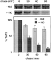
Starvation induces turnover of TAT2. Logarithmically growing wild-type cells expressing HA-TAT2 (JK9-3d/pAS55) were labeled with 35S-methionine. A chase was performed by adding an excess of cold methionine, either alone or in combination with rapamycin (200 ng/ml). Aliquots of the untreated and rapamycin-treated cultures were removed at the indicated times of chase, and cell extracts were prepared. HA-TAT2 was immunoprecipitated from these extracts, eluted from the washed protein G–Sepharose beads at 37°C, subjected to SDS-PAGE, and visualized by fluorography (see Materials and Methods). Bars represent the average value from two independent experiments.
Figure 3
Figure 3
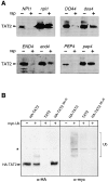
Turnover of TAT2 requires ubiquitination, endocytosis, and vacuolar proteases. (A) Parental wild-type strains (NPI1, 23344c; DOA4, 23344c; PEP4, JK9-3d) and npi1 (27038a), doa4 (27002d), and pep4 (MH684) mutants expressing HA-TAT2 (from pAS55 or, in the case of PEP4 and pep4, from pAS64), were grown in SC medium at 30°C to early logarithmic phase. Rapamycin (rap) or empty drug vehicle was added. The parental wild-type END4 strain (RH1602) and the conditional end4 mutant (RH1597) were grown at 24°C and shifted to 37°C for 5 min before the addition of rapamycin or empty drug vehicle. After 60 min in the presence of rapamycin, cells were harvested and processed for Western analysis of HA-TAT2 (TAT2). (B) Wild-type cells (JK9-3d) expressing either HA-TAT2 (pAS55), untagged TAT2 (pAS8), or HA-TAT25K>R (pTB355), and containing a plasmid for copper-inducible expression of myc-tagged ubiquitin (YEp105) were grown in SC medium. Expression of myc-ub was not induced (−) or induced (+) with copper sulfate (100 μM) for 3 h. Before harvesting, cells were treated for 15 min with rapamycin (200 ng/ml). Cells were collected and processed for immunoprecipitation with anti-HA (from 1 mg of total protein, see Materials and Methods). Immunoprecipitated protein was subjected to Western analysis. The blot was probed with anti-HA antibody (α-HA), stripped and reprobed with anti-myc antibody (α-myc). The asterisk denotes a nonspecific band.
Figure 4
Figure 4
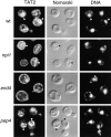
Cellular distribution of HA-TAT2 by indirect immunofluorescence. Wild-type (wt, JK9-3d) and npi1 (27038a) and pep4 (MH684) mutant strains transformed with pHA-TAT2 (pAS55 or, in the case of pep4, pAS64) were grown in SC medium at 30°C to early logarithmic phase. The shown wild-type cells were not treated with rapamycin. All other cells shown were treated with rapamcyin for 60 min. The conditional end4 mutant (RH1597/pAS55) was grown at 24°C and shifted to 37°C for 5 min before the addition of rapamycin. Cells were fixed and processed for detection of HA-TAT2 by immunofluorescence (see Materials and Methods). The cellular distribution of HA-TAT2 is shown in the left column (TAT2). The same field of cells is shown in the middle (Normarski) and right columns (DNA). The right column shows the cells stained with the DNA specific stain DAPI. The endoplasmic reticulum corresponds to the area encircling the DAPI-staining nucleus (see arrowheads in wt cells). The vacuole appears as a large crater in cells visualized by Nomarski optics (see arrowheads in npi1 and pep4 cells). The exposure time to visualize HA-TAT2 in end4 cells was approximately sixfold longer than for other strains shown.
Figure 5
Figure 5
The NH2 terminus of TAT2 is required for degradation. (A) Cells expressing HA-TAT2 containing NH2-terminal mutations were grown to early logarithmic phase in SC medium (JK9-3d containing either pHA-TAT2Δ10, pHA-TAT2Δ17, pHA-TAT2Δ20, pHA-TAT2Δ29, pHA-TAT2Δ29K31R, pHA-TAT2Δ31, pHA-TAT2Δ17-31, or pHA-TAT25K>R). Rapamycin was added to one half of each culture. After 60 min of further incubation, extracts were prepared and assayed for HA-TAT2 by Western analysis. The levels of the mutant forms of HA-TAT2 are not normalized to the levels of wild-type HA-TAT2 (due to different exposure times), and thus an increased abundance for the stabilized proteins, as described in the text, is not reflected in these data. The numbering of amino acids refers to untagged TAT2. (B) TAT25K>R accumulates in the plasma membrane. Wild-type cells (JK9-3d) expressing HA-TAT25K>R were grown in SC medium to early logarithmic phase, treated with rapamycin for 60 min, and processed for visualization of HA-TAT25K>R by immunofluorescence (see Fig. 4 legend).
Figure 5
Figure 5
The NH2 terminus of TAT2 is required for degradation. (A) Cells expressing HA-TAT2 containing NH2-terminal mutations were grown to early logarithmic phase in SC medium (JK9-3d containing either pHA-TAT2Δ10, pHA-TAT2Δ17, pHA-TAT2Δ20, pHA-TAT2Δ29, pHA-TAT2Δ29K31R, pHA-TAT2Δ31, pHA-TAT2Δ17-31, or pHA-TAT25K>R). Rapamycin was added to one half of each culture. After 60 min of further incubation, extracts were prepared and assayed for HA-TAT2 by Western analysis. The levels of the mutant forms of HA-TAT2 are not normalized to the levels of wild-type HA-TAT2 (due to different exposure times), and thus an increased abundance for the stabilized proteins, as described in the text, is not reflected in these data. The numbering of amino acids refers to untagged TAT2. (B) TAT25K>R accumulates in the plasma membrane. Wild-type cells (JK9-3d) expressing HA-TAT25K>R were grown in SC medium to early logarithmic phase, treated with rapamycin for 60 min, and processed for visualization of HA-TAT25K>R by immunofluorescence (see Fig. 4 legend).
Figure 6
Figure 6
Upon starvation, TAT2 is sorted from internal compartments to the vacuole independently of the plasma membrane. A parental wild-type strain (RSY255), and sec4 (RH1552), sec18 (RSY271), sec23 (RSY281), apg1 (MT16-9B), pep12 (CBY9/5C), and vps27 (RH2379) mutants expressing HA-TAT2 (pAS55) were grown at 24°C to early logarithmic phase and shifted to 37°C for 5 min (10 min for the pep12 mutant). Rapamycin was added to half of each culture. After 60 min of further incubation, cells were harvested and processed for Western analysis.
Figure 7
Figure 7
NH2-terminal lysine residues of TAT2 are required for rapamycin-induced degradation of newly made TAT2. Temperature-sensitive sec4 mutant cells expressing HA-TAT2 (pAS55) or HA-TAT25K>R (pTB355) were grown in SC medium at 24°C to early logarithmic phase, labeled with 35S-methionine for 3 min and shifted to 37°C for 5 min. Labeled cells were harvested and chased at 37°C in SC medium containing 200 ng/ml of rapamycin. Aliquots were taken at the indicated times of chase, and cell extracts were prepared. HA-TAT2 or HA-TAT25K>R was immunoprecipitated, eluted from the washed protein G–Sepharose beads at 37°C, subjected to SDS-PAGE, and visualized by fluorography (for details, see Materials and Methods).
Figure 8
Figure 8
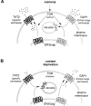
Model for inverse regulation of specific (TAT2) and broad-range (GAP1) amino acid permeases by TOR in response to nutrients. (A) In the presence of nutrients, active TOR mediates the sorting of TAT2 to the plasma membrane and the routing of GAP1 to the vacuole for degradation. (B) Upon nutrient deprivation, TOR is inactive and TAT2 is targeted to the vacuole for degradation whereas GAP1 is routed to the plasma membrane. TOR is associated with the plasma membrane.

Similar articles

Cited by

References

    1. Ausubel F.M., Brent R., Kingston R.E., Moore D.D., Seidman J.G., Smith J.A., Struhl K. Current Protocols in Molecular Biology. John Wiley & Sons, Inc; New York: 1998.
    1. Barbet N.C., Schneider U., Helliwell S.B., Stansfield I., Tuite M.F., Hall M.N. TOR controls translation initiation and early G1 progression in yeast. Mol. Biol. Cell. 1996;7:25–42. - PMC - PubMed
    1. Becherer K.A., Rieder S.E., Emr S.D., Jones E.W. Novel syntaxin homologue, Pep12p, required for the sorting of lumenal hydrolases to the lysosome-like vacuole in yeast. Mol. Biol. Cell. 1996;7:579–594. - PMC - PubMed
    1. Berset C., Trachsel H., Altmann M. The TOR (target of rapamycin) signal transduction pathway regulates the stability of translation initiation factor eIF4G in the yeast Saccharomyces cerevisiae . Proc. Natl. Acad. Sci. USA. 1998;95:4264–4269. - PMC - PubMed
    1. Bickle M., Delley P.A., Schmidt A., Hall M.N. Cell wall integrity modulates RHO1 activity via the exchange factor ROM2. EMBO J. 1998;17:2235–2245. - PMC - PubMed

Publication types

MeSH terms