Comparative Study
. 1999 Sep 20;146(6):1255-64.
doi: 10.1083/jcb.146.6.1255.

Regulation of p21(cip1) expression by growth factors and the extracellular matrix reveals a role for transient ERK activity in G1 phase

Affiliations
Comparative Study

Regulation of p21(cip1) expression by growth factors and the extracellular matrix reveals a role for transient ERK activity in G1 phase

M E Bottazzi et al. J Cell Biol. .

Abstract

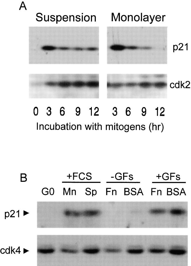
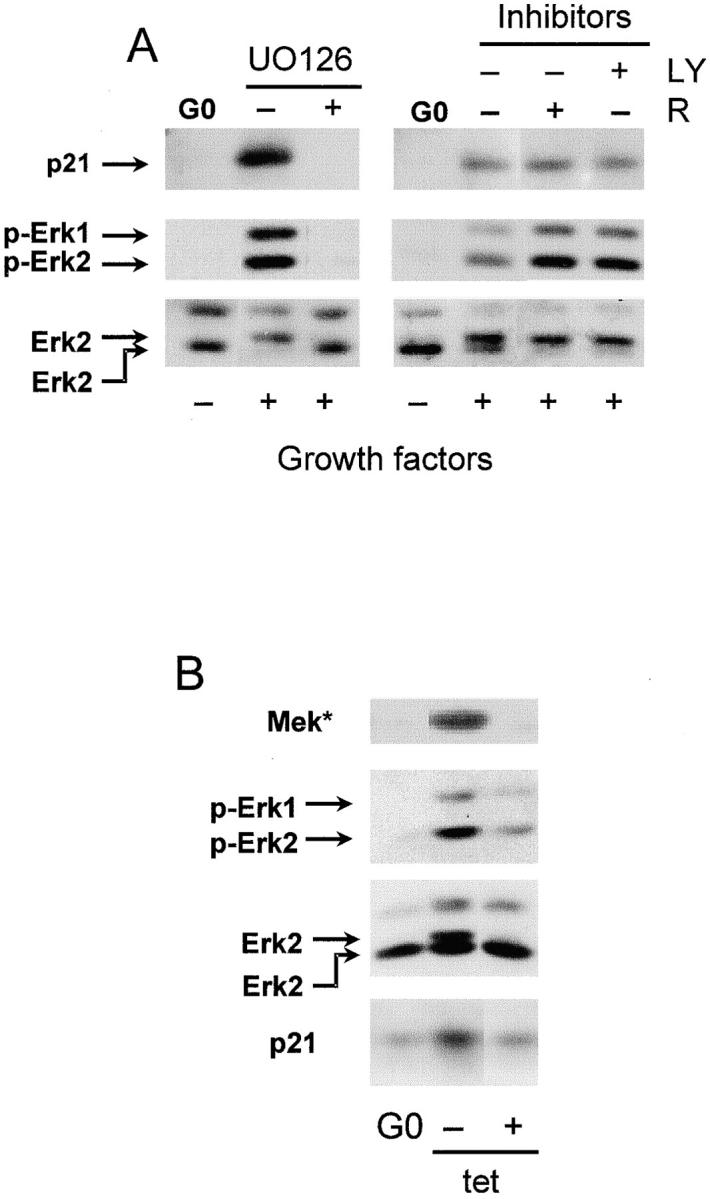
We have examined the regulation of p21(cip1) by soluble mitogens and cell anchorage as well as the relationship between the expression of p21(cip1) and activation of the ERK subfamily of MAP kinases. We find that p21(cip1) expression in G1 phase can be divided into two discrete phases: an initial induction that requires growth factors and the activation of ERK, and then a subsequent decline that is enhanced by cell anchorage in an ERK-independent manner. In contrast to the induction of cyclin D1, the induction of p21(cip1) is mediated by transient ERK activity. Comparative studies with wild-type and p21(cip1)-null fibroblasts indicate that adhesion-dependent regulation of p21(cip1) is important for proper control of cyclin E-cdk2 activity. These data lead to a model in which mitogens and anchorage act in a parallel fashion to regulate G1 phase expression of p21(cip1). They also show that (a) growth factors and growth factor/extracellular matrix cooperation can have different roles in regulating G1 phase ERK activity and (b) both transient and sustained ERK signals have functionally significant roles in controlling cell cycle progression through G1 phase.


Figures

Figure 1
Figure 1
Biphasic regulation of p21cip1 expression by growth factors and the extracellular matrix. In A, G0-synchronized MEFs were seeded in suspension and monolayer in 10% FCS-DME and incubated for 0–12 h. Cell extracts were prepared and equal amounts of protein were fractionated on reducing SDS gels and analyzed by immunoblotting and enhanced chemiluminescence (ECL) using antibodies against p21cip1 and cdk2 (a control for loading). [Note: In this experiment the downregulation of p21 was essentially complete in 12 h, but other experiments indicate that strong downregulation usually occurs over a 12–18-h period.] In B, quiescent MEFs (G0) were cultured for 3 h in defined medium on dishes coated with BSA or fibronectin (Fn). The incubations were also performed in the absence (−GF) or presence (+GF) of purified growth factors. Duplicate cultures were incubated in monolayer (Mn) and suspension (Sp) in 10% FCS-DME for comparison. Collected cells were analyzed by immunoblotting with anti-p21cip1 and anti-cdk4 (a control for loading).
Figure 2
Figure 2
Activation of ERKs controls induction of p21cip1 by mitogens. In A, G0-synchronized MEFs (G0) were washed and preincubated for 30 min in 10 ml defined medium with 100 μM U0126, 5 nM rapamycin (R), 25 μM LY294002 (LY), or solvent (dimethylsulfoxide) as control (–). Purified growth factors were directly added to the cultures. The cells were incubated for 1 h before collection and lysis. Equal amounts of cell extracts were analyzed by immunoblotting using antibodies against p21cip1, phospho-ERK (p-ERK), and ERK. Controls (not shown) demonstrated that the concentrations of LY294002 and rapamycin we used completely inhibited phosphorylation of p70S6 kinase. In B, tet-MEK*-3T3 cells were grown to confluence in 10% FCS-DME with 2 μg/ml tetracycline. The medium was removed, replaced with serum-free DME, and the cultures were serum-starved for 1 d in the absence of tetracycline (G0). Serum-starved cells were trypsinized and reseeded (2 × 10⁶ in 10 ml 0.5% FCS) in 100-mm agarose-coated dishes in the absence and presence of tetracycline (tet). Cells were collected after 9 h and lysed. Equal amounts of protein were fractionated on an SDS gel and analyzed by immunoblotting with Mek-1, phospho-ERK (p-ERK), ERK and p21cip1 antibodies. For both panels, upper and lower arrows in the ERK blots show the phosphorylated and unphosphorylated ERK2, respectively.
Figure 3
Figure 3
Anchorage-independent induction of p21cip1 is associated with a short-term activation of ERK. In A, quiescent MEFs were trypsinized, suspended in defined medium, and reseeded on BSA- or fibronectin (FN)-coated dishes for 1 and 3 h in the presence of purified growth factors. Cells were collected, lysed, and analyzed by immunoblotting with anti-ERK (top), anti-phospho-ERK (middle panel), and anti-p21cip1. In B, MEFs were rendered quiescent, trypsinized, suspended in 10% FCS-DME, and reseeded in monolayer and suspension for 0–12 h. Cells were collected, lysed, and analyzed by immunoblotting using antibodies to ERK and cyclin D1. In C, quiescent MEFs were trypsinized, suspended in defined medium, and reseeded on BSA- or fibronectin-coated dishes for 1 h in the presence of increasing concentrations of purified growth factors (the triangles represent a dose-response curve of a PDGF-insulin-EGF cocktail used at 25, 50, and 100% of the concentration described in Materials and Methods). For all panels, upper and lower arrowheads in the ERK blots show the phosphorylated and unphosphorylated ERK, respectively.
Figure 4
Figure 4
A transient ERK signal induces p21cip1. MEFs were G0-synchronized in defined medium and stimulated with purified growth factors for the times shown. In A, cells were collected, lysed, and analyzed by immunoblotting with anti-ERK (ERK), anti-phospho-ERK (p-ERK), anti-p21cip1, and anti-cdk4 (loading control). In B, the cells were pretreated with purified growth factors for 10 min (time 0) before the addition of U0126 (100 μM final concentration). After incubation with UO126 for 10–60 min, cells were collected, lysed, and the degree of ERK activation was determined by gel-shift and direct assessment of dually phosphorylated ERK by immunoblotting with anti-ERK (ERK) and anti-phospho-ERK (p-ERK), respectively. For A and B, the upper and lower arrowheads in the ERK blots show the phosphorylated and unphosphorylated ERK2, respectively. In C, the G0-synchronized cells were stimulated with purified growth factors, and U0126 was added to the cultures 10, 20, and 40 min after the stimulation with growth factors. All cells were collected at 60 min (a time sufficient for p21cip1 induction in cells lacking UO126), lysed, and analyzed for the expression of p21cip1 by immunoblotting. MEFs stimulated in the absence of U0126 for 60 min were used as a positive control (C) for the induction of p21cip1. In D, G0-synchronized MEFs were stimulated with purified growth factors and U0126 was added to the cells 10, 20, 40, and 60 min after stimulation with growth factors. All cultures were collected at 8 h (a time sufficient for cyclin D1 induction in cells lacking UO126), lysed and analyzed for the expression of cyclin D1 by immunoblotting. MEFs stimulated in the absence of U0126 for 8 h were used as a positive control (C) for the expression of cyclin D1 (detected as the doublet migrating slightly above cdk4). In C and D, immunoblotting with anti-cdk4 was used to control for protein loading.
Figure 5
Figure 5
Turnover of p21cip1 in adherent and nonadherent cells. G0-synchronized MEFs were trypsinized and replated in suspension or monolayer for 3 h with 10% FCS-DME before the addition of cycloheximide (10 μg/ml final concentration). Cells were collected at the indicated times spanning 120 min and lysed. Equal amounts of protein were fractionated on SDS gels and immunoblotted using anti-p21cip1 and anti-cdk2 (loading control).
Figure 6
Figure 6
Full downregulation of p21cip1 gene expression requires cell adhesion. In A, MEFs were rendered quiescent, trypsinized, and then replated in monolayer (Mn) and suspension (Sp) in the presence of 10% FCS-DME. At the times shown, cells were collected and lysed for collection of RNA. Northern blotting was performed using a 32P-labeled, random-primed probe for murine p21cip1 and equal loading of samples was confirmed by ethidium-bromide staining of rRNA (not shown). In B, MEFs were transiently transfected with a p21cip1 promoter-luciferase construct as described in Materials and Methods. Transfected cells were G0-synchronized and stimulated with 10% FCS-DME in monolayer and suspension for the times shown. B shows fold-induction of luciferase activity, relative to G0-synchronized cells and normalized to constant activity of the Renilla luciferase control reporter vector.
Figure 7
Figure 7
Downregulation of p21cip1 by ECM does not require p53 and is independent of ERK. In A, serum-starved p53-null MEFs were cultured in monolayer (Mn) and suspension (Sp) and analyzed by Northern blotting as described in the legend to Fig. 6 A. Equal RNA loading was confirmed by ethidium-bromide staining of rRNA (not shown). In B, asynchronous tet-mek*-3T3 cells (2 × 10⁶ cells per 100-mm dish) were incubated with 10 ml of 10% FCS-DME in suspension (Sp) and monolayer (Mn) for 24 h in the absence (−) and presence (+) of tetracycline (tet). Cells were collected and lysed for immunoblotting analysis using antibodies to mek-1, ERK, phospho-ERK (p-ERK), and p21cip1. In this experiment, lysates were fractionated on a 5–15% gradient SDS gel that does not permit complete resolution of unphosphorylated and phosphorylated ERKs by gel-shift.
Figure 8
Figure 8
p21cip1 is important for the anchorage dependency of cyclin E–cdk2 activity. G0-synchronized (G0) MEFs from wild-type (p21+/+) and p21cip1-null (p21−/−) mice were trypsinized, reseeded in suspension (Sp) and monolayer (Mn), and incubated in 10% FCS-DME for 18 h. The cells were collected and lysed. Equal amounts of cellular protein (200 μg) were loaded on a reducing SDS gradient gel (5–15% acrylamide) and analyzed by immunoblotting with antibodies to cyclin E, p21cip1, p27kip1, cdk2, cyclin D1, pRb, cyclin A, and cdk4. The immunoreactive proteins were visualized by ECL. The upper and lower arrows on the pRb blot show the phosphorylated and hypophosphorylated pRb, respectively. Duplicate aliquots of 200 μg from the same experiment were incubated with anti-cyclin E in order to collect cyclin E–cdk2 complexes for determination of cyclin E–cdk2 kinase activity in vitro using histone H1 as substrate as described (Zhu et al. 1996). p21−/− MEFs progressed through G1 phase several hours faster than p21+/+ MEFs (data not shown), and the difference in cyclin E–cdk2 kinase activity detected in monolayer cultures of p21+/+ cells vs. p21−/− cells most likely reflects the fact that the two lines were not in identical positions within G1 phase when assayed 18 h after exposure to mitogens.
Figure 9
Figure 9
Cooperative effects of growth factors and the ECM in G1 phase. The model shows that the induction of p21cip1 and cyclin D1 in G1 phase are differentially regulated by growth factors and the ECM largely because p21cip1 induction occurs in response to transient ERK activation while cyclin D1 induction requires sustained ERK activation. The figure also shows that the ECM-enhanced downregulation of p21cip1 in mid-late G1 phase is independent of ERK activity.

Similar articles

Cited by

References

    1. Albanese C., Johnson J., Watanabe G., Eklund N., Vu D., Arnold A., Pestell R.G. Transforming p21ras mutants and c-Ets-2 activate the cyclin D1 promoter through distinguishable regions. J. Biol. Chem. 1995;270:23589–23597. - PubMed
    1. Aplin A.E., Juliano R.L. Integrin and cytoskeletal regulation of growth factor signaling to the MAP kinase pathway. J. Cell Sci. 1999;112:695–706. - PubMed
    1. Assoian R.K. Anchorage-dependent cell cycle progression. J. Cell Biol. 1997;136:1–4. - PMC - PubMed
    1. Auer K.L., Park J.-S., Seth P., Coffey R.J., Darlingtons G., Abo A., McMahon M., DePinho R.A., Fisher P.B., Dent P. Prolonged activation of the mitogen-activated protein kinase pathway promotes DNA synthesis in primary hepatocytes from p21cip1/waf1-null mice, but not in hepatocytes from p16INK4a-null mice. Biochem. J. 1998;336:551–560. - PMC - PubMed
    1. Böhmer R.M., Scharf E., Assoian R.K. Cytoskeletal integrity is required throughout the mitogen stimulation phase of the cell cycle and mediates the anchorage-dependent expression of cyclin D1. Mol. Biol. Cell. 1996;7:101–111. - PMC - PubMed

Publication types

MeSH terms