. 1999 May-Jun;6(3):216-31.

Activity-dependent activation of TrkB neurotrophin receptors in the adult CNS

Affiliations

Activity-dependent activation of TrkB neurotrophin receptors in the adult CNS

R Aloyz et al. Learn Mem. 1999 May-Jun.

Abstract

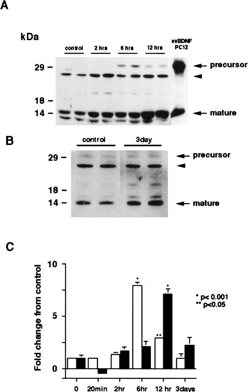
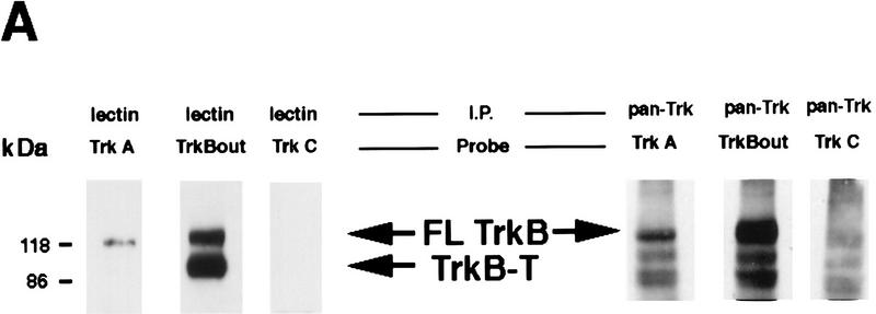
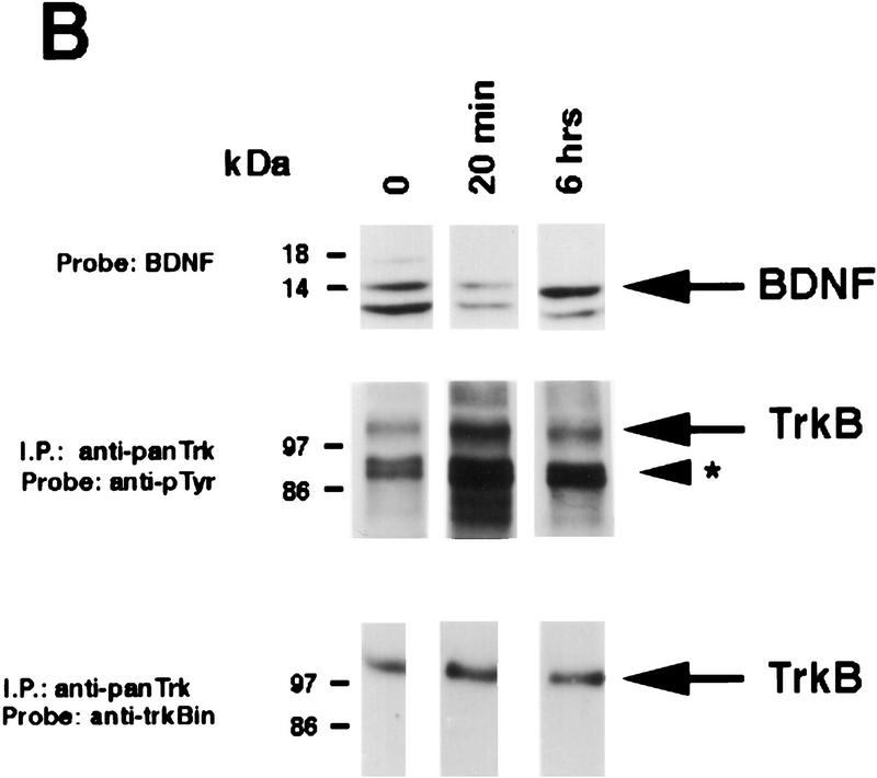
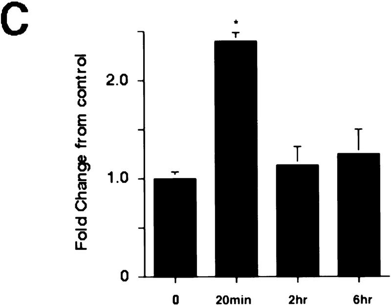
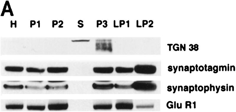
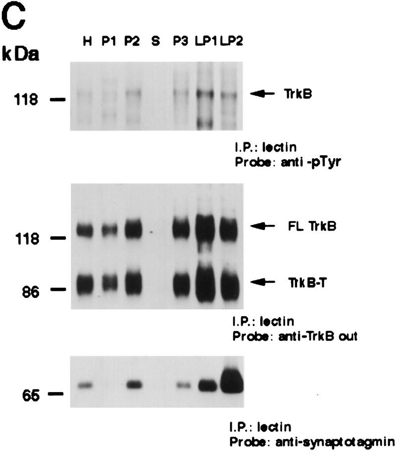
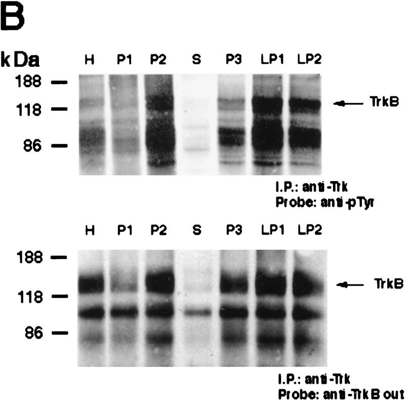
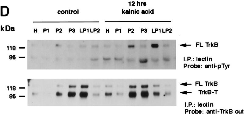
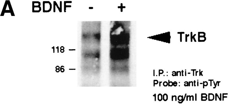
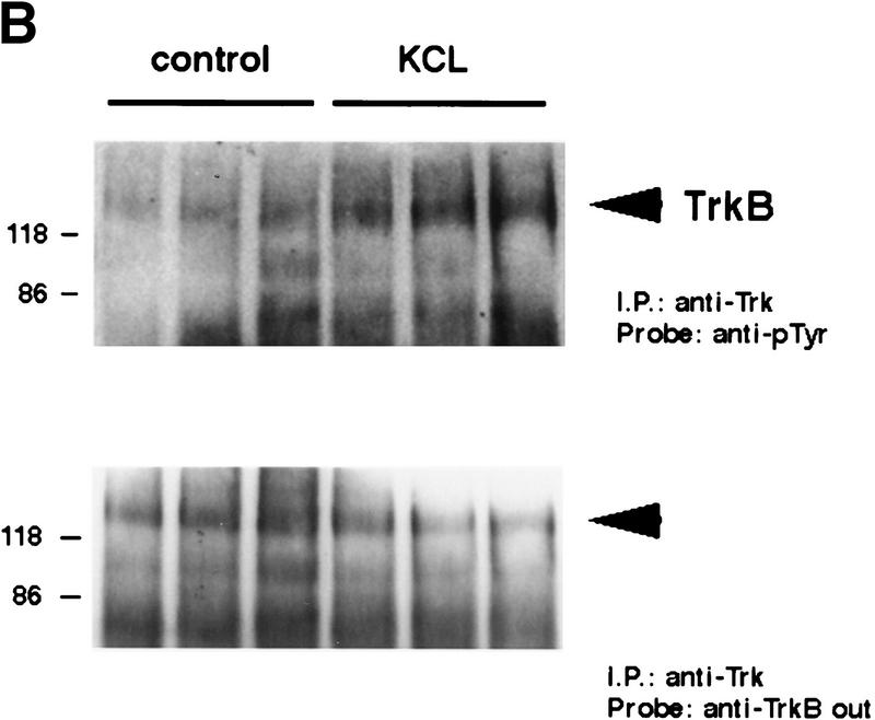
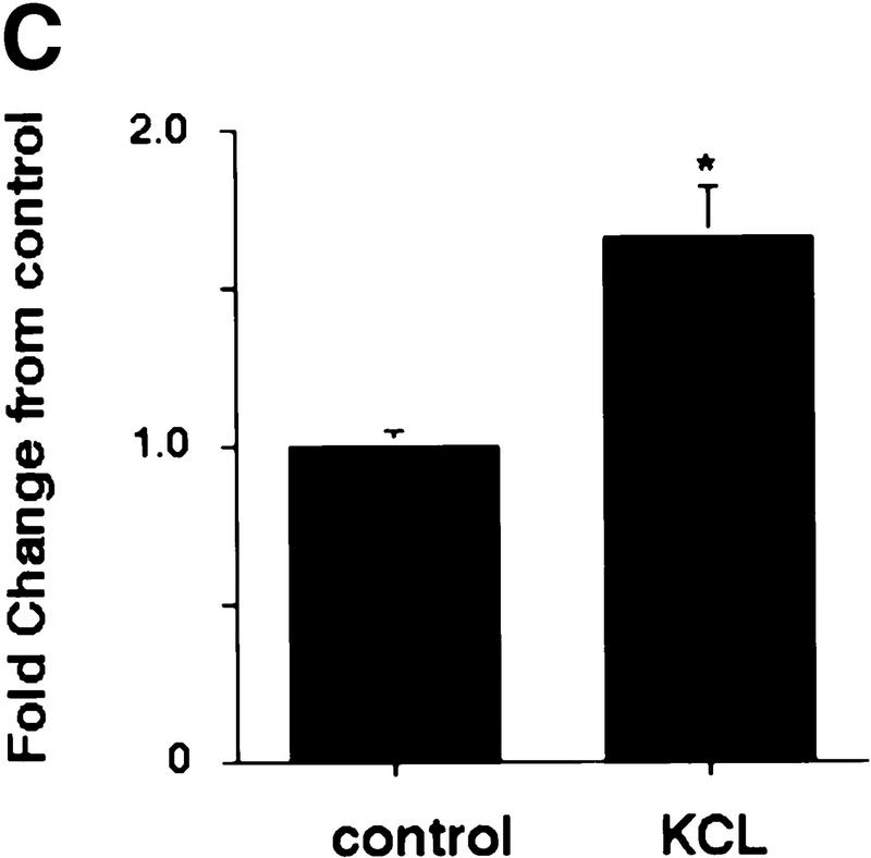
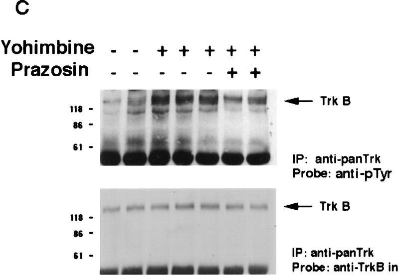
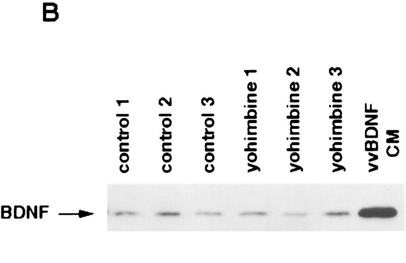
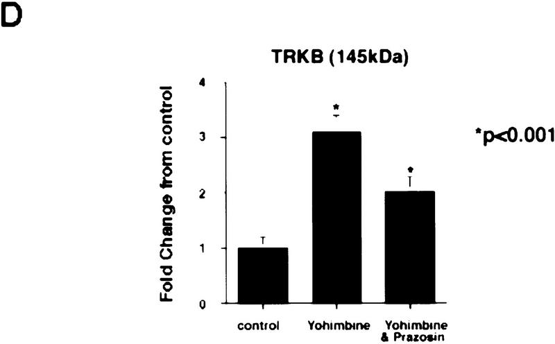
In this paper we have investigated the hypothesis that neural activity causes rapid activation of TrkB neurotrophin receptors in the adult mammalian CNS. These studies demonstrate that kainic acid-induced seizures led to a rapid and transient activation of TrkB receptors in the cortex. Subcellular fractionation demonstrated that these activated Trk receptors were preferentially enriched in the synaptosomal membrane fraction that also contained postsynaptic glutamate receptors. The fast activation of synaptic TrkB receptors could be duplicated in isolated cortical synaptosomes with KCl, presumably as a consequence of depolarization-induced BDNF release. Importantly, TrkB activation was also observed following pharmacological activation of brain-stem noradrenergic neurons, which synthesize and anterogradely transport BDNF; treatment with yohimbine led to activation of cortical TrkB receptors within 30 min. Pharmacological blockade of the postsynaptic alpha1-adrenergic receptors with prazosin only partially inhibited this effect, suggesting that the TrkB activation was partially due to a direct effect on postsynaptic cortical neurons. Together, these data support the hypothesis that activity causes release of BDNF from presynaptic terminals, resulting in a rapid activation of postsynaptic TrkB receptors. This activity-dependent TrkB activation could play a major role in morphological growth and remodelling in both the developing and mature nervous systems.


Figures

Figure 1
Figure 1
Increased synthesis of BDNF in the cortex of kainic acid-treated animals. (A,B) Western blot analysis of equal amounts of protein derived from the cortex of individual control animals or of animals 2 hr (2 hrs), 6 hr (6 hrs), 12 hr (12 hrs) (A), or 3 days (B) following kainic acid treatment. As a control, lysates of PC12 cells infected with a vaccinia virus encoding BDNF (vv BDNF PC12) (Fawcett et al. 1997; Mowla et al. 1999) were also run. The numbers at left indicate the molecular mass markers; the arrows at right indicate the 32-kD BDNF precursor and mature, processed BDNF. The arrowhead indicates a nonspecific, cross-reactive band (see text). (C) Quantitation of experiments similar to those shown in A and B. Western blots were scanned, the intensity of the bands quantitated by image analysis, and these numbers were then normalized to the numbers obtained from control animals analyzed on the same Western blots. The graph represents the mean fold change from controls at time points ranging from 20 min to 3 days, and the error bars indicate the s.e.m. (Open bars) BDNF precursor; (solid bars) mature BDNF. For each time point, n = 4 animals. Statistical significance was determined using ANOVA, and results that are significantly different from controls are indicated by the asterisks (*) P < 0.001; (**) P < 0.05.
Figure 2
Figure 2
Rapid activation of cortical Trk receptors following kainic acid treatment. (A) Western blot analysis of cortical tissue derived from adult rats precipitated with either WGA (lectin) or anti-panTrk (pan-Trk) and then probed with antibodies specific to TrkA, TrkB [both full-length (FL-TrkB) and truncated (TrkB-T) forms], or TrkC. Note that TrkB is the most abundant Trk in the adult cortex and that although TrkC is not detectable in the WGA precipitates, it can be detected at low levels when cortical tissue is immunoprecipitated with anti-panTrk. The numbers at left of the blots indicate the molecular mass markers. (B) Western blot analysis of equal amounts of protein isolated from the cortex of naive, control animals (0) or of adult rats at 20 min and 6 hr following kainic acid treatment. In the case of BDNF (top), total lysates were probed with a BDNF-specific antibody. The same samples were also immunoprecipitated with anti-panTrk, which recognizes all Trk receptors, and the immunoprecipitates were probed with anti-phosphotyrosine (anti-pTyr) (middle) or with an antibody specific for the full-length form of TrkB (anti-trkBin). In both cases, the arrows indicate the size of full-lengthTrkB, whereas in the middle panel, the arrowhead indicates a tyrosine phosphorylated protein that coimmunoprecipitates with Trk and whose identity is currently unknown. The numbers at left of the blots indicate the molecular mass markers. Note that although similar amounts of TrkB are present at all time points, the level of Trk receptor autophosphorylation is increased at 20 min following kainic acid treatment. (C) Quantitation of three separate experiments similar to that shown in B. Western blots were scanned, the intensity of the bands quantitated by image analysis, and the data were expressed as the ratio of phosphoTrk to full-length TrkB levels. These ratios were then normalized to the ratio for control animals analyzed on the same blots. The graph represents the mean fold change from controls at 20 min, 2 hr, and 6 hr, and the error bars indicate the s.e.m. For each time point, n = 4 animals. Asterisks (*) indicate those points that are statistically different from the controls using ANOVA (P < 0.01).
Figure 2
Figure 2
Rapid activation of cortical Trk receptors following kainic acid treatment. (A) Western blot analysis of cortical tissue derived from adult rats precipitated with either WGA (lectin) or anti-panTrk (pan-Trk) and then probed with antibodies specific to TrkA, TrkB [both full-length (FL-TrkB) and truncated (TrkB-T) forms], or TrkC. Note that TrkB is the most abundant Trk in the adult cortex and that although TrkC is not detectable in the WGA precipitates, it can be detected at low levels when cortical tissue is immunoprecipitated with anti-panTrk. The numbers at left of the blots indicate the molecular mass markers. (B) Western blot analysis of equal amounts of protein isolated from the cortex of naive, control animals (0) or of adult rats at 20 min and 6 hr following kainic acid treatment. In the case of BDNF (top), total lysates were probed with a BDNF-specific antibody. The same samples were also immunoprecipitated with anti-panTrk, which recognizes all Trk receptors, and the immunoprecipitates were probed with anti-phosphotyrosine (anti-pTyr) (middle) or with an antibody specific for the full-length form of TrkB (anti-trkBin). In both cases, the arrows indicate the size of full-lengthTrkB, whereas in the middle panel, the arrowhead indicates a tyrosine phosphorylated protein that coimmunoprecipitates with Trk and whose identity is currently unknown. The numbers at left of the blots indicate the molecular mass markers. Note that although similar amounts of TrkB are present at all time points, the level of Trk receptor autophosphorylation is increased at 20 min following kainic acid treatment. (C) Quantitation of three separate experiments similar to that shown in B. Western blots were scanned, the intensity of the bands quantitated by image analysis, and the data were expressed as the ratio of phosphoTrk to full-length TrkB levels. These ratios were then normalized to the ratio for control animals analyzed on the same blots. The graph represents the mean fold change from controls at 20 min, 2 hr, and 6 hr, and the error bars indicate the s.e.m. For each time point, n = 4 animals. Asterisks (*) indicate those points that are statistically different from the controls using ANOVA (P < 0.01).
Figure 3
Figure 3
Activated Trk is enriched in synaptic fractions following kainic acid treatment. (A) Adult cortical tissue was fractionated, and Western blot analysis was performed for a variety of marker proteins to ensure the accuracy of the subcellular fractionation. Equal amounts of protein from each fraction were probed with antibodies specific for TGN 38, a marker for the Golgi apparatus that is enriched in P3, for synaptotagmin and synaptophysin, two synaptic vesicle markers that are enriched in LP2, and for GluR1, the postsynaptic glutamate receptor that is enriched in LP1 (a fraction containing synaptic membranes) and in P2 (the fraction from which LP1 and LP2 are derived). H indicates the original tissue homogenate, and S is a cytosolic fraction. (B) Equal amounts of protein from the cortex of animals that were kainic acid treated for 20 min were immunoprecipitated with anti-panTrk, and the immunoprecipitates were analyzed by Western blot analysis with anti-phosphotyrosine (top) or anti-TrkBout (bottom). The arrows at right of both panels indicate the size of full-length TrkB, and the numbers at left indicate the molecular mass markers. Note that although TrkB is present in most fractions, tyrosine phosphorylated Trk is most enriched in LP1. (C) Equal amounts of protein from subcellular fractions derived from the cortex of animals that were kainic acid treated for 20 min were precipitated with WGA (lectin) and then probed with antibodies against phosphotyrosine (top), against all forms of TrkB (middle), or against synaptotagmin, a marker for synaptic vesicles (bottom). (Top) The arrow indicates a tyrosine phosphorylated band the size of full-length TrkB. (Middle) One arrow indicates the full-length TrkB receptor (FL TrkB), whereas the second indicates the truncated TrkB receptor (TrkB-T). For all panels, numbers at left of the blots indicate the molecular mass markers. (D) Equal amounts of protein from subcellular fractions of the cortex of control animals or of animals that were treated with kainic acid for 12 hr were precipitated with WGA (lectin) and then probed with antibodies against phosphotyrosine (top) or against the TrkB receptor (bottom). At top, the arrow indicates a tyrosine phosphorylated protein of the same size as full-length TrkB; at bottom, the arrows indicate both the full-length (FL TrkB) and truncated (TrkB-T) forms of TrkB.
Figure 3
Figure 3
Activated Trk is enriched in synaptic fractions following kainic acid treatment. (A) Adult cortical tissue was fractionated, and Western blot analysis was performed for a variety of marker proteins to ensure the accuracy of the subcellular fractionation. Equal amounts of protein from each fraction were probed with antibodies specific for TGN 38, a marker for the Golgi apparatus that is enriched in P3, for synaptotagmin and synaptophysin, two synaptic vesicle markers that are enriched in LP2, and for GluR1, the postsynaptic glutamate receptor that is enriched in LP1 (a fraction containing synaptic membranes) and in P2 (the fraction from which LP1 and LP2 are derived). H indicates the original tissue homogenate, and S is a cytosolic fraction. (B) Equal amounts of protein from the cortex of animals that were kainic acid treated for 20 min were immunoprecipitated with anti-panTrk, and the immunoprecipitates were analyzed by Western blot analysis with anti-phosphotyrosine (top) or anti-TrkBout (bottom). The arrows at right of both panels indicate the size of full-length TrkB, and the numbers at left indicate the molecular mass markers. Note that although TrkB is present in most fractions, tyrosine phosphorylated Trk is most enriched in LP1. (C) Equal amounts of protein from subcellular fractions derived from the cortex of animals that were kainic acid treated for 20 min were precipitated with WGA (lectin) and then probed with antibodies against phosphotyrosine (top), against all forms of TrkB (middle), or against synaptotagmin, a marker for synaptic vesicles (bottom). (Top) The arrow indicates a tyrosine phosphorylated band the size of full-length TrkB. (Middle) One arrow indicates the full-length TrkB receptor (FL TrkB), whereas the second indicates the truncated TrkB receptor (TrkB-T). For all panels, numbers at left of the blots indicate the molecular mass markers. (D) Equal amounts of protein from subcellular fractions of the cortex of control animals or of animals that were treated with kainic acid for 12 hr were precipitated with WGA (lectin) and then probed with antibodies against phosphotyrosine (top) or against the TrkB receptor (bottom). At top, the arrow indicates a tyrosine phosphorylated protein of the same size as full-length TrkB; at bottom, the arrows indicate both the full-length (FL TrkB) and truncated (TrkB-T) forms of TrkB.
Figure 4
Figure 4
Depolarization-induced activation of Trk receptors in isolated cortical synaptosomes. (A) Western blot analysis of anti-panTrk immunoprecipitates of equal amounts of protein from synaptosomes treated with (+) or without (−) 100 ng/ml BDNF and probed with anti-phosphotyrosine. The numbers at left of the blot indicate the molecular mass markers, whereas the arrowhead indicates the size of full-length TrkB. (B) Western blot analysis of anti-panTrk immunoprecipitates of equal amounts of protein from synaptosomes treated with (KCL) or without (control) 117 mm KCl for 10 min and probed with anti-phosphotyrosine (top). To ensure that similar amounts of TrkB are present in each sample, the same blot was reprobed with anti-TrkBout (bottom). In both panels, the numbers at left of the blot indicate the molecular mass markers, whereas the arrowhead indicates the size of full-length TrkB. (C) Quantitation of experiments similar to that shown in B (n = 8 for controls, n = 9 for KCl treated). Western blots were scanned, the intensity of the bands quantitated by image analysis, and the data were expressed as a ratio of phosphoTrk to full-length TrkB levels. These ratios were then normalized to the ratio for the control synaptosomes analyzed on the same Western blots. Significance was determined by Student’s t-test (P = 0.0081). (*) P < 0.05.
Figure 4
Figure 4
Depolarization-induced activation of Trk receptors in isolated cortical synaptosomes. (A) Western blot analysis of anti-panTrk immunoprecipitates of equal amounts of protein from synaptosomes treated with (+) or without (−) 100 ng/ml BDNF and probed with anti-phosphotyrosine. The numbers at left of the blot indicate the molecular mass markers, whereas the arrowhead indicates the size of full-length TrkB. (B) Western blot analysis of anti-panTrk immunoprecipitates of equal amounts of protein from synaptosomes treated with (KCL) or without (control) 117 mm KCl for 10 min and probed with anti-phosphotyrosine (top). To ensure that similar amounts of TrkB are present in each sample, the same blot was reprobed with anti-TrkBout (bottom). In both panels, the numbers at left of the blot indicate the molecular mass markers, whereas the arrowhead indicates the size of full-length TrkB. (C) Quantitation of experiments similar to that shown in B (n = 8 for controls, n = 9 for KCl treated). Western blots were scanned, the intensity of the bands quantitated by image analysis, and the data were expressed as a ratio of phosphoTrk to full-length TrkB levels. These ratios were then normalized to the ratio for the control synaptosomes analyzed on the same Western blots. Significance was determined by Student’s t-test (P = 0.0081). (*) P < 0.05.
Figure 5
Figure 5
Activation of noradrenergic neurons with yohimbine leads to rapid autophosphorylation of postsynaptic Trk receptors in the cortex. (A) Yohimbine treatment increases tyrosine phosphorylation of TrkB. Cortical lysates from mice treated for 30 or 60 min with 2 mg/kg yohimbine were immunoprecipitated with anti-panTrk, and Western blots of the immunoprecipitates were probed with anti-phosphotyrosine to monitor Trk receptor activation (top) and then reprobed with anti-TrkBin to monitor total TrkB levels (bottom). The numbers at left of the panels indicate the molecular mass markers. (B) BDNF levels in the cortex do not increase after yohimbine treatment. Cortical lysates from individual control and yohimbine-treated (30 min) animals were analyzed by SDS-PAGE, and the resultant Western blot was probed with an antibody specific to BDNF. Each lane represents lysates from one animal. BDNF produced by PC12 cells infected with a BDNF-expressing vaccinia virus (Fawcett et al. 1997) was used as a control. (C) Prazosin partially reverses yohimbine-induced activation of Trk receptors in the cortex. Cortical lysates from mice treated for 30 min with 2.5 mg/kg yohimbine were immunoprecipitated with anti-panTrk and then probed with anti-phosphotyrosine (top) and reprobed with anti-TrkBin (bottom). Some of these animals were pretreated with 1 mg/kg prazosin 40 min prior to yohimbine injection. Each lane represents lysates from one animal. The arrows indicate the size of full-length TrkB, and the numbers at left of the panels indicate the molecular mass markers. Note that although there is some animal-to-animal variation, in all cases the highest levels of Trk tyrosine phosphorylation are observed in the yohimbine-treated animals and that this is somewhat diminished in animals that were also treated with prazosin. (D) Quantitation of experiments similar to those shown in C (n = 3 for controls, n = 4 for yohimbine, and n = 3 for yohimbine + prazosin). Western blots were scanned, the intensity of the bands quantitated by image analysis, and the data were expressed as the ratio of phosphoTrk to full-length TrkB levels. These ratios were then normalized to the ratio for control animals analyzed on the same blots. The graphs represent the mean fold change from controls, and the error bars indicate the s.e.m. Asterisks (*) indicate those values that are significantly different from the controls as assessed using ANOVA (P < 0.01).
Figure 5
Figure 5
Activation of noradrenergic neurons with yohimbine leads to rapid autophosphorylation of postsynaptic Trk receptors in the cortex. (A) Yohimbine treatment increases tyrosine phosphorylation of TrkB. Cortical lysates from mice treated for 30 or 60 min with 2 mg/kg yohimbine were immunoprecipitated with anti-panTrk, and Western blots of the immunoprecipitates were probed with anti-phosphotyrosine to monitor Trk receptor activation (top) and then reprobed with anti-TrkBin to monitor total TrkB levels (bottom). The numbers at left of the panels indicate the molecular mass markers. (B) BDNF levels in the cortex do not increase after yohimbine treatment. Cortical lysates from individual control and yohimbine-treated (30 min) animals were analyzed by SDS-PAGE, and the resultant Western blot was probed with an antibody specific to BDNF. Each lane represents lysates from one animal. BDNF produced by PC12 cells infected with a BDNF-expressing vaccinia virus (Fawcett et al. 1997) was used as a control. (C) Prazosin partially reverses yohimbine-induced activation of Trk receptors in the cortex. Cortical lysates from mice treated for 30 min with 2.5 mg/kg yohimbine were immunoprecipitated with anti-panTrk and then probed with anti-phosphotyrosine (top) and reprobed with anti-TrkBin (bottom). Some of these animals were pretreated with 1 mg/kg prazosin 40 min prior to yohimbine injection. Each lane represents lysates from one animal. The arrows indicate the size of full-length TrkB, and the numbers at left of the panels indicate the molecular mass markers. Note that although there is some animal-to-animal variation, in all cases the highest levels of Trk tyrosine phosphorylation are observed in the yohimbine-treated animals and that this is somewhat diminished in animals that were also treated with prazosin. (D) Quantitation of experiments similar to those shown in C (n = 3 for controls, n = 4 for yohimbine, and n = 3 for yohimbine + prazosin). Western blots were scanned, the intensity of the bands quantitated by image analysis, and the data were expressed as the ratio of phosphoTrk to full-length TrkB levels. These ratios were then normalized to the ratio for control animals analyzed on the same blots. The graphs represent the mean fold change from controls, and the error bars indicate the s.e.m. Asterisks (*) indicate those values that are significantly different from the controls as assessed using ANOVA (P < 0.01).

Similar articles

Cited by

References

    1. Androutsellis-Theotokis A, McCormack WJ, Bradford HF, Stern GM, Pliego-Rivero FB. The depolarisation-induced release of [125I]BDNF from brain tissue. Brain Res. 1996;743:40–48. - PubMed
    1. Altar CA, Cai N, Bliven T, Juhasz M, Conner JM, Acheson AL, Lindsay RM, Wiegand SJ. Anterograde transport of brain-derived neurotrophic factor and its role in the brain. Nature. 1997;389:856–860. - PubMed
    1. Barde Y-A, Edgar D, Thoenen H. Purification of a new neurotrophic factor from mammalian brain. EMBO J. 1982;1:549–553. - PMC - PubMed
    1. Belliveau DJ, Krivko I, Kohn J, Lachance C, Pozniak C, Rusakov D, Kaplan D, Miller FD. NGF and NT-3 both activate TrkA on sympathetic neurons but differentially regulate survival and neuritogenesis. J Cell Biol. 1997;136:374–388. - PMC - PubMed
    1. Binder DK, Routbort MJ, McNamara JO. Immunohistochemical evidence of seizure-induced activation of trk receptors in the mossy fiber pathway of adult rat hippocampus. J Neurosci. 1999;19:4616–4626. - PMC - PubMed

Publication types

MeSH terms

Substances

LinkOut - more resources