. 1999 Oct 1;19(19):8163-71.
doi: 10.1523/JNEUROSCI.19-19-08163.1999.

Arachidonic acid reciprocally alters the availability of transient and sustained dendritic K(+) channels in hippocampal CA1 pyramidal neurons

Affiliations

Arachidonic acid reciprocally alters the availability of transient and sustained dendritic K(+) channels in hippocampal CA1 pyramidal neurons

C M Colbert et al. J Neurosci. .

Abstract

The dendrites of hippocampal CA1 pyramidal cell dendrites express a high density of transient A-type K(+) channels, which play a critical role in the back-propagation of action potentials and in the determination of dendritic excitability. Recently, arachidonic acid and its nonmetabolizable analogue 5,8,11,14-eicosatetraynoic acid (ETYA) were shown to block transient K(+) channels in the somata of these cells (), but to have little effect on the somatic action potential. In the present study we have investigated the effects of arachidonic acid and ETYA on the gating of channels and the excitability of the apical dendrites of CA1 pyramidal neurons. We found not only a block of transient K(+) channels, but also an enhancement of sustained outward currents. The sustained currents consisted of at least two distinct channel types. The larger conductance channel (>50 pS) was identified as a K(+) channel. Arachidonic acid greatly enhanced the amplitude of back-propagating dendritic action potentials (>200 micrometer from the soma) but did not result in sustained depolarizations of the dendrites similar to those seen with 4-aminopyridine (4-AP) application. In fact, arachidonic acid reduced dendritic excitability when applied after 4-AP. Thus, arachidonic acid appears to cause a shift of available channels from the fast, transient type to the slower, sustained types. The net effect appears to be an enhancement of dendritic action potential amplitude that occurs without compromising the electrical stability of the dendrites.


Figures
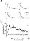
Fig. 1.
Fig. 1.
Bath application of ETYA reversibly increases the amplitude of the back-propagating action potential. Whole-cell recording from the apical dendrite 200 μm from the soma. Action potentials were evoked by a stimulating electrode in the alveus.A, a, Control dendritic action potential. In the remaining panels, this waveform is shown as a dotted linefor comparison. b, ETYA (40 μm) increases the amplitude of the dendritic action potential. c, Dendritic action potential before washout begins. d, After washout the dendritic action potential has similar amplitude to the control action potential. Bovine serum albumin (1 mg/ml) was added to the bath to aid washout. B, Time course of bath application of ETYA experiment. Graph plots the amplitude of the dendritic action potential during the experiment. Letters above the plot identify the points corresponding to the waveforms in A. After the action potential increased, the electrode series resistance began to increase. The electrode was cleared (asterisk), and a new baseline was achieved before washout began. Membrane potential was held at −65 mV throughout the experiment.
Fig. 2.
Fig. 2.
Bath application of ETYA increases dendritic action potentials but not maximum rate of rise. A, Results from two cells showing that ETYA (40 μm) can increase the amplitude of the back-propagating dendritic action potential to nearly that of a somatic action potential (90–100 mV). Note, however, that there was no Ca2+ spike triggered by these large dendritic spikes, as seen when dendritic action potential amplitude is increased by 4-AP (compare Fig. 10) B, Despite the increase in amplitude, the maximum rate of rise of the action potential (dV/dt) does not increase. Rates of rise were computed by subtraction of successive data samples. C, Summary of bath application of ETYA (n = 5 cells). Significant changes are marked with an asterisk.
Fig. 3.
Fig. 3.
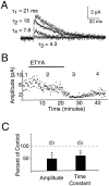
Bath application of ETYA (40 μm) decreases the amplitude of fast, transient A-type K+channel currents in cell-attached dendritic patches. Cell-attached patch from apical dendrite ∼200 μm from the soma. Sustained and slowly inactivating K+ currents were blocked with 4-AP (1 mm) and TEA (10 mm) in the patch pipette solution. Transient K+ currents were evoked by voltage steps from −90 to +30 mV. A, Kinetics during progressive block by ETYA. Ensemble averages of 10 sweeps are shown superimposed. Solid lines are exponential fits of the data. Time constants given are single exponential time constants. Data were well-fit by single exponentials. The time constant decreases with block of the channels. B, Time course of bath application experiment. The graph plots the peak amplitude of the transient current throughout the experiment. ETYA was switched off as soon as an effect was detected to aid washout. Numbers above the plot correspond to the ensemble averages and time constants shown inA. C, Summary of ETYA effect on transient current (n = 5 cells).
Fig. 4.
Fig. 4.
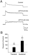
Bath application of ETYA (40 μm) enhances sustained current while decreasing transient current.A, Top panel, K+ currents were evoked by voltage steps from −90 to +30 mV. No K+channel blockers were included in the patch pipette. Top panel, Waveform is ensemble average of 30 sweeps in control solution, showing transient and sustained currents. Middle panel, Solid waveform is ensemble average of 15 sweeps beginning 8 min after start of bath application of ETYA, showing an increase in the sustained current and a small decrease in the transient current.Bottom panel, Solid waveform is ensemble average of 15 sweeps beginning 24 min after the start of ETYA. The 8 min waveform is shown as a dotted line for comparison. There is no further increase of the sustained current, but the transient current continues to decrease in amplitude. B, Summary of the ETYA effect on transient and sustained outward currents after 20–30 min exposure.
Fig. 5.
Fig. 5.
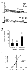
Bath application of arachidonic acid (30 μm) enhances sustained current while decreasing transient current. A, Top panel, Ensemble averages of 12 sweeps in control conditions for steps from −90 mV to command potentials from −30 to +50 mV. Bottom panel, Ensemble averages of 15 sweeps after application of arachidonic acid.B, Summary of experiment. Arachidonic acid decreased the amplitude of the transient current and the inactivation time constant (τ), while increasing the amplitude of the sustained current.C, Activation curves for the transient currents before (open squares) and after (filled dots) application of arachidonic acid. Block of the transient current was not associated with a significant shift in the activation curve. Error bars were approximately the size of the symbols. Lines represent least-squares fit to Boltzmann (see Materials and Methods).
Fig. 6.
Fig. 6.
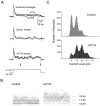
Enhancement of sustained current by a small conductance channel. A, Top panel, K+ currents in a cell-attached patch ∼200 μm from the soma evoked by a step from −87 to +17 mV. Ensemble averages of 25 sweeps before and after application of ETYA showing enhancement of sustained current. Middle and Bottom panels, Individual sweeps show clear openings and closings of sustained channels. B, Plotting the sample points beginning 80 msec after the step depolarization (A, between the arrows) demonstrates two favored levels of current for the sustained channels, and a shift in the favored level after ETYA application. C, Plotting these data as all-points histograms indicates clear peaks at multiples of 1.9 pA (∼15 pS). After ETYA there is a shift in the favored level, but the same peaks are maintained. Thus, the additional conductance may be caused by increased opening of the same channels or activation of additional channels of similar conductance.
Fig. 7.
Fig. 7.
Enhancement of sustained current by a large conductance channel. A, K+ currents in a cell-attached patch ∼200 μm from the soma evoked by a step from −90 to +30 mV. A, Control, 10 superimposed consecutive sweeps and their ensemble average showing transient and sustained currents. B, 10 superimposed consecutive sweeps and their ensemble average after bath application of arachidonic acid. Note that transient currents decrease and a new favored level appears, enhancing the sustained current in the ensemble average. C, Individual sweeps from B show the favored level results from the opening of a larger conductance channel (∼55 pS).
Fig. 8.
Fig. 8.
Reversal potential of large-conductance channel activated by ETYA. Currents in an inside-out patch from ∼150 μm from soma. Pipette solution included high K+ to decrease rectification and allow an estimate of the reversal potential.A, The patch was held at −30 mV to inactivate transient currents and stepped to a command potential of +30 mV. Waveforms are 15 consecutive individual sweeps before (control) and after (ETYA) application of ETYA (40 μm) to the bath. Data are not leak-subtracted. B, Representative traces at various potentials to show single-channel openings. Patch was held at −90 mV and stepped to potentials from −60 to +60 mV as indicated. Waveforms begin ∼50 msec after the step. C,I–V plot constructed from unitary currents as inB. From the best fit line, the slope yields a conductance of 89 pS, and the current reverses near −40, near the calculated equilibrium potential for K+.
Fig. 9.
Fig. 9.
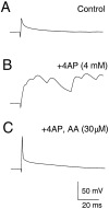
Arachidonic acid decreases the sustained depolarizations and bursts resulting from K+ channel blockade by 4-AP. Whole-cell recording from apical dendrite ∼200 μm from the soma. Back-propagating action potentials were evoked by a stimulating electrode in the alveus. A, Antidromic stimulation evokes a back-propagating action potential in the dendrites. B, Bath application of 4-AP (4 mm) increases the amplitude of the dendritic action potential, but results in large, sustained Ca+ spikes. C, Approximately 15 min after application of arachidonic acid (30 μm), the sustained depolarizations were completely blocked, leaving a single back-propagating action potential nearly twice the amplitude it had in the initial control condition.
Fig. 10.
Fig. 10.
Block of sustained currents produces sustained depolarizations. Whole-cell recording from an apical dendrite ∼200 μm from the soma. Current was injected through the dendritic recording electrode to depolarize the dendrite. A, In the control condition the current injection was below the threshold for initiating action potentials. B, Bath application of arachidonic acid lowered the threshold for initiating action potentials. Note that there was some tendency for spikes to cluster, but no sustained bursts. C, Subsequent addition of TEA (4 mm) to the bath to block sustained currents allowed sustained depolarizations to occur. Thus, sustained currents contribute to membrane stability when transient currents are reduced. These data also demonstrate that Ca2+ currents, which may be reduced by arachidonic acid, are still sufficient under these conditions to produce a sustained depolarization.

References

    1. Breukel AI, Besselsen E, Lopes da Silva FH, Lopes da Silva FH, Ghijsen WE. Arachidonic acid inhibits uptake of amino acids and potentiates PKC effects on glutamate, but not GABA, exocytosis in isolated hippocampal nerve terminals. Brain Res. 1997;773:90–97. - PubMed
    1. Buzsáki G, Kandel A. Somadendritic backpropagation of action potentials in cortical pyramidal cells of the awake rat. J Neurophysiol. 1998;79:1587–1591. - PubMed
    1. Buzsáki G, Penttonen M, Nádasdy Z, Bragin A. Pattern and inhibition-dependent invasion of pyramidal cell dendrites by fast spikes in the hippocampus in vivo. Proc Natl Acad Sci USA. 1996;93:9921–9925. - PMC - PubMed
    1. Callaway JC, Ross WN. Frequency-dependent propagation of sodium action potentials in dendrites of hippocampal CA1 pyramidal neurons. J Neurophysiol. 1995;74:1395–1403. - PubMed
    1. Colbert CM, Magee JC, Hoffman DA, Johnston D. Slow recovery from inactivation of Na+ channels underlies the activity-dependent attenuation of dendritic action potentials in hippocampal CA1 pyramidal neurons. J Neurosci. 1997;17:6512–6521. - PMC - PubMed

Publication types

MeSH terms

LinkOut - more resources