. 1999 Oct 1;19(19):8207-18.
doi: 10.1523/JNEUROSCI.19-19-08207.1999.

Characterization of an NGF-P-TrkA retrograde-signaling complex and age-dependent regulation of TrkA phosphorylation in sympathetic neurons

Affiliations

Characterization of an NGF-P-TrkA retrograde-signaling complex and age-dependent regulation of TrkA phosphorylation in sympathetic neurons

B A Tsui-Pierchala et al. J Neurosci. .

Abstract

Nerve growth factor (NGF) is a target-derived trophic factor for developing sympathetic and cutaneous sensory neurons. NGF promotes growth and survival of neurons via activation of the receptor tyrosine kinase TrkA. We used compartmentalized cultures of sympathetic neurons to address the mechanism of NGF signaling from distal axons and terminals to proximal axons and cell bodies. Our results demonstrate that an NGF-phospho-TrkA (NGF-P-TrkA)-signaling complex forms in distal axons and is retrogradely transported as a complex to cell bodies of sympathetic neurons. Although a minor fraction of both NGF and TrkA is retrogradely transported, a large fraction of the NGF that is retrogradely transported is found complexed with retrogradely transported TrkA. Interestingly, the metabolism of the P-TrkA complex is dramatically different in young, NGF-dependent sympathetic neurons as compared to older, NGF-independent sympathetic neurons. After withdrawal of NGF from distal axons of young neurons, P-TrkA within distal axons, as well as within proximal axons and cell bodies, dephosphorylates rapidly. In contrast, after withdrawal of NGF from distal axons of older neurons, P-TrkA within distal axons dephosphorylates completely, although more slowly than that in young neurons, whereas dephosphorylation of P-TrkA within proximal axons and cell bodies occurs markedly more slowly, with at least one-half of the level of P-TrkA remaining 2 d after NGF withdrawal. Thus, P-TrkA within the cell bodies of young, NGF-dependent sympathetic neurons is derived from distal axons. A more stable P-TrkA complex within cell bodies of mature sympathetic neurons may contribute to the acquisition of NGF independence for survival of mature sympathetic neurons.


Figures
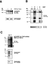
Fig. 1.
Fig. 1.
Tyrosine phosphorylation of TrkA and downstream effectors in cell bodies and distal axons and terminals of sympathetic neurons. A, The distal axons and terminals of 28 DIV sympathetic neurons grown in biochemistry chambers were placed in medium containing low NGF (2 ng/ml) for 2 d before treatment with medium alone (−) or medium containing NGF (200 ng/ml; +) for 1 hr. TrkA was immunoprecipitated from cell lysates prepared from individual compartments, and the immunoprecipitates were subjected to phosphotyrosine immunoblotting (top). α-Tubulin immunoblots were performed on the supernatants from the immunoprecipitates (bottom) to demonstrate equal amounts of total protein in the extracts. B, Thirty-five DIV sympathetic neurons grown in biochemistry chambers were placed in medium containing low NGF (2 ng/ml) for 2 d before treatment with either control medium (−) or 200 ng/ml NGF (+) directly on the cell bodies (CB) or distal axons and terminals (T) for 10 min. Extracts were subjected to the same analysis described in A. To demonstrate equal amounts of proteins in the two cell body compartments and in the two distal axon compartments, the phosphotyrosine immunoblot (top) was stripped and reprobed with an antibody against Trk (bottom). C, Thirty-three 12 DIV cultures of compartmentalized neurons were maintained for 2 d in medium containing anti-NGF in the cell body compartment and in medium containing NGF (200 ng/ml) in the terminal compartment. Lysates prepared from individual compartments of all 33 cultures were pooled and subjected to Trk immunoprecipitation and phosphotyrosine immunoblotting analysis (top). Proteins in supernatants of immunoprecipitates were subjected to anti-phosphotyrosine (middle) and α-tubulin (bottom) immunoblotting. IP, Immunoprecipitate;Term, terminals; W, Western blot.
Fig. 2.
Fig. 2.
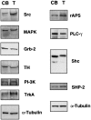
Distribution of TrkA and some of its effector molecules in sympathetic neurons. Thirty-five DIV sympathetic neurons grown in biochemistry chambers were maintained with NGF present only in the medium in distal axons and terminal compartments for 2 weeks before lysis. Cellular extracts prepared from the CB andT compartments were subjected to immunoblot analysis using the several different antibodies shown in the figure and described in Materials and Methods. Each columnrepresents the entire lysate prepared from sympathetic neurons from 8 to 10 biochemistry chambers, and the proteins analyzed are listed on the right of the immunoblot.
Fig. 3.
Fig. 3.
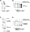
Phosphorylated TrkA is retrogradely transported in sympathetic neurons. A, Mass cultures of sympathetic neurons maintained for 12 DIV were incubated with 0, 0.5, or 2 mm NHS-LC-biotin for 30 min at 4°C to biotinylate cell surface proteins. The neurons were then washed and treated with medium alone (−) or medium containing NGF (200 ng/ml; +) for 10 min. Biotinylated proteins were precipitated (P) with immobilized streptavidin (top), and the supernatants of this precipitation were next subjected to Trk immunoprecipitation (middle). Precipitates were then electrophoresed on 8% SDS-polyacrylamide gels and subjected to phosphotyrosine immunoblotting. α-Tubulin immunoblotting performed on the supernatants of the immunoprecipitates denotes equal protein loading (bottom). B, Cell surface proteins of mass cultures of sympathetic neurons grown in medium containing anti-NGF for 7 hr were biotinylated as described inA and treated with medium alone (−) or medium containing NGF (200 ng/ml; +). Lysates from these treatments were first subjected to a nonstoichiometric Trk immunoprecipitation followed by a precipitation with immobilized streptavidin, as labeledaboveeachlane. The precipitates (top) as well as the supernatants from this analysis (middle) were subjected to phosphotyrosine immunoblotting, and supernatants were subjected to α-tubulin immunoblotting (bottom) to normalize for protein amounts in the lysates. C, Compartmentalized cultures were chilled to 4°C, and plasma membrane proteins of distal axons and terminals were subjected to biotinylation, while the cell body compartments were left untreated. Compartmentalized cultures were then washed extensively and warmed to 37°C, and distal axons and terminals were treated with either medium alone (−) or medium containing NGF (200 ng/ml; +) for 2 hr. Extracts prepared from both the cell body and distal axons and terminal compartments were subjected to immobilized streptavidin precipitations, and the precipitants were subjected to phosphotyrosine immunoblot analysis (top). α-Tubulin immunoblot analysis performed on supernatants of the precipitation is shown (bottom). This experiment was performed three times with independent cultures with similar results. D,Membrane proteins of cell bodies were biotinylated, while distal axons and terminals were left untreated. Then, cultures were warmed to 37°C, and either the cell bodies were treated with medium alone, the cell bodies were treated with medium containing NGF (200 ng/ml), or the distal axons and terminals were treated with medium containing NGF (200 ng/ml). Biotinylated proteins were precipitated first using immobilized streptavidin (top), and the supernatants subsequently were subjected to immunoprecipitation using anti-Trk C14 (middle); both precipitates were then subjected to phosphotyrosine immunoblotting. The top immunoblot was reprobed with a Trk antibody to demonstrate equal precipitation of TrkA in all three conditions (bottom). Only precipitations from cell body compartments are shown.
Fig. 4.
Fig. 4.
NGF and TrkA are cotransported from distal axons and terminals to cell bodies after NGF treatment of distal axons and terminals. A, Mass cultures of sympathetic neurons were treated with 125I-NGF (40 ng/ml) for 30 min at 37°C, washed, and subjected to cross-linking in lysis buffer containing 2 mm BS3. Immunoprecipitations were next performed using either C14 Trk (1:200) or CBP (1:200) rabbit polyclonal antibodies under identical conditions. The immunoprecipitates were resolved on 15% SDS-polyacrylamide gels and next analyzed for the presence of 125I-NGF by autoradiography (top). Equal protein loading was determined by probing immunoblots of supernatants of the immunoprecipitates with anti-MAPK (bottom). B, Distal axons and terminals of neurons grown in biochemistry chambers were treated with125I-NGF (40 ng/ml) or with 125I-NGF (40 ng/ml) and excess unlabeled NGF (500 ng/ml) for 8 hr. Extracts prepared from both the cell body and distal axon and terminal compartments were subjected to cross-linking and Trk immunoprecipitation, and the immune complexes were analyzed as described in A(top). Anti-MAPK immunoblots were performed on supernatants of the immunoprecipitates to confirm that equal amounts of protein were present in the extracts (bottom). This experiment was performed four times using independent cultures with similar results. C, Cell surface proteins of neurons grown in mass culture were biotinylated as described in Figure 3. Then neurons were treated with 40 ng/ml 125I-NGF for 30 min, washed, and subjected to immobilized streptavidin precipitation (top) or Trk immunoprecipitation (middle) without previous cross-linking. Precipitates were analyzed as described in A, and equal protein loading was determined by α-tubulin immunoblot analysis on supernatants of the precipitates (bottom). D, Distal axons and terminals of neurons grown in biochemistry chambers were biotinylated and then treated with either 40 ng/ml 125I-NGF or 40 ng/ml125I-NGF with an excess of unlabeled NGF (500 ng/ml) for 8 hr. Extracts prepared from the cell body and distal axon and terminal compartments were subjected to immobilized streptavidin precipitation followed by 15% SDS–PAGE and autoradiography (top). Identical results were obtained from two independent cultures. Equal protein loading was determined with anti-MAPK immunoblot analysis (bottom). P, precipitation;W, Western blot.
Fig. 5.
Fig. 5.
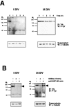
TrkA dephosphorylation is markedly faster in young, NGF-dependent neurons than in older, NGF-independent neurons after NGF withdrawal. A, Medium containing NGF was replaced with medium containing anti-NGF in NGF-dependent (5 DIV;left panels) or NGF-independent (35 DIV; right panels) sympathetic neurons grown in mass culture. Then, TrkA was immunoprecipitated at the times indicated above thepanels, and immune complexes were subjected to phosphotyrosine immunoblotting (top). Supernatants of immunoprecipitations were subjected to α-tubulin immunoblotting (bottom). B, Five DIV (left panels) or 35 DIV (right panels) mass cultures of sympathetic neurons were treated with control growth medium (leftlanes), growth medium containing K252a (200 nm) for 15 min (middle lanes), or medium containing anti-NGF for 90 min (right lanes). Then, TrkA was immunoprecipitated and subjected to phosphotyrosine immunoblotting as above.
Fig. 6.
Fig. 6.
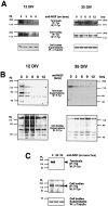
Tyrosine phosphorylation of TrkA localized in cell bodies of mature, NGF-independent sympathetic neurons in the absence of NGF. A, Twelve DIV NGF-dependent neurons (left) and 35 DIV NGF-independent neurons (right) were grown in compartmentalized cultures with medium containing anti-NGF in cell body compartments and with medium containing NGF (200 ng/ml) in distal axon and terminal compartments for several days. Media in distal axon and terminal compartments were replaced with media containing anti-NGF for the indicated times (in hours). Then, TrkA was immunoprecipitated from extracts prepared from either cell bodies (middle) or distal axons and terminals (top), and immune complexes and supernatants of immunoprecipitates were subjected to phosphotyrosine immunoblot analysis. Similar results were obtained from three to four independent experiments. B, Supernatants of the immunoprecipitates from A were subjected to phosphotyrosine immunoblotting.C, Thirty-five DIV NGF-independent neurons were grown in compartmentalized cultures with medium containing anti-NGF in cell body compartments and with medium containing NGF (200 ng/ml) in distal axon and terminal compartments for several days. Medium in distal axon and terminal compartments was replaced with media containing anti-NGF for 0, 24, or 52 hr. Then, TrkA was immunoprecipitated from extracts prepared from either cell bodies (middle) or distal axons and terminals (top), and immune complexes were subjected to phosphotyrosine immunoblot analysis.

Similar articles

Cited by

References

    1. Angeletti PU, Levi-Montalcini R, Caramia F. Analysis of the effects of the antiserum to the nerve growth factor in adult mice. Brain Res. 1971;27:343–355. - PubMed
    1. Barde Y-A. Trophic factors and neuronal survival. Neuron. 1989;2:1525–1534. - PubMed
    1. Berg MM, Sternberg DW, Parada LF, Chao MV. K-252a inhibits nerve growth factor-induced trk proto-oncogene tyrosine phosphorylation and kinase activity. J Biol Chem. 1992;267:13–16. - PubMed
    1. Bhattacharyya A, Watson F, Bradlee T, Pomeroy S, Stiles C, Segal R. Trk receptors function as rapid retrograde signal carriers in the adult nervous system. J Neurosci. 1997;17:7007–7016. - PMC - PubMed
    1. Bjette B, Wilkund L, Edwards DC. A study of the de- and regenerative changes in the sympathetic nervous system of the adult mouse after treatment with antisera to NGF. Brain Res. 1975;92:257–278. - PubMed

Publication types

MeSH terms