. 1999 Oct 1;19(19):8337-48.
doi: 10.1523/JNEUROSCI.19-19-08337.1999.

delta opioid receptor modulation of several voltage-dependent Ca(2+) currents in rat sensory neurons

Affiliations

delta opioid receptor modulation of several voltage-dependent Ca(2+) currents in rat sensory neurons

C G Acosta et al. J Neurosci. .

Abstract

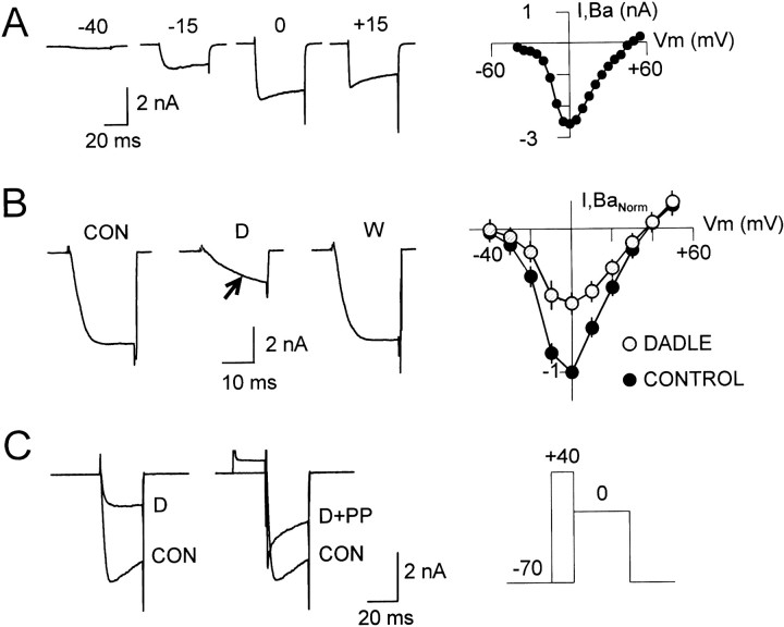
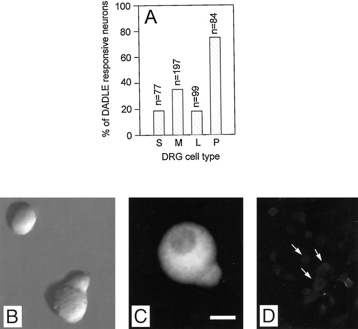
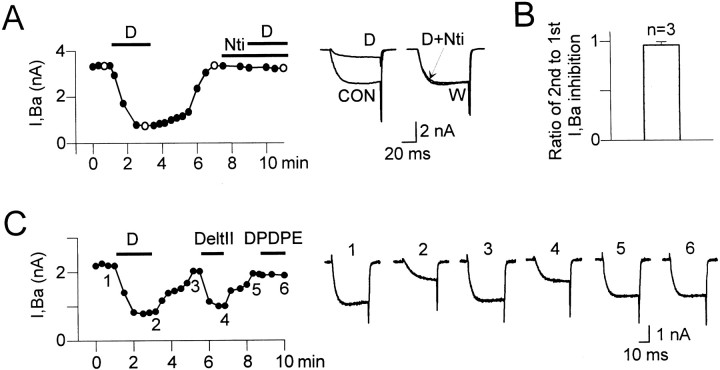
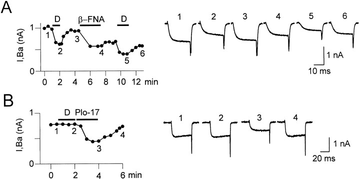
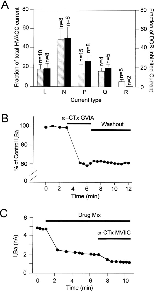
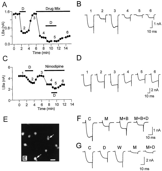
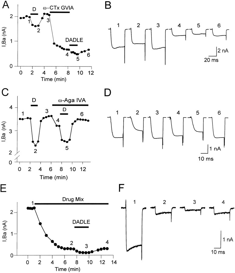
Endogenous enkephalins and delta opiates affect sensory function and pain sensation by inhibiting synaptic transmission in sensory circuits via delta opioid receptors (DORs). DORs have long been suspected of mediating these effects by modulating voltage-dependent Ca(2+) entry in primary sensory neurons. However, not only has this hypothesis never been validated in these cells, but in fact several previous studies have only turned up negative results. By using whole-cell current recordings, we show that the delta enkephalin analog [D-Ala(2), D-Leu(5)]-enkephalin (DADLE) inhibits, via DORs, L-, N-, P-, and Q-high voltage-activated Ca(2+) channel currents in cultured rat dorsal root ganglion (DRG) neurons. The percentage of responding cells was remarkably high (75%) within a novel subpopulation of substance P-containing neurons compared with the other cells (18-35%). DADLE (1 microM) inhibited 32% of the total barium current through calcium channels (I(Ba)). A delta (naltrindole, 1 microM), but not a mu (beta-funaltrexamine, 5 microM), antagonist prevented the DADLE response, whereas a DOR-2 subtype (deltorphin-II, 100 nM), but not a DOR-1 (DPDPE, 1 microM), agonist mimicked the response. L-, N-, P-, and Q-type currents contributed, on average, 18, 48, 14, and 16% to the total I(Ba) and 19, 50, 26, and 20% to the DADLE-sensitive current, respectively. The drug-insensitive R-type current component was not affected by the agonist. This work represents the first demonstration that DORs modulate Ca(2+) entry in sensory neurons and suggests that delta opioids could affect diverse Ca(2+)-dependent processes linked to Ca(2+) influx through different high-voltage-activated channel types.


Figures

Fig. 1.
Fig. 1.
HVACC currents in cultured postnatal DRG neurons and characteristics of their inhibition by DADLE.A, Whole-cell Ba2+ current (IBa) evoked with 40 msec voltage pulses to −40, −15, 0, and 15 mV, from aVH of −70 mV (left) andI–V plot of IBa at the end of 40 msec voltage test pulses (right). The voltage pulses, delivered every 15 sec, ranged from −40 to 55 mV, with 5 mV intervals, from a VH of −70 mV.B, Left shows the preagonist controlIBa current (traceCON) and its inhibition by 1 μmDADLE (trace D), obtained at 0 mV from aVH of −70 mV. The agonist effect fully reversed upon agonist washout (trace W). The inhibited current typically showed a slowing down of activation (arrow in trace D). TheI–V relationship (right) of the normalized IBa (data from 3 neurons) measured 25 msec after the onset of the test pulse, before (filled circles), and during (hatched circles) DADLE (1 μm) inhibition illustrates the current reduction in a wide range of test voltages (−40 to 50 mV).C, Voltage dependence of the agonist-mediatedIBa inhibition. The inhibition of the control IBa current observed at 0 mV (left panel, traces CON and D) was partially removed by 20 msec prepulse to 40 mV, which preceded the test pulse to 0 mV (right panel, trace D+PP; for comparison, the preagonist control current was also included). The agonist was present throughout. The voltage pulse protocol is shown on the right. On average, the prepulse depolarization reverted 39% of the inhibition.
Fig. 2.
Fig. 2.
Percentage of DADLE-responding cells in different neuron subpopulations. A, The percentages of DADLE-responsive neurons in the subpopulations of small (S), medium (M), and large (L) neurons and medium-sized P-neurons (P, from pear-shaped) were 18.7, 35, 18.2, and 75%, respectively. The number of cells tested appear ontop of the bars. The relative fractional contribution of each cell type to the total population in culture was 58 (S), 26 (M), 9 (L), and 7% (P).B, P-neuron under Nomarski phase contrast cultured for 20 hr. A small round neuron is also visible in the field.C, D, An antibody against substance P labeled all P-neurons (an example is shown in C), but only a fraction of the other cells types (arrows inD show examples of nonlabeled cells). Cells after 24 hr in culture. Scale bar: B, C, 10 μm;D, 40 μm.
Fig. 3.
Fig. 3.
DOR involvement in the DADLE-induced inhibition of IBa.A, Plot of time course (left) andIBa current records (right) corresponding to open symbols of an experiment testing the effect of the highly selective DOR antagonist naltrindole (Nti) on the DADLE-induced inhibition.IBa was elicited with 60 msec pulses to 0 mV from a VH of −70 mV. The data points in the plot correspond to IBa values at the end of the test pulses. The control current (trace CON) was powerfully inhibited by 1 μm DADLE (trace D), and the inhibition fully reversed upon agonist washout (trace W). DADLE was completely ineffective when subsequently coapplied with 1 μm naltrindole (trace D+Nti). The lack of DADLE effect in the presence of naltrindole was not attributable to response desensitization (B). B, The responses to short (1.5–2 min) applications of DADLE, as used in our experiments, were not affected by desensitization. Two applications of DADLE of 2 min each, separated by an interval of 3–5 min, caused a similar amount ofIBa inhibition in control cells (n = 3), as illustrated by the plot of the ratio of the magnitudes of the second to the firstIBa inhibition (essentially 1). Ratios in the individual cells were 0.93, 0.95, and 0.99. C, Time course (left) and current records (right) of an experiment in which the DOR-2-selective agonist deltorphin-II (100 nm) mimicked the DADLE-induced inhibition (traces 1–3) of IBa in a reversible way (trace 3–5). Current records correspond to similarly numbered data points in the time course plot. As in other four cells, 1 μm DPDPE (a DOR-1-selective agonist) had no effect on IBa (trace 6). In this cell, DADLE and deltorphin-II (Delt-II) reduced 1.4 and 1.02 nA ofIBa, respectively.
Fig. 4.
Fig. 4.
The DADLE response does not involve MOR.A, Time course (left) and corresponding traces (right) showing the reversible response to DADLE before (traces 1–3) and after (traces4–6) the irreversible block of MOR with 5 μm β-FNA, a MOR-selective antagonist. In this and in other four cells, β-FNA had no effect on the reversible DADLE-induced inhibition. The reduction of IBa during β-FNA application results from the κ opioid activity of this compound. B, The MOR-selective agonist PLO-17 (1 μm) reversibly inhibited IBa(traces 2–4) in a cell in which DADLE (1 μm) was ineffective (traces 1,2). Records from a P-neuron. Similar results were found in six cells. The MOR-mediated IBainhibition did not show desensitization. In all experiments,VH was −70 mV, and the test voltage pulses were 0 mV. Current records in A and Bcorrespond to similarly numbered data points in their respective time course plots.
Fig. 5.
Fig. 5.
HVACC currents inhibited by toxins and targeted by DORs. A,Striped bars show the average ± SEM contributions of L-, N-, P-, and Q-type HVACC currents to the whole-cell IBa. These values were taken as equal to the percentages ofIBa inhibited by 10 μmnimodipine (L), 2 μm ω-CTx-GVIA (N), 50 nm ω-Aga-IVA (P), and 0.5–1 μm ω-CTx-MVIIC (Q), respectively. Because of its lack of selectivity, ω-CTx-MVIIC was applied after N-, P-, and L-type currents were all previously blocked. The R-type was defined as the fraction that remained unblocked in the presence of all above drugs (R) and was fully blocked by Cd2+. Solid bars show the average ± SEM contributions of each current type to the DADLE-sensitive current (ID). Notice the large contribution of the P-type current toID despite its relatively small contribution to IBa. B, Plot ofIBa versus time before and after the application of 2 μm ω-CTx-GVIA. The data points represent values of IBa at the end of 40 msec depolarizing test pulses to 0 mV from aVH of −70 mV. The ω-CTx-GVIA-induced inhibition stabilized within 2 min and was essentially irreversible (tested up to 20 min). In this neuron, the N-type current contributed 40% of IBa. To better visualize the toxin effect, the ordinate axis was truncated. Data from a medium-sized, round neuron. C, Time course from an experiment testing the contribution of the Q-type current toIBa, which was evoked with 20 msec test pulses. The L, N, and P currents contributing to the initialIBa were blocked by a combination (Drug Mix) of 10 μm nimodipine, 2 μm ω-CTx-GVIA, and 50 nm ω-Aga-IVA. The fraction subsequently inhibited by 0.5 or 1 μmω-CTx-MVIIC (1 μm in this cell) was defined as Q-type. The time course graph plots the values ofIBa at the end of the test pulses. The current remaining unblocked was defined as R-type. Data from a P-neuron. All measures made at the end of voltage test pulses to 0 mV from a VH of −70 mV.
Fig. 6.
Fig. 6.
DOR-mediated inhibition of the dihydropyridine-sensitive L-type current. Data points inA and C represent values ofIBa at the end of 25 msec test pulses to 0 mV from a VH of −70 mV. A,Time course of an experiment testing the L-type current contribution toID with protocol-1. The first DADLE application reversibly inhibited 63% ofIBa. Subsequently, the block of N-, P-, and Q-type currents with 2 μm ω-CTx-GVIA, 50 nmω-Aga-IVA, and 1 μm ω-CTx-MVIIC (Drug Mix) inhibited 81% of IBa. The contribution of the L-type current to ID was revealed by a second DADLE application, which inhibited 53% of the non-N, non-P, and non-Q currents in a fully reversible way.B, Current records corresponding to similarly numbered data points in A. C, Time course of an experiment testing the L-type contribution toID with protocol-2. The block of the L-type current fraction (36% in this cell) with 10 μmnimodipine reduced the amount of IBainhibited by 1 μm DADLE. The second DADLE application was done in the continuous presence of nimodipine. D,Current records corresponding to similarly numbered data points inC. The current reversibly inhibited by DADLE decreased from 1.3 to 0.74 nA after blocking the L-type current with 10 μm nimodipine. E, All sensory neurons in culture, regardless of cell subpopulation, were immunoreactive for the α1C subunit of neuronal Ca2+ channels, indicating the presence of L-type HVACC in these cells. Neurons obtained from 5-d-old postnatal animals. P-neurons in the culture are labeled with arrows. Scale bar, 20 μm.F, The control HVACC current (trace C), activated by voltage pulses to 0 mV from aVH of −70 mV, was partially blocked by a mixture of 2 μm ω-CTx-GVIA, 50 nmω-Aga-IVA, and 1 μm ω-CTx-MVIIC, which was kept in the bath for the rest of the experiment (trace M). S-(±)-Bay-K 8644 (10 μm) increased the amplitude of the drug mixture-resistant current (trace M+B), and the subsequent application of 1 μm DADLE in the continuous presence of Bay-K 8644 reduced the enhanced current (M+B+D). The magnitude of the inward current in the presence of DADLE was smaller than that beforeS-(±)-Bay-K 8644 application. G, The R-type current, defined as the current remaining unblocked in the presence of a mixture of 10 μm nimodipine, 2 μm ω-CTx-GVIA, 50 nm ω-Aga-IVA, and 1 μm ω-CTx-MVIIC (trace M), was completely insensitive to DADLE (trace M+D). The agonist reversibly inhibited IBa before the application of the drug mixture (traces C, D, W). Data from a P-neuron.
Fig. 7.
Fig. 7.
DOR-mediated inhibition of N-, P-, and Q-type currents. A, Time course of an experiment testing the N-type current contribution to ID. The block of the N-type current fraction by 2 μm ω-CTx-GVIA reduced the amount of IBa inhibited by 1 μm DADLE. B, Current records corresponding to similarly numbered data point of the experiment in A. DADLE reversibly inhibited 0.35 nA of the initialIBa (traces 1–3). After the application of 2 μm ω-CTx-GVIA, which inhibited 1.4 nA of IBa (trace 4), a second DADLE application reversibly inhibited 0.18 nA of the remaining non-N current (traces4–6).C, Time course of an experiment testing the P-type current contribution to ID. The data points represent values of IBa at the end of 25 msec voltage test pulses to 0 mV from a VHof −70 mV. The block of the P-type current fraction (11.8% in this cell) with 50 nm ω-Aga-IVA reduced the amount ofIBa inhibited by 1 μm DADLE. The toxin was present throughout the second DADLE application because its effect was partially reversible. D, Current records corresponding to similarly numbered data points in C. The first DADLE application reversibly inhibited 1.2 nA of the totalIBa (traces1–3), whereas the second agonist application, after the block of the P-type current, suppressed only 0.72 nA of the non-PIBa (traces4–6). Because the magnitude of the inhibitedIBa was larger during the second (trace 5) than during the first DADLE application (trace 2), the effect of DADLE deceptively appeared to be smaller than predicted from the loss of target, ω-Aga-IVA-sensitive P-type channels. This occurred because the second DADLE application was tested on the larger preagonist current (trace 3 vs trace 1) resulting from the over-recovery of IBa during reversal of the first DADLE-induced inhibition (trace 3; see data points in C). When allowances are made for this change in the basal IBa, the reduction in DADLE-induced inhibition can be entirely accounted for by the suppression of P-type target channels. E,F, Time course and corresponding traces from a representative experiment in a round neuron, using protocol-1 to test the contribution of Q-type HVACC current toID. The numbered points inE correspond to similarly numbered tracesin F. N-, P-, and L-type HVACC currents were inhibited by perfusing the cell with a solution containing 2 μm ω-CTx-GVIA, 50 nm ω-Aga-IVA, and 10 μm nimodipine (Drug Mix; traces 1, 2 in F). DADLE reversibly inhibited a fraction of the remaining non-N, non-L, and non-P currents (traces 2, 3 in F). This inhibition was fully reversible (trace 4 inF). The data points are the values ofIBa at the end of 20 msec voltage test pulses to 0 mV from a VH of −70 mV.

References

    1. Albillos A, Carbone E, Gandia L, Garcia A, Pollo A. Opioid inhibition of Ca2+ channel subtypes in bovine chromaffin cells: selectivity of action and voltage-dependence. Eur J Neurosci. 1996;8:1561–1570. - PubMed
    1. Al Zein M, Lutz-Bucher B, Koch B. Modulation by leu-enkephalin of peptide release from perifused neurointermediate pituitary. I. Selective effect on potassium-, veratridine- and isoproterenol-stimulated secretion of vasopressin. Neuroendocrinology. 1984;39:392–396. - PubMed
    1. Amico C, Marchetti C, Nobile M, Usai C. Pharmacological types of calcium channels and their modulation by baclofen in cerebellar granules. J Neurosci. 1995;15:2839–2848. - PMC - PubMed
    1. Aronin N, DiFiglia M, Liotta AS, Martin JB. Ultarstructural localization and biochemical features of immunoreactive leu-enkephalin in monkey dorsal horn. J Neurosci. 1981;1:561–577. - PMC - PubMed
    1. Bean BP. Neurotransmitter inhibition of neuronal calcium currents by changes in channel voltage dependence. Nature. 1989;340:153–156. - PubMed

Publication types

MeSH terms

LinkOut - more resources