. 1999 Oct 1;19(19):8349-57.
doi: 10.1523/JNEUROSCI.19-19-08349.1999.

Identification of a new exon in the myelin proteolipid protein gene encoding novel protein isoforms that are restricted to the somata of oligodendrocytes and neurons

Affiliations

Identification of a new exon in the myelin proteolipid protein gene encoding novel protein isoforms that are restricted to the somata of oligodendrocytes and neurons

E R Bongarzone et al. J Neurosci. .

Abstract

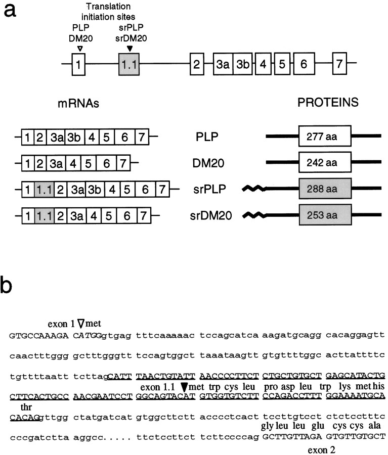
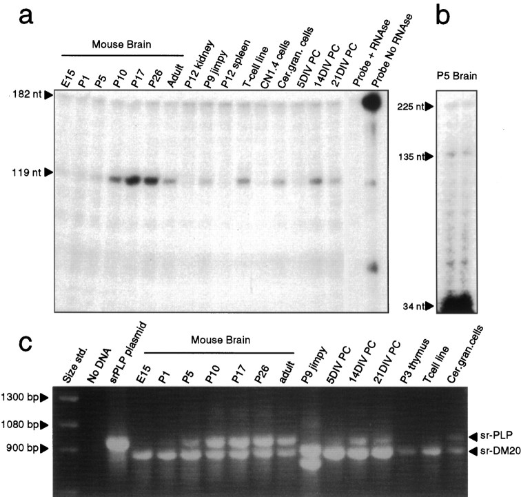
The myelin proteolipid protein (PLP) gene (i.e., the PLP/DM20 gene) has been of some interest because of its role in certain human demyelinating diseases, such as Pelizaeus-Merzbacher disease. A substantial amount of evidence, including neuronal pathology in knock-out and transgenic animals, suggests the gene also has functions unrelated to myelin structure, but the products of the gene responsible for these putative functions have not yet been identified. Here we report the identification of a new exon of the PLP/DM20 gene and at least two new products of the gene that contain this exon. The new exon, located between exons 1 and 2, is spliced into PLP and DM20 mRNAs creating a new translation initiation site that generates PLP and DM20 proteins with a 12 amino acid leader sequence. This leader sequence appears to target these proteins to a different cellular compartment within the cell bodies of oligodendrocytes and away from the myelin membranes. Furthermore, these new products are also expressed in a number of neuronal populations within the postnatal mouse brain, including the cerebellum, hippocampus, and olfactory system. We term these products somal-restricted PLP and DM20 proteins to distinguish them from the classic PLP and DM20 proteolipids. They represent putative candidates for some of the nonmyelin-related functions of the PLP/DM20 gene.


Figures

Fig. 1.
Fig. 1.
Organization of the myelin proteolipid protein gene and the sequence of the new exon and the N-terminal peptide encoded by this exon. a, Gene structure showing the location of the new exon 1.1 between exons 1 and 2.Below the gene to the left are shown the exonic compositions of the srPLP and srDM20 mRNAs containing exon 1.1 compared with those of the classic PLP and DM20 mRNAs. The translation initiation sites for the classic PLP and DM20 and for the srPLP and srDM20 proteins are indicated in the gene structure within exons 1 and 1.1, respectively. To the right of the mRNAs arediagrams of the protein products corresponding to each of the mRNAs and the number of amino acids (aa) in the protein. The 12 amino acid N-terminal sequences encoded by exon 1.1 are indicated on thediagrams for srPLP and srDM20 (wavylines). The sizes and locations of the exons are not drawn to scale. b, The sequence of the new 109 bp exon 1.1, underlined in capitalletters, given along with the complete intronic sequence between exons 1 and 1.1 as well as some intronic sequence between exons 1.1 and 2 (in lowercase). The translation start site (filledarrowhead) for srPLP and srDM20 proteins and the new N-terminal amino acid sequence contributed by exon 1.1 are shown above the sequence. The translation initiation site used in the classic PLP and DM20 mRNAs (openarrowhead) at the 3′ end of exon 1 is shown in italics.
Fig. 2.
Fig. 2.
Invitrotranscription–translation of srDM20 and Northern blot of srPLP and srDM20 mRNAs. a, Plasmids containing the coding regions of srDM20 and the classic DM20 and PLP isoforms were translated in anin vitro transcription–translation system and analyzed by SDS-PAGE. Autoradiograms of the total products of synthesis indicated that the principal radiolabeled bands corresponded in size to the new srDM20 (filledarrowhead,middlelane) and the classic DM20 and PLP (open and hatchedarrowheads, left and rightlanes, respectively). The individuallanes were part of the same transcription–translation experiment, analyzed in the same gel. Molecular weight standards were also included in the gel to assign the size for each band but are not shown. b, Total RNA from P18 mouse brain (Br) and from enriched OL cultures (OL) was analyzed by Northern blot with a 32P-riboprobe specific to exon 1.1. A 3.4 kb band was identified in the brain sample. In enriched OL RNA, the 3.4 kb band appeared as a doublet with a minor band at ∼3.5 kb. In both samples, a 12 kb band, presumably corresponding to heterogeneous nuclear RNA, was observed. Migration of molecular weight standards (data not shown) was used to assign the size for each band.
Fig. 3.
Fig. 3.
Distribution of srPLP and srDM20 in various tissues and cells. a, The expression of srPLP and srDM20 was examined by RPA using an exon 1.1–specific sequence. In mouse brain, peak expression of the srPLP and srDM20 mRNAs was observed between P17 and P26, closely resembling the developmental expression of the classic PLP and DM20 mRNAs. b, The steady levels of both sr and classic mRNAs in P5 mouse brain were examined by RPA using a riboprobe that contained 101 nt of exon 1.1 and 34 nt of exon 2. The level of protection of the classic fragment (34 nt) was significantly higher than that of the sr fragment (135 nt).c, The expression of either srPLP or srDM20 mRNAs was analyzed by RT-PCR using primers 114S and 3NPLP. The expression of srDM20 was found to precede that of srPLP during the development of the mouse brain and in mouse brain mixed glial primary cultures (PC). Truncated forms of srPLP and srDM20 mRNAs, reflecting the deletion of exon 5, were expressed in jimpy brains. Interestingly, the srPLP and srDM20 mRNAs were also expressed in cerebellar granule neurons as well as in thymus and a mouse T-cell line (LBRM). Cer. gran., Cerebellar granule;CN1.4, a conditionally immortalized cortical neuronal cell line; DIV, days in vitro;nt, nucleotide.
Fig. 4.
Fig. 4.
Detection of srPLP and srDM20 proteins in proteolipid extracts by Western blot. Extracts of total proteolipid proteins were prepared from mouse P14 brains, separated by SDS-PAGE, and analyzed on Western blots with a polyclonal antiserum specific for the N terminal of srPLP and srDM20. The anti-srPLP and -srDM20 recognized several bands, the most intense of which migrated with an apparent molecular weight of 27 kDa (lanea). Other lower molecular weight bands were evident at 21, 20, and 11 kDa. The blot was stripped and reprobed with AA3, a monoclonal antibody that recognizes the C terminal of PLP and DM20 (lane b). Very heavy bands of PLP and DM20 were evident, and these migrated with apparent molecular weights of 25 and 20 kDa. Because of the differences in the levels of srPLP and srDM20 and of classic PLP and DM20, laneb is an adjacent lane on the reprobed blot, loaded with less protein. Molecular weight standards were included to assign the relative sizes (data not shown). On the basis of the relative sizes and mobilities of the bands in both preparations, the srPLP and srDM20 were assigned to the 27 and 21 kDa bands, respectively.
Fig. 5.
Fig. 5.
Expression of srPLP and srDM20 in OLs is localized in the cell bodies but not in the myelin-like membranes. Top Panels, Cells within the OL lineage were found to coexpress srPLP and srDM20 (redfluorescence) with markers of different oligodendroglial developmental stages (greenfluorescence) including A2B5 (precursors), O4 (immature/mature), and O1 (mature). The combined immunofluorescence for srPLP and srDM20 with these markers is shown.Middle,Bottom Panels, Single confocal sections of mature, enriched OLs double stained for srPLP and srDM20 (redfluorescence) and either O1, an antibody that stains primarily galactocerebrosides, or AA3, an antibody that stains classic PLP and DM20 (greenfluorescence), are shown. In all cases examined the srPLP and srDM20 staining was localized primarily in the cell soma and major processes but not in the membranes elaborated by the OLs.
Fig. 6.
Fig. 6.
Expression of srPLP and srDM20 is primarily in white matter regions of the mouse brain but is present in some neuronal populations. a,In situ hybridization of a sagittal section of a P14 mouse brain with a33P-riboprobe specific for exon 1.1 showing expression of the srPLP and srDM20 mRNAs. b,In situ hybridization of a section adjacent toa using a sense control. c, Higher magnification of a different section (counterstained with cresyl violet) showing portions of the corpus callosum and hippocampus and illustrating the punctate labeling throughout the corpus callosum and in the hippocampus below it. d, Immunohistochemical detection of srPLP and srDM20 in the corpus callosum and the hippocampus. Arrows in c andd point to cells of the size, location, and morphology of OLs that contain srPLP and srDM20 mRNAs and proteins, respectively. Unlike classic PLP and DM20 staining in the corpus callosum, srPLP and srDM20 labeling is confined to the cell somas. Also labeled are the CA2 and CA3 regions of the hippocampus.e, Higher magnification of a cerebellar region showing punctate labeling of srPLP and srDM20 mRNAs in cells throughout the white matter (redarrows) and the internal granular cell layer (whitearrows) as well as the Purkinje cells (yellowarrows). f,Immunohistochemical detection of srPLP and srDM20 in a region of the cerebellum similar to that in e. Cell bodies of OLs are indicated with redarrows, whereas Purkinje cells are indicated with blackarrows. Granule neurons located within theIGL were readily immunostained with the antibody.g, Higher magnification of the immunohistochemical detection of srPLP and srDM20 in the internal granule cell layer (openarrows) and Purkinje cells (filledarrows) from the mouse cerebellum. h, Immunohistochemical detection of srPLP and srDM20 proteins in medium-sized neurons within the anterior olfactory nucleus of the P14 mouse brain. i,Immunohistochemical control for background staining in the cerebellum with preimmune serum to the srPLP and srDM20 antibody.j–l, Primary granular neuron cultures doubly stained for srPLP and srDM20 (j;redfluorescence) and for tubulin III (k;green). l is an overlay showing that srPLP and srDM20 proteins remain associated with the neuronal cell bodies. CC, Corpus callosum; DCWM, deep cerebellar white matter; Hp, hippocampus;IC, internal capsule; IGL, internal granule cell layer.

References

    1. Anderson TJ, Schneider A, Barrie JA, Klugmann M, McCulloch MC, Kirkham D, Kyriakides E, Nave KA, Griffiths IR. Late-onset neurodegeneration in mice with increased dosage of the proteolipid protein gene. J Comp Neurol. 1998;394:506–519. - PubMed
    1. Bizzozero OA, Besio-Moreno M, Pasquini JM, Soto EF, Gomez CJ. An electrophoretic analysis of proteolipids from different rat brain subcellular fractions. Biochim Biophys Acta. 1982;691:281–292. - PubMed
    1. Bongarzone ER, Foster LM, Byravan S, Verity AN, Landry CF, Schonmann V, Amur-Umarjee S, Campagnoni AT. Conditionally immortalized neural cell lines: potential models for the study of neural cell function. Methods. 1996;10:489–500. - PubMed
    1. Bongarzone ER, Howard SG, Schonmann V, Campagnoni AT. Identification of the dopamine D3 receptor in oligodendrocyte precursors: potential role in regulating differentiation and myelin formation. J Neurosci. 1998a;18:5344–5353. - PMC - PubMed
    1. Bongarzone ER, Foster L, Byravan S, Casaccia-Bonnefil P, Schonmann V, Campagnoni AT. Two neuronal cell lines expressing the Golli-MBP gene display differences in their in vitro survival and in their interaction with glia. J Neurosci Res. 1998b;54:309–319. - PubMed

Publication types

MeSH terms

LinkOut - more resources