. 1999 Oct 1;19(19):8367-76.
doi: 10.1523/JNEUROSCI.19-19-08367.1999.

Optic nerve crush: axonal responses in wild-type and bcl-2 transgenic mice

Affiliations

Optic nerve crush: axonal responses in wild-type and bcl-2 transgenic mice

S Chierzi et al. J Neurosci. .

Abstract

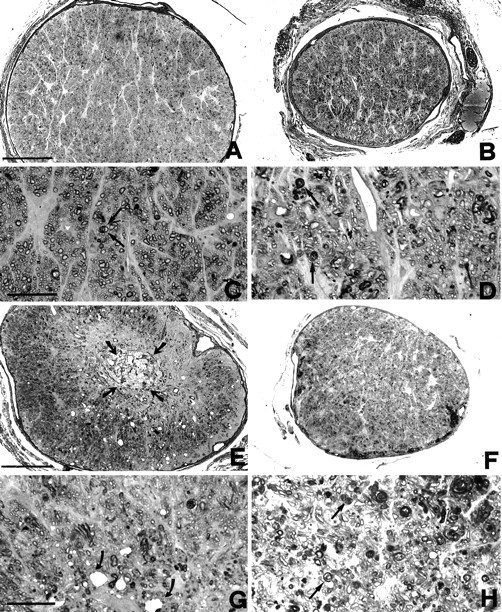
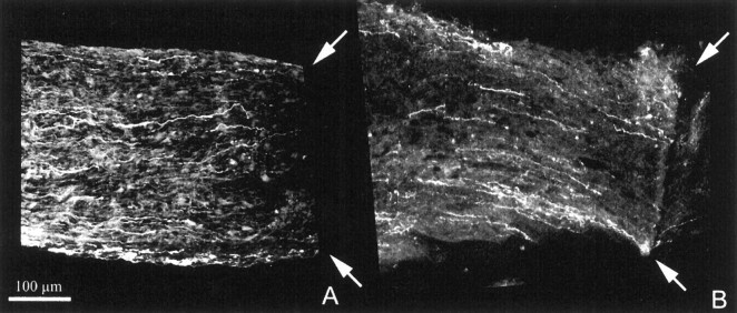
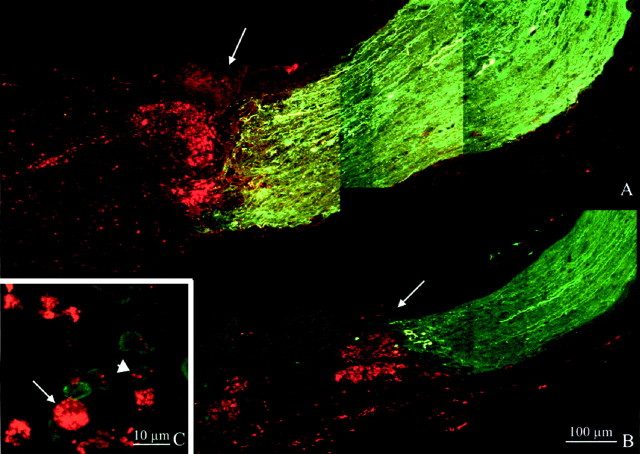
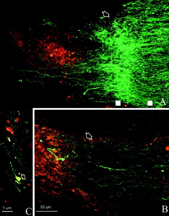
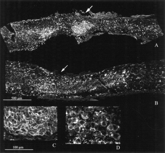
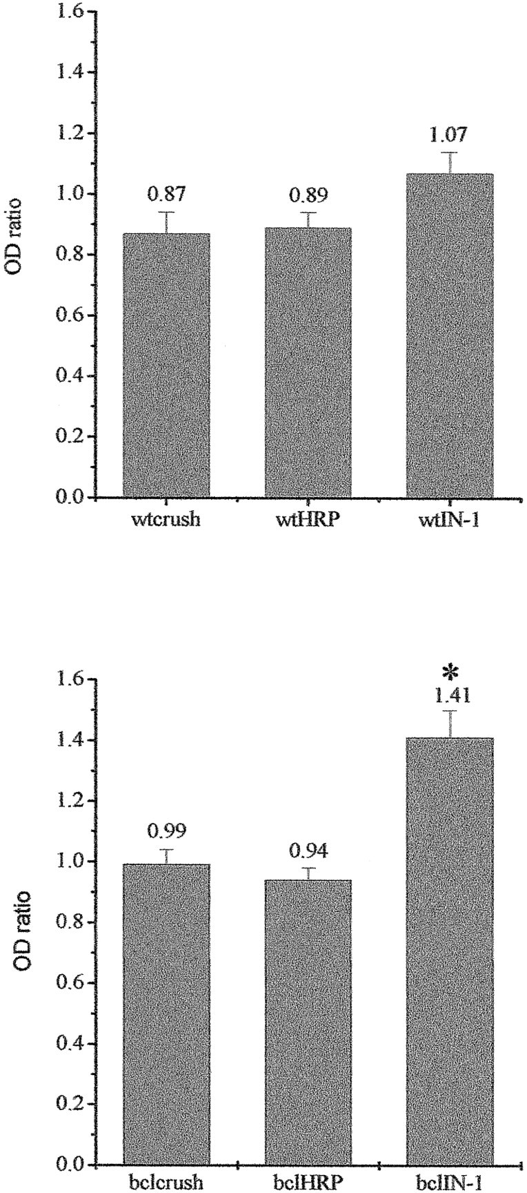
Retinal ganglion cells of transgenic mice overexpressing the anti-apoptotic protein Bcl-2 in neurons show a dramatic increase of survival rate after axotomy. We used this experimental system to test the regenerative potentials of central neurons after reduction of nonpermissive environmental factors. Survival of retinal ganglion cells 1 month after intracranial crush of the optic nerve was found to be 100% in adult bcl-2 mice and 44% in matched wild-type (wt) mice. In the optic nerve, and particularly at the crush site, fibers regrowing spontaneously or simply sprouting were absent in both wt and bcl-2 mice. We attempted to stimulate regeneration implanting in the crushed nerves hybridoma cells secreting antibodies that neutralize central myelin proteins, shown to inhibit regeneration (IN-1 antibodies) (Caroni and Schwab, 1988). Again, we found that regeneration of fibers beyond the site of crush was virtually absent in the optic nerves of both wt and bcl-2 mice. However, in bcl-2 animals treated with IN-1 antibodies, fibers showed sprouting in the proximity of the hybridoma implant. These results suggest that neurons overexpressing bcl-2 are capable of surviving axotomy and sprout when faced with an environment in which inhibition of regeneration has been reduced. Nevertheless, extensive regeneration does not occur, possibly because other factors act by preventing it.


Figures

Fig. 1.
Fig. 1.
Survival rate of retinal ganglion cells 1 month after intracranial crush of the left optic nerve.Columns are percentage average survivals with error bars. The first column refers to wild-type retinas (n = 3) and the second column to bcl-2 retinas (n = 3). Notice that survival is 100% in bcl-2 animals. The last column shows the survival rate of ganglion cells in wt retinas (n = 3) after injection of IN-1 hybridoma cells at the site of crush.
Fig. 2.
Fig. 2.
Light micrographs of the optic nerves of bcl-2 (left) and wild-type (right) mice 1 month after intracranial crush. A, B, Sections obtained in the proximity of the optic nerve head. C,D, High magnifications of A andB, respectively. The general morphology is well preserved in both wt and bcl-2 (A, B); degenerating fibers, characterized by a disorganized myelin sheath, are rare in the bcl-2 and more abundant in the wt (arrows inC and D). E,F, Sections obtained near the crush site and proximal to it. At low magnification, a central core of degeneration is evident in the bcl-2 nerve (arrows in E). At higher magnification, vacuoles are visible in the nerve core of the bcl-2 (arrows in G); however, numerous fibers appear still viable at more superficial positions (topof G). In the nerve of the wt (F,H) degeneration of fibers is widespread, as clearly illustrated in H; arrows point to altered myelin. Scale bars: (in A, E)A, B, E, F, 100 μm; (in C, G) C,D, G, H, 20 μm.
Fig. 3.
Fig. 3.
Longitudinal sections of wt (A) and bcl-2 (B) nerves shown near the crush site (arrows) after neurobiotin injection in the corresponding eyes. This and the following images have been obtained at the confocal microscope as explained in Materials and Methods. The optic nerve head is located to the left. Note that, in both cases, resilient fibers fail to pass beyond the crush site and do not sprout.
Fig. 4.
Fig. 4.
Longitudinal sections of crushed optic nerves of bcl-2 (A) and wt (B) mice 1 month after ON crush and implantation of IN-1 hybridoma cells. Fibers are labeled green by neurobiotin injected into the eye and revealed with FITC-avidin. Hybridoma cells, stained with DiI, appear red. Arrows point to the crush site. C, Staining with F4/80 antibody, specific for mouse microglia and macrophages. Double-labeled cells (short arrow) are immunopositive cells that have engulfed DiI, and thered ones are hybridoma cells (long arrow).
Fig. 5.
Fig. 5.
High magnification of bcl-2 (A) and wt (B) nerves illustrated at the site of crush. Arrows indicate the lesion site. Note the wealth of sprouting profiles in the bcl-2 (A) compared with the paucity of fibers present in the wild type (B). White squares are examples of the points intersected by lines perpendicular to the nerve major axes and used to calculate the density values of fluorescence staining. The square on theleft is at the sprouting site, and theright one is located more proximally. C, The tip of a regenerating fiber from A exhibits the typical morphology of a growth cone (arrow).
Fig. 6.
Fig. 6.
Longitudinal sections of wt (A) and bcl-2 (B) nerves that have received an injection of HRP hybridoma cells at the site of crush. Fibers stop at the lesion site, indicated byarrows.
Fig. 7.
Fig. 7.
F4/80 staining in crushed nerves of wt (A) and bcl-2 (B) that had received an injection of HRP cells. The crush site is indicated byarrows. Stained cells are dense at the lesion site;C and D (enlargements of the fields shown as rectangles in A and B) illustrate large size cells having a round morphology, typical of macrophages. Notice that there are not obvious differences in the staining between A and B.
Fig. 8.
Fig. 8.
Semiquantitative analysis of fiber responses in different experimental conditions. Top shows the wt groups; bottom shows the bcl-2. Columnsrepresent average OD ratios with standard errors. Theasterisk points out the significant difference among the bcl-2 IN-1 group and the remaining two.

References

    1. Adams JM, Cory S. The bcl-2 protein family: arbiters of cell survival. Science. 1998;281:1322–1326. - PubMed
    1. Allsopp TE, Wyatt S, Patterson HF, Davies AM. The proto-oncogene bcl-2 can selectively rescue neurotrophic factor-dependent neurons from apoptosis. Cell. 1993;73:295–307. - PubMed
    1. Austyn JM, Gordon S. F4/80, a monoclonal antibody directed specifically against the mouse macrophage. Eur J Immunol. 1981;11:805–815. - PubMed
    1. Bähr M, Przyrembel C, Bastmeyer M. Astrocytes from adult rat optic nerves are nonpermissive for regenerating retinal ganglion cell axons. Exp Neurol. 1995;131:211–220. - PubMed
    1. Bandtlow CE, Schmidt MF, Hassinger TD, Schwab ME, Kater SB. Role of intracellular calcium in NI-35-evoked collapse of neuronal growth cones. Science. 1993;259:80–83. - PubMed

Publication types

Substances

Grants and funding

LinkOut - more resources