. 1999 Oct 1;19(19):8435-42.
doi: 10.1523/JNEUROSCI.19-19-08435.1999.

Drosophila presenilin is required for neuronal differentiation and affects notch subcellular localization and signaling

Affiliations

Drosophila presenilin is required for neuronal differentiation and affects notch subcellular localization and signaling

Y Guo et al. J Neurosci. .

Abstract

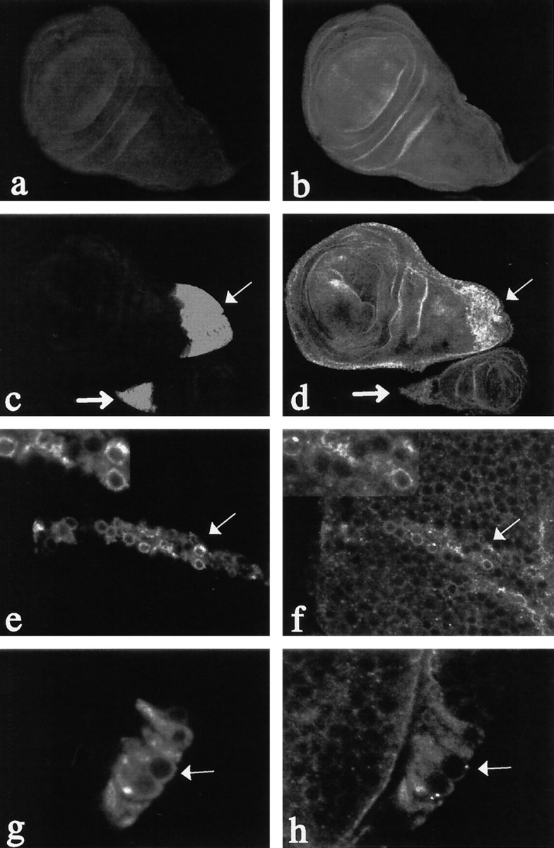
Presenilins are a highly conserved family of proteins first identified as causative genes in early onset familial Alzheimer's disease. Recent studies have suggested a role for presenilins in the Notch-signaling pathway, but their specific function within this pathway remains unclear. Here, we have characterized the Drosophila presenilin gene and protein and studied their interaction with Notch in both mutants and transgenics. We find that the Drosophila presenilin protein is proteolytically cleaved and broadly expressed during development with the highest levels in neurons within the larval CNS. We also show that mutations in Drosophila presenilin (Dps) genetically interact with Notch and result in an early pupal-lethal phenotype characterized by defects in eye and wing development and incomplete neuronal differentiation within the larval CNS. Moreover, we find that processing of Notch in the Golgi by the furin protease is unaffected in Dps mutants and that Notch is present and may even accumulate on the plasma membrane of neuroblasts in the larval CNS of Dps mutants. In contrast, overexpression of Dps in transgenics causes Notch to accumulate in the cytoplasm. Taken together, these results indicate that Drosophila presenilin is required for proper neuronal differentiation and may regulate the subcellular localization of Notch proteins within cells, necessary for their accumulation and subsequent signaling capabilities.


Figures

Fig. 1.
Fig. 1.
Genomic organization and deletion analysis of theDps locus. The genomic organization of theDps gene was determined by sequencing genomic DNA obtained from a P1 clone (#DS03069; Berkeley DrosophilaGenome Project) and from genomic DNA obtained after plasmid rescue from the P4 insertion line (GenBank accession number, AF093402) as described (Pirrotta, 1986). The Dps gene is flanked on either side by the genes encoding lipoic acid synthase (accession numbers, AA820940and AA201873) and 50 S ribosomal protein L15 (clone name, GM01846).Dps is comprised of nine exons (boxes) separated by eight introns. The location of an alternative splice site that gives rise to two dps (dpsa1 and dpsa2) isoforms is shown (hatchedbox). The P-element insertion P4 used to generate Dps deletions is shown. Overlapping deletions in Dps were generated by imprecise excision of the P-element P4 and are indicated by dashedlines. The 5′ and 3′ deletion break points were determined by a combination of Southern blot analysis using several restriction enzyme sites and sequencing of the break points from genomic DNA obtained from the deletion mutants, W20, W6, and W11.
Fig. 2.
Fig. 2.
Dps is proteolytically cleaved. Immunoblot analysis of second instar larval extracts from wild type (wt) and mutants indicates that Dps is proteolytically cleaved to give rise to a 25 kDa N-terminal fragment (a) and a 35 kDa C-terminal fragment (b) that are absent from the deletion mutants W6 and W11. Full-length protein can only be detected in third instar larval extracts of transgenic flies that overexpress Dpsfrom a heatshock promoter (hs-Dps) or use a daughterless-GAL4 driver (da-Dps) (c). In the absence of GAL4, only the N-terminal fragment is detected with the N-terminal-specific Dps antibody.
Fig. 3.
Fig. 3.
Dps mutants fail to undergo proper neuronal differentiation within the optic lobe anlagen and the eye imaginal disk. a, Wild-type CNS and disks derived from late third larval instar and double-immunostained for CUT (green) and ELAV (red). Note the precise laminar array organization of the optic lobe anlagen (OL) and the regular array of neurons within the developing eye imaginal disk (longarrow). At this stage, CUT expression is also abundant in developing antennal disk (shortarrow). b, DAPI staining of disks demonstrating the overall morphology of wild-type eye (arrow) and antennae (arrowhead) disks. c, Wild-type wing imaginal disks immunostained for WINGLESS. Note the normal distribution of wingless in the dorsal region of the wing disk in regions that will give rise to the adult notum and hinge (h) and the ventral staining in regions of the wing blade (wb) that will give rise to the wing margin. d, CNS and imaginal disks fromDps46/DpsW11 late third larval instar. Note the general disorganization of the optic lobe anlagen (OL) and the absence of ELAV expression in the eye imaginal disk (longarrow). In contrast, expression of CUT in the antennal disk appears normal (shortarrow). e, DAPI staining of disks fromDps46/DpsW11mutants. Although the antennae disk (arrowhead) appears normal, the eye disk (arrow) is small and disorganized. f, Wing imaginal disks fromDps46/DpsW11. WINGLESS expression appears normal in the dorsal compartment in regions that will give rise to the notum and hinge (h) but is absent from ventral regions that give rise to the wing blade and margin (wb).
Fig. 4.
Fig. 4.
Dps genetically interacts withNotch and Delta. Dps mutants can modify the phenotype of both Notch and Deltaalleles in transheterozygotes. a, Wild type.b,Dps46.c,DpsW11.d,Df(1)N-8. Note the mild notching at the wing tip (arrow). e, Df(1)N-8;Dps46. The Dpsmutations (e, f) enhance the notching at the wing tip (arrow). f,Df(1)N-8; DpsW11.g,Nnd-3. Note the thickening of wing vein L3 (arrow). h,Nnd-3;Dps46. The Dpsmutations (h, i) enhance the phenotype at the wing veins (thinarrows) and cause notching at the wing tips (thickarrow).i,Nnd-3;DpsW11. j,NAx-1. Note the interruption of wing vein L5 and the absence of L2 (arrows).k,NAx-1;Dps46. Note that the Dpsmutation suppresses partially the interruption of L5 and restores L2 (arrows). l,NAx-1;DpsW11. m,Dl[7]. Note the thickening of the wing veins and the slight delta between L4 and L5 (arrows).n,Dl[7];Dps46. Dps mutants also enhance the wing vein phenotype of Delta mutants in transheterozygotes. Note the enhanced thickening of the wing veins and deltas (thinarrows,thickarrow). o,Dl[7];DpsW11.
Fig. 5.
Fig. 5.
Notch staining is increased on the plasma membrane in the third larval instar CNS of Dps mutants. a, c, The distribution of Notch protein in the CNS of wild-type third instar larvae is shown at low (a) and high (c) magnification. Notch is expressed at high levels in the optic lobe anlagen and imaginal disks (a). Within the CNS, Notch protein is detected within the cytoplasm and the plasma membrane (c).b, d, In the CNS of Dps mutants, the overall pattern of Notch expression is affected (b), and the protein levels are reduced in the cytoplasm and increased on the plasma membrane (d). e, f, In contrast, the distribution of HRP on the plasma membrane is unaffected inDps mutants. In wild type (e), HRP is found on the entire surface of neuroblasts in the CNS, and the same pattern of expression can be observed in Dps mutants (f).
Fig. 6.
Fig. 6.
Notch staining is increased on the plasma membrane in the second larval instar CNS of Dps mutants. a, In wild-type second larval instar CNS, Notch expression is observed throughout the cytoplasm of neuroblasts and at high levels at the plasma membrane in regions of contact between the neuroblast and its progeny (arrow). b, c, In the CNS of Dps mutants, Notch expression is increased on the entire surface of the plasma membrane (thickarrows) and at the regions of contact between cells (thinarrow). d, Processing of Notch to give rise to a functional heterodimeric receptor is unaffected inDps mutants. Protein extracts were obtained from isolated CNS and imaginal disks from wild-type and Dps homozygous mutant third larval instars. No significant differences were observed in the levels or processing of Notch in Dps mutants compared with that of wild type.
Fig. 7.
Fig. 7.
Notch staining is increased in the cytoplasm ofDps-overexpressing cells. The distribution of Notch and Dps in third instar larval wing disks is shown. a, In wild-type disks, Dps protein is expressed at very low levels.b, Notch is expressed ubiquitously throughout the larval wing disk. c, d, Dps protein can be induced in wing (thinarrow) and haltere (thickarrow) disks using apnr-GAL4 line to drive expression of UAS-Dps in transgenic lines (c). Notch protein specifically accumulates in cells overexpressing Dps (d).e, f,cut-GAL4 was used to overexpressDps at the developing wing margin. Note the high levels of Dps protein that accumulate in the cytoplasm of cells at the wing margin (e, arrow). Overexpression ofDps at the margin causes Notch to accumulate within the cytoplasm of these cells (f, arrow).g, h, Overexpression of Dps in a subset of neurons within the developing eye disk (g, arrow) also causes increases in Notch expression within the cytoplasm of these cells (h, arrow).

Similar articles

Cited by

References

    1. Baumeister R, Leimer U, Zweckbronner I, Jakubek C, Grunberg J, Haass C. Human presenilin-1, but not familial Alzheimer’s disease (FAD) mutants, facilitate Caenorhabditis elegans Notch signalling independently of proteolytic processing. Genes Funct. 1997;1:149–159. - PubMed
    1. Berezovska O, Xia M, Page K, Wasco W, Tanzi R, Hyman B. Developmental regulation of presenilin mRNA expression parallels Notch expression. J Neuropathol Exp Neurol. 1997;56:40–44. - PubMed
    1. Blaumueller CM, Qi H, Zagouras P, Artavanis-Tsakonas S. Intracellular cleavage of Notch leads to a heterodimeric receptor on the plasma membrane. Cell. 1997;90:281–291. - PubMed
    1. Blochlinger K, Jan LY, Jan YN. Postembryonic patterns of expression of cut, a locus regulating sensory organ identity in Drosophila. Development. 1993;117:441–450. - PubMed
    1. Borchelt DR, Thinakaran G, Eckman CB, Lee MK, Davenport F, Ratovitsky T, Prada C-M, Kim G, Seekins S, Yager D, Slunt HH, Wang R, Seeger M, Levey AI, Gandy SE, Copeland NG, Jenkins NA, Price DL, Younkin SG, Sisodia SS. Familial Alzheimer’s disease-linked presenilin 1 variants elevate Aβ1–42/1–40 ratio in vitro and in vivo. Neuron. 1996;17:1005–1013. - PubMed

Publication types

MeSH terms

Associated data

LinkOut - more resources