. 1999 Oct 1;19(19):8589-96.
doi: 10.1523/JNEUROSCI.19-19-08589.1999.

Membrane potential oscillations in dorsal root ganglion neurons: role in normal electrogenesis and neuropathic pain

Affiliations

Membrane potential oscillations in dorsal root ganglion neurons: role in normal electrogenesis and neuropathic pain

R Amir et al. J Neurosci. .

Abstract

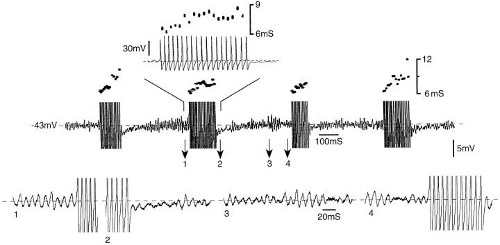
Abnormal afferent discharge originating at ectopic sites in injured primary sensory neurons is thought to be an important generator of paraesthesias, dysaesthesias, and chronic neuropathic pain. We report here that the ability of these neurons to sustain repetitive discharge depends on intrinsic resonant properties of the cell membrane and that the prevalence of this characteristic increases after nerve injury. Recording from primary sensory neurons in excised rat dorsal root ganglia, we found that some cells show subthreshold oscillations in their membrane potential. The amplitude, frequency, and coherence of these oscillations were voltage sensitive. Oscillations gave rise to action potentials when they reached threshold. Indeed, the presence of oscillations proved to be a necessary condition for sustained spiking both at resting membrane potential and on depolarization; neurons without them were incapable of sustained discharge even on deep depolarization. Previous nerve injury increased the proportion of neurons sampled that had subthreshold oscillations, and hence the proportion that generated ectopic spike discharge. Oscillatory behavior and ectopic spiking were eliminated by [Na(+)](o) substitution or bath application of lidocaine or tetrodotoxin (TTX), under conditions that preserved axonal spike propagation. This suggests that a TTX-sensitive Na(+) conductance contributes to the oscillations. Selective pharmacological suppression of subthreshold oscillations may offer a means of controlling neuropathic paraesthesias and pain without blocking afferent nerve conduction.


Figures

Fig. 1.
Fig. 1.
Membrane potential oscillations recorded from DRG A- and C-neurons. A, Subthreshold oscillations recorded from an A0-neuron in an immature rat with nerves intact. Oscillation amplitude was increased when the cell was depolarized from rest (Vr) to −35 mV and subsequently decreased with still deeper depolarization (left). This neuron did not fire action potentials. The FFT profile in this cell illustrates oscillation coherence and amplitude (power) peak (103 Hz) at −35 mV and the increase in oscillation frequency with depolarization (right). Power scale was normalized relative to the maximal power recorded at −35 mV. Theinset shows the autocorrelogram at −35 mV.B, Membrane oscillations recorded from a C-neuron in a mature rat 15 d after sciatic injury. At −36 mV, oscillations occasionally triggered action potentials [spike amplitude is truncated (left)]. The FFT profile at different membrane potentials in this cell, and its autocorrelogram (inset), illustrates a dominant oscillation frequency of 20 Hz at −39 mV (right). The power scale was normalized as in A.
Fig. 2.
Fig. 2.
Spontaneous repetitive bursty discharge in a DRG A0-neuron from an immature rat. Spikes are truncated except in the inset at top, which shows one of the bursts on a faster timebase. The interspike interval dot display above each of the four spike bursts shown illustrates the gradual deceleration of discharge during the course of the burst. A similar firing pattern is seen in in vivo recordings of ectopic burst discharge originating in the DRG. Segments of the record at time points 1–4 are shown below on a still faster timebase to illustrate the triggering of spike bursts by membrane potential oscillations.
Fig. 3.
Fig. 3.
Subthreshold membrane potential oscillations, and resulting spike bursting, depend on voltage-sensitive Na+ conductance. A illustrates a period of subthreshold oscillations that lead up to a spike burst in an A0-neuron from an adult rat whose ipsilateral sciatic nerve had been cut 7 d previously (control). Replacing NaCl in the bath solution with choline-Cl, thus reducing the bath Na+ concentration from 151 to 27 mm, abolished the oscillations and the resulting spikes within 2 min. They could not be restored by further depolarization. Washout of choline and return to the control bath solution restored both the oscillations and bursting within 17 min (recovery). All three traces were recorded at −62 mV.Top traces show (left) the intracellularly recorded spike before, during, and after choline application [at resting potential (Vr) = −69 mV], and (right) the simultaneously recorded DR compound action potential. Both spike and compound action potential persisted, if with a reduced amplitude, when oscillations and ectopic spiking were eliminated. Propagation distance from the sciatic nerve stimulation site was 37 mm to the cell soma and 50 mm to the compound action potential recording electrode. B, In this A0-neuron from an adult rat DRG, depolarization from rest (Vr = −69 mV) to −57 mV yielded subthreshold oscillations that triggered spike bursting (control trace; spikes truncated). Bath application of 1 μm TTX abolished the oscillations and spikes within 2 min (TTX 2′). Spike propagation in response to nerve stimulation at a distance of 7 mm persisted at this time, although it failed 4 min later (TTX 6′). Further depolarization failed to restore oscillations or spiking, but TTX washout frequently did (data not shown).

Similar articles

Cited by

References

    1. Akopian AN, Sivilotti L, Wood JN. A tetrodotoxin-resistant voltage-gated sodium channel expressed by sensory neurons. Nature. 1996;379:257–262. - PubMed
    1. Alonzo A, Klink R. Differential electroresponsiveness of stellate and pyramidal-like cells of medial entorhinal cortex layer II. J Neurophysiol. 1993;70:144–157. - PubMed
    1. Amir R, Devor M. Chemically mediated cross-excitation in rat dorsal root ganglia. J Neurosci. 1996;16:4733–4741. - PMC - PubMed
    1. Amir R, Devor M. Spike-evoked suppression and burst patterning in dorsal root ganglion neurons. J Physiol (Lond) 1997;501:183–196. - PMC - PubMed
    1. Baker MD, Bostock H. Low-threshold, persistent sodium current in large dorsal root ganglion neurons in culture. J Neurophysiol. 1997;77:1503–1513. - PubMed

Publication types

MeSH terms