. 1999 Oct 1;19(19):8637-45.
doi: 10.1523/JNEUROSCI.19-19-08637.1999.

Cholinergic induction of theta-frequency oscillations in hippocampal inhibitory interneurons and pacing of pyramidal cell firing

Affiliations

Cholinergic induction of theta-frequency oscillations in hippocampal inhibitory interneurons and pacing of pyramidal cell firing

C A Chapman et al. J Neurosci. .

Abstract

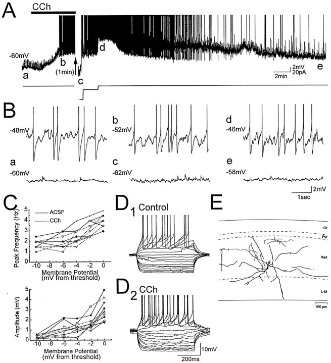
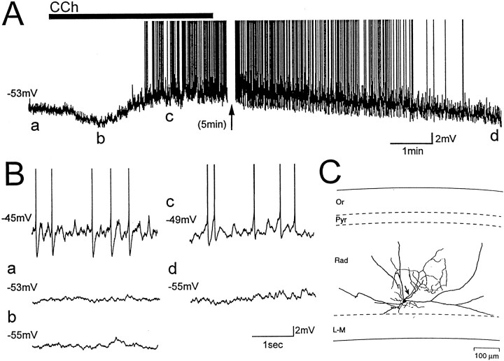
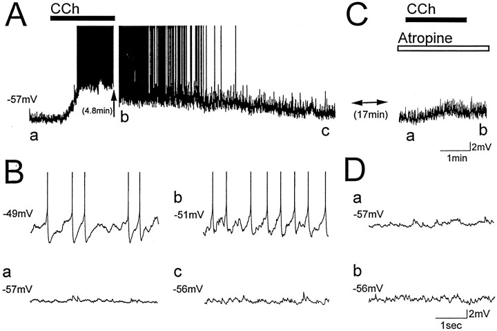
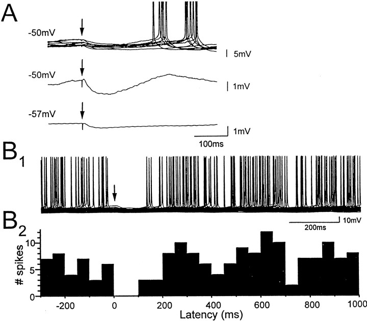
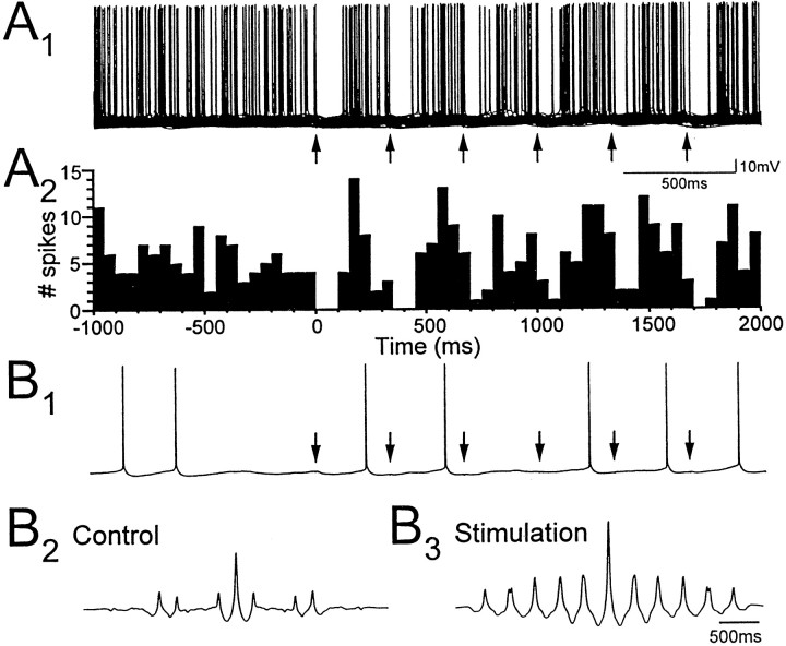
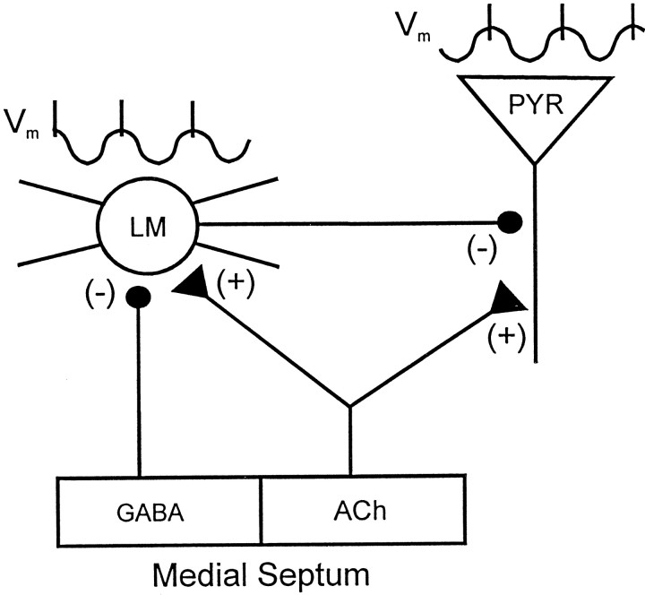
Cholinergic and GABAergic medial septal afferents contribute to hippocampal theta activity in part by actions on local interneurons. Interneurons near the border between stratum radiatum and stratum lacunosum-moleculare (LM) display intrinsic membrane potential oscillations at theta frequency when depolarized near threshold. First, whole-cell current-clamp recordings in rat hippocampal slices were used to examine effects of the cholinergic agonist carbachol on biocytin-labeled LM interneurons. At resting membrane potential, cells were depolarized by bath application of 25 microM carbachol, and the depolarization was sufficient to induce membrane potential oscillations (2.4 +/- 0.2 mV) that paced cell firing. Carbachol also depolarized LM interneurons in the presence of 6-cyano-7-nitroquinoxaline-2,3-dione, (+/-)-2-amino-5-phosphonopentanoic acid, and bicuculline, indicating that cholinergic depolarization of LM cells does not depend on ionotropic glutamate or GABA(A) synaptic transmission in local circuits. Atropine blocked the depolarization, indicating that muscarinic receptors were involved. Minimal stimulation applied to visually identified LM interneurons was then used to determine if spontaneous activity in CA1 pyramidal cells can be paced by rhythmic inhibition generated by LM cells at theta frequency. Inhibitory postsynaptic potentials evoked in pyramidal cells by single minimal stimulations were followed by rebound depolarizations and action potentials. When trains of minimal stimulation were delivered, membrane potential oscillations of depolarized pyramidal cells followed the stimulation frequency. Minimal stimulation led pyramidal cell firing with an average phase of 177 degrees. Thus, muscarinic induction of theta-frequency membrane potential oscillations in LM interneurons may contribute to the generation of rhythmic inhibition that paces intrinsically generated theta activity in CA1 pyramidal cells.


Figures

Fig. 1.
Fig. 1.
Carbachol depolarizes LM interneurons and induces voltage-dependent membrane potential oscillations.A,B, Membrane potential recording from an LM interneuron exposed to the cholinergic agonist carbachol (CCh,solid bar; 25 μm). Letters in A indicate times from which expanded traces in B were taken, and action potentials are truncated in this and subsequent figures. Voltage-dependent oscillations were induced in the LM cell with positive current injection before carbachol application (B, top left trace). Application of carbachol at resting membrane potential (a) was followed by a depolarization that induced oscillations that paced action potentials (b, d). The depolarization was preceded by a small transient hyperpolarization (see also Fig. 2A). Oscillations induced by carbachol were eliminated when the cell was hyperpolarized with steady negative current injection (c) or when the cell repolarized after washout of carbachol (e), indicating that the oscillations are voltage-dependent. C, Power spectral analysis of oscillations in six LM cells in which membrane potential relative to spike threshold was varied using steady current injection. Frequency and amplitude of oscillations, and their voltage dependence, were not significantly different in carbachol (dashed lines) and in normal ACSF (solid lines). D, Voltage responses to positive and negative current pulses that ranged from −100 to 60 pA in 10 pA steps. For this cell, carbachol increased input resistance and decreased afterhyperpolarization amplitude.E, Camera lucida tracing of the LM interneuron from which recordings in A, B, andD were obtained. The axon is indicated by anarrow. Abbreviations in this figure and in Figure 2:Or, stratum oriens; Pyr, stratum pyramidale; Rad, stratum radiatum; L-M, stratum lacunosum-moleculare.
Fig. 2.
Fig. 2.
Glutamate and GABAA synaptic activity are not necessary for the depolarization of LM interneurons induced by carbachol. A, B, Membrane potential of an LM interneuron exposed to bath application of 25 μm carbachol (CCh, solid bar) during blockade of ionotropic glutamate and GABAA synaptic transmission with CNQX (20 μm), AP-5 (50 μm), and bicuculline (25 μm). Letters indicate times from which expanded traces in B were taken. Steady current injection induced voltage-dependent oscillations before carbachol application (B, top left trace). Application of carbachol at resting membrane potential (a) was followed by a transient hyperpolarization (b), and then a depolarization that induced oscillations and cell firing (c). C, Camera lucida tracing of the LM interneuron from which recordings were obtained. The axon is indicated by an arrow.
Fig. 3.
Fig. 3.
Muscarinic receptors mediate the depolarization of LM interneurons induced by carbachol. Membrane potential of an LM interneuron exposed to a 2 min bath application of 25 μmcarbachol (CCh, solid bar) in normal ACSF (A, B) and in the presence of the muscarinic receptor antagonist atropine (C, D,open bar; 1 μm). Letters in A andC indicate times from which the corresponding traces inB and D were taken. Steady current injection induced voltage-dependent oscillations in normal ACSF before carbachol application (B, top left trace). In normal ACSF, carbachol depolarized the cell from resting membrane potential and induced voltage-dependent oscillations and cell firing (A, a vs b). Oscillations were eliminated during washout of carbachol as membrane potential repolarized (c). After exposure to atropine for at least 15 min (C,D), carbachol did not induce oscillations or cell firing (C, a vsb).
Fig. 4.
Fig. 4.
Minimal stimulation in stratum lacunosum-moleculare evokes IPSPs, followed by rebound depolarization and spiking in CA1 pyramidal neurons. A, IPSPs evoked in pyramidal cells by minimal stimulation in stratum lacunosum-moleculare (arrows) at membrane potentials indicated atleft. Eleven superimposed traces (A, top) show minimal IPSPs that were followed by an action potential. Rebound depolarizations at the same latencies as the action potentials were also observed when the cell was held just below spike threshold (A, middle, mean of 12 traces), but not at more hyperpolarized levels (A,bottom, mean of 61 traces).B1, Fifty superimposed traces showing the effect of minimally evoked IPSPs (arrow) on firing of the CA1 pyramidal cell. B2, The corresponding spike frequency histogram shows peaks at latencies near 250 and 600 msec, suggesting that intrinsic mechanisms generating rhythmic membrane potential oscillations in the pyramidal cell are reset by minimal IPSPs generated from stratum lacunosum-moleculare.
Fig. 5.
Fig. 5.
Low-frequency minimal stimulation of stratum lacunosum-moleculare paces repetitive firing of pyramidal neurons.A1, Seventy superimposed traces showing the effect of a 3 Hz train of minimal stimulation (arrows) on the firing of a CA1 pyramidal cell depolarized to threshold by steady current injection.A2, The corresponding spike frequency histogram shows that the timing of low-frequency spiking in the pyramidal cell is paced throughout the train of minimal stimulation in stratum lacunosum-moleculare. On average, the firing of pyramidal cells was ∼180° out of phase with the minimal stimulation at 3 Hz.B, Increased rhythmicity of pyramidal cell firing (B1) is reflected in autocorrelation functions of 2 sec recordings of membrane potential obtained just before (B2) and during (B3) the train of minimal stimulation.
Fig. 6.
Fig. 6.
Schematic diagram indicating how LM interneurons can contribute to the pacing of theta activity in CA1 pyramidal neurons. CA1 pyramidal cells (PYR) are shown to receive excitatory cholinergic inputs (+) from the medial septum, whereas LM interneurons receive both cholinergic and inhibitory GABAergic (−) septal inputs. The proposed relationship between membrane potential (Vm) in LM cells and pyramidal cells during theta activity is indicated by the traces above each cell. In the model, cholinergic septal afferents depolarize both LM cells and pyramidal cells and induce intrinsic theta-frequency membrane potential oscillations in LM cells that pace action potential generation. Generation of dendritic IPSPs at theta frequency by LM interneurons paces the intrinsic oscillatory activity of the pyramidal cell such that it is ∼180° out of phase with the firing of LM interneurons. Furthermore, because each LM interneuron contacts many pyramidal cells, rhythmic firing of single LM interneurons may synchronize theta activity among large numbers of pyramidal cells. Synchronization of theta-frequency oscillations among LM interneurons may be accomplished in part by GABAergic inputs from the medial septum through a mechanism similar to that proposed for the pacing of theta activity in pyramidal cells by LM interneurons.

Similar articles

Cited by

References

    1. Atzori M. Pyramidal cells and stratum lacunosum-moleculare interneurons in the CA1 hippocampal region share a GABAergic spontaneous input. Hippocampus. 1996;6:72–78. - PubMed
    1. Banks MI, Li T-B, Pearce RA. The synaptic basis of GABAA,slow. J Neurosci. 1998;18:1305–1317. - PMC - PubMed
    1. Behrends JC, ten Bruggencate G. Cholinergic modulation of synaptic inhibition in the guinea pig hippocampus in vitro: excitation of GABAergic interneurons and inhibition of GABA-release. J Neurophysiol. 1993;69:626–629. - PubMed
    1. Bland BH, Colom LV. Extrinsic and intrinsic properties underlying oscillation and synchrony in limbic cortex. Prog Neurobiol. 1993;41:157–208. - PubMed
    1. Bland BH, Oddie SD, Colom LV. Mechanisms of neural synchrony in the septohippocampal pathways underlying hippocampal theta generation. J Neurosci. 1999;19:3223–3237. - PMC - PubMed

Publication types

MeSH terms

LinkOut - more resources