. 1999 Oct 1;19(19):8646-55.
doi: 10.1523/JNEUROSCI.19-19-08646.1999.

Extinction of behavior in infant rats: development of functional coupling between septal, hippocampal, and ventral tegmental regions

Affiliations

Extinction of behavior in infant rats: development of functional coupling between septal, hippocampal, and ventral tegmental regions

H P Nair et al. J Neurosci. .

Abstract

Learning of a behavior at a particular age during the postnatal period presumably occurs when the functional brain circuit mediating the behavior matures. The inability to express a learned behavior, such as inhibition, may be accounted for by the functional dissociation of brain regions comprising the circuit. In this study we tested this hypothesis by measuring brain metabolic activity, as revealed by fluorodeoxyglucose (FDG) autoradiography, during behavioral extinction in 12- and 17-d-old rat pups. Subjects were first trained on a straight alley runway task known as patterned single alternation (PSA), wherein reward and nonreward trials alternate successively. They were then injected with FDG and given 50 trials of continuous nonreward (i.e., extinction). Pups at postnatal day 12 (P12) demonstrated significantly slower extinction rates compared to their P17 counterparts, despite the fact that both reliably demonstrated the PSA effect, i.e., both age groups distinguished between reward and nonreward trials during acquisition. Covariance analysis revealed that the dentate gyrus, hippocampal fields CA1-3, subiculum, and lateral septal area were significantly correlated in P17 but not P12 pups. Significant correlations were also found between the lateral septal area, ventral tegmental area, and the medial septal nucleus in P17 pups. Similar correlative patterns were not found in P12 and P17 handled control animals. Taken together, these results suggest that septal, hippocampal, and mesencephalic regions may be functionally dissociated at P12, and the subsequent maturation of functional connectivity between these regions allows for the more rapid expression of behavioral inhibition during extinction at P17.


Figures

Fig. 1.
Fig. 1.
Schematic view of pup runway. Photocells are placed at positions 1,2, and 3, to time start, run, and goal speeds and intertrial intervals. D1, Door from startbox to alley; D2, door from alley to goalbox;D3, automated door, dividing goalbox into two compartments, which is controlled by signals from Photocell 3. An anesthetized dam placed in the rear compartment (R) is accessible on rewarded trials when D3 is raised. On nonrewarded trials, pups remain in the front compartment (F) with D3 in lowered position. On reward-milk trials, milk is delivered via an infusion pump (not shown) into the oral cannula of the pup while it is attaching to the nipple and suckling. The length of the runway is adjusted depending on age group. For 16- to 17-d-old pups, the length is 60 cm from start door to goal box. The length is shortened to 45 cm for 11- to 12-d-old pups.
Fig. 2.
Fig. 2.
Sampled regions. Regions of interest were from six different anatomical levels. The distance from interaural line for each age group is as follows (in millimeters): A, P12, 7.0; P17, 9.0; B, P12, 5.3; P17, 6.5; C, P12, 2.6; P17, 3.2; D, P12, 1.2; P17, 1.6; E,P12, 0.8; P17, 1.2; F, P12, 0.4; P17, 1.2.PMC, Primary motor cortex; MS, medial septum; LS, lateral septum; LH, lateral hypothalamus; Mam, mammillary bodies;Sub, subiculum; DG, dentate gyrus;EC, entorhinal cortex; VTA, ventral tegmental area. (Section diagrams were reproduced with permission fromThe Rat Brain in Stereotaxic Coordinates, G. Paxinos and C. Watson, New York: Academic, 1997, CD-ROM.)
Fig. 3.
Fig. 3.
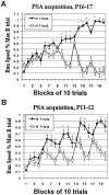
Acquisition data. The faster run speeds (presented as a percentage of maximum run speed on R trials) on rewarded trials by the end of training indicate that both P17 (A) and P12 (B) successfully discriminated between reward (R) and nonreward (N) trials. *Indicates significant difference (p < 0.05) between reward and nonreward trials.
Fig. 4.
Fig. 4.
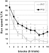
Extinction data. P12 pups demonstrated attenuated extinction rates (presented as percentage of maximum run speed on the last block of rewarded trials during acquisition) on trials 3 and 5–10. Asterisk indicates significant difference (p < 0.05) between P12 and P17 groups.
Fig. 5.
Fig. 5.
Pooled FDG uptake data. Of the eleven regions sampled, the CA1 and CA3 fields of the hippocampus, subiculum (Sub), mammillary bodies (Mam), medial septum (MS), and ventral tegmental area (VTA), were significantly higher in P17 animals compared with P12.
Fig. 6.
Fig. 6.
Autoradiographic images demonstrating changes in FDG uptake in CA1, CA3, and subiculum between (A) P17 and (B) P12 pups. A schematic of the region imaged is presented in C.
Fig. 7.
Fig. 7.
Covariance patterns of FDG uptake across groups.Black arrows indicate reliable pairwise correlations significantly different from zero (p < 0.05). Note changes in covariance patterns between PSA 17 (A) and PSA12 (C). The same correlations did not appear between handled control groups (B, D), indicating the altered covariance patterns across PSA groups were related to extinction training rather than handling or developmental effects.
Fig. 8.
Fig. 8.
Scatter plots of correlations between CA3 and dentate gyrus (DG), CA3 and lateral septum (LS), and CA3 and CA1. FDG ratio, or whole brain ratio, is the raw FDG value divided by the whole brain average. Note the linearity of values in the PSA17 group (A) relative to those found within HC17 (B), PSA12 (C), and HC12 (D) groups.

References

    1. Amaral DG, Dent JA. Development of the mossy fibers of the dentate gyrus: a light and electron microscopic study of the mossy fibers and their expansions. J Comp Neurol. 1981;195:51–86. - PubMed
    1. Amsel A. Frustration theory. Cambridge UP; New York: 1992.
    1. Bowe MA, Nadler JV. Developmental increase in the sensitivity to magnesium of NMDA receptors on CA1 hippocampal pyramidal cells. Dev Brain Res. 1990;56:55–61. - PubMed
    1. Bronstein PM, Neiman H, Wolkoff FD, Levine MJ. The development of habituation in the rat. Anim Learn Behav. 1971;2:92–96.
    1. Chugani HT, Phelps ME. Maturational changes in cerebral function in infants determined by [18]FDG positron emission tomography. Science. 1986;231:840–843. - PubMed

Publication types

Substances