. 1999 Oct;67(10):5083-90.
doi: 10.1128/IAI.67.10.5083-5090.1999.

Clostridium botulinum C2 toxin delays entry into mitosis and activation of p34cdc2 kinase and cdc25-C phosphatase in HeLa cells

Affiliations

Clostridium botulinum C2 toxin delays entry into mitosis and activation of p34cdc2 kinase and cdc25-C phosphatase in HeLa cells

H Barth et al. Infect Immun. 1999 Oct.

Abstract

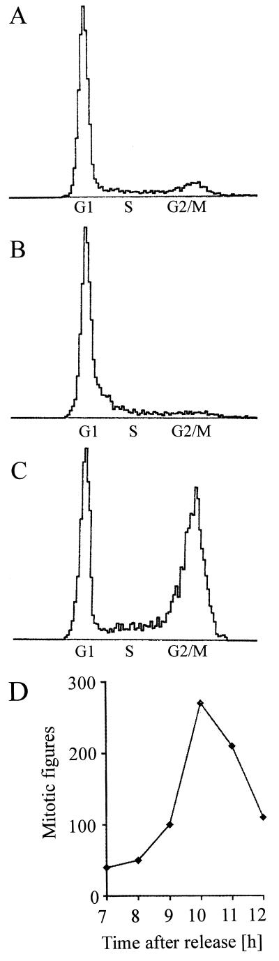
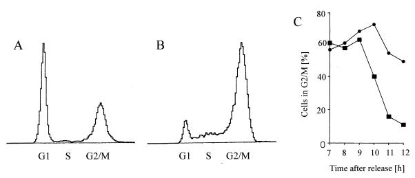
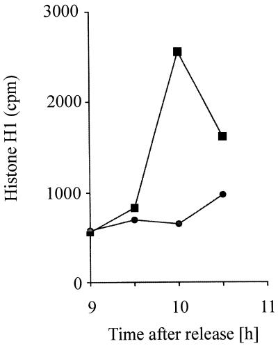
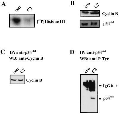
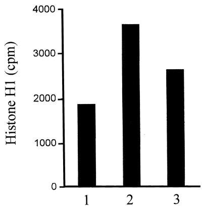
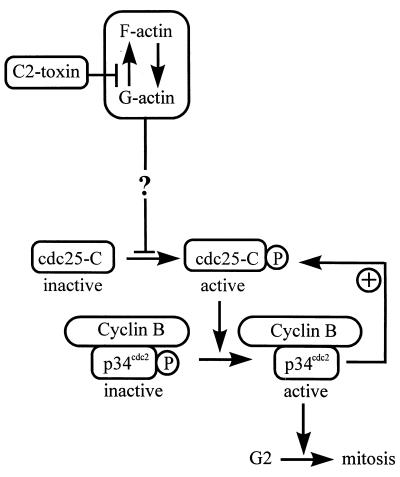
The Clostridium botulinum C2 toxin ADP-ribosylates monomeric actin, thereby inducing disassembly of actin filaments, alteration of focal adhesions, and rounding of cells. After treatment with C2 toxin, cells stop to proliferate but remain viable for about 2 days. In view of reported correlations between the structure of the actin cytoskeleton and cell cycle transition, the effects of C2 toxin on the G(2)/M phase transition of the cell division cycle were studied. Since C2 toxin delayed entry into mitosis in HeLa cells, those enzymes which control entry into mitosis, the cyclin-dependent protein kinase mitosis-promoting factor (MPF) and the phosphatase cdc25-C were examined after treatment of synchronized cells with C2 toxin. MPF is composed of the regulatory cyclin B and the enzymatic p34cdc2 kinase subunits. For its activation at the G2/M border, p34cdc2 needs to be associated with cyclin B and additionally dephosphorylated at Tyr-15 by the specific phosphatase cdc25-C. Treatment of synchronized cells in S or G2 phase with C. botulinum C2 toxin prevented p34cdc2 protein kinase activation by inhibiting its tyrosine dephosphorylation at the G2/M border. Furthermore, the activity of cdc25-C phosphatase was decreased after treatment of cells with C2 toxin. Our results suggest that the prevented activation of the mitotic inducers p34cdc2 kinase and cdc25-C phosphatase represents the final downstream events in the action of C2 toxin resulting in a G(2) phase cell cycle delay in synchronized HeLa cells.


Figures

FIG. 1
FIG. 1
Cell cycle phase distribution of synchronized HeLa cells. HeLa cells were synchronized with amethopterin and thymidine as described in the text. At the indicated times, cells were fixed and analyzed by flow cytometry. DNA histograms represent asynchronous cells (A), amethopterin-blocked cells (16 h; B) and cells 9 h after release from the amethopterin block (C). Abscissa, relative fluorescence; ordinate, relative cell number. (D) Time course of mitotic figures of amethopterin-thymidine-synchronized HeLa cells starting 7 h after release from the block.
FIG. 2
FIG. 2
Cytotoxic effect of C. botulinum C2 toxin on synchronous HeLa cells. (A) Time course of C2 toxin-induced actin ADP-ribosylation. At 6 h after release from the amethopterin block, C2 toxin (200 ng of C2II and 100 ng of C2I per ml) was added to synchronized HeLa cells. Cells were incubated at 37°C; immediately and every 30 min after toxin addition, cells were lysed and lysate proteins (100 μg) were subjected to an in vitro ADP-ribosylation assay with C2I. The autoradiogram of [32P]ADP-ribosylated actin is shown. Lane 1, control (without C2 toxin); lanes 2 to 8, incubation for 30 min with C2 toxin, 60 min with C2, 90 min with C2, 120 min with C2, 150 min with C2, 180 min with C2, and 210 min with C2, respectively. (B) C2 toxin-induced morphological changes and F-actin redistribution. Synchronized control cells (8 h after release from the amethopterin block) as well as synchronized cells treated with C2 toxin for 2 h (6 to 8 h after release from the block; 200 ng of C2II and 100 ng of C2I per ml) were fixed, and F-actin was stained with phalloidin-rhodamine.
FIG. 3
FIG. 3
G2 delay of synchronous HeLa cells induced by the C. botulinum C2 toxin. Four hours after release from the amethopterin block, the cells were treated with C2 toxin (200 ng of C2II and 100 ng of C2I per ml). Starting at 3 h after addition of the C2 toxin (i.e., 7 h after release from the block), cells were fixed and analyzed by flow cytometry. DNA histograms represent control cells (A) and C2 toxin-treated cells (B) at 11.5 h after release from the block (i.e., after 7.5 h of C2 toxin treatment). (C) Time course of the percentage of control (■) and C2-treated (●) cells in G2/M phase, determined by flow cytometry.
FIG. 4
FIG. 4
Time course of p34cdc2 protein kinase activity of synchronized HeLa cells untreated (control) or treated with C. botulinum C2 toxin. At 6 h after release from the amethopterin block, C2 toxin (200 ng of C2II and 100 ng of C2I per ml) was added to the medium, and the cells were further incubated at 37°C. Starting at 9 h after release from the block, every 30 min control cells (■) and cells treated with C2 toxin (●) were lysed, and p34cdc2 was immunoprecipitated and analyzed for histone H1 kinase activity.
FIG. 5
FIG. 5
Effect of C. botulinum C2 toxin on the MPF complex of synchronized HeLa cells. At 6 h after release from the amethopterin block, C2 toxin (200 ng of C2II and 100 ng of C2I per ml) was added to the cells. After 4 h at 37°C, cells were lysed and p34cdc2 was immunoprecipitated for determination of histone kinase (A). Lysate proteins (100 μg) were subjected to immunoblot analysis with anti-p34cdc2 antibody and anti-cyclin B antibody (B). Anti-p34cdc2 immunoprecipitates (IP) from the same lysates were probed by Western blotting (WB) with anti-cyclin B antibody (C). The influence of C2 toxin on the tyrosine phosphorylation of p34cdc2 was analyzed by immunoblot analysis of p34cdc2 immunoprecipitates with anti-P-Tyr (D). con, control; IgG h. c., immunoglobulin G heavy chain.
FIG. 6
FIG. 6
Effect of C. botulinum C2 toxin on cdc25-C phosphatase of synchronized HeLa cells. At 6 h after release from the amethopterin block, C2 toxin (200 ng of C2II and 100 ng of C2I per ml) was given to the cells (for control without toxin). After further 4 h at 37°C, i.e., at 10 h after release, cells were lysed and cdc25-C was immunoprecipitated for determination of phosphatase activity. The cdc25-C immunoprecipitates were incubated for 15 min at 30°C with inactive p34cdc2 immunoprecipitated from 500 μg of S-phase HeLa lysate protein. Activity of p34cdc2 kinase was measured by histone H1 phosphorylation assay. Histone H1 bands were measured by scintillation counting. Bar 1, S-phase p34cdc2 kinase (without cdc25-C); bar 2, S-phase p34cdc2 kinase plus cdc25-C from control cells; bar 3, S-phase p34cdc2 kinase plus cdc25-C from C2 toxin-treated cells.
FIG. 7
FIG. 7
Model for the G2 phase cell cycle arrest induced by C. botulinum C2 toxin. Cyclin B/p34cdc2 is activated at the G2/M border by dephosphorylation of p34cdc2 by the phosphatase cdc25-C. Active p34cdc2 kinase drives the cell into mitosis. Furthermore, active p34cdc2 kinase activates cdc25-C phosphatase via an autocatalytic loop. Treatment of HeLa cells with C2 toxin delays the G2/M transition by preventing tyrosine dephosphorylation and thereby activation of the p34cdc2 protein kinase. C2 toxin treatment also prevents activation of cdc25-C phosphatase, which indicates that C2 toxin may affect intracellular signal cascades upstream of the mitotic inducers p34cdc2 and cdc25-C.

References

    1. Aktories K, Bärmann M, Ohishi I, Tsuyama S, Jakobs K H, Habermann E. Botulinum C2 toxin ADP-ribosylates actin. Nature. 1986;322:390–392. - PubMed
    1. Aragon V, Chao K, Dreyfus L A. Effect of cytolethal distending toxin on F-actin assembly and cell division in Chinese hamster ovary cells. Infect Immun. 1997;65:3774–3780. - PMC - PubMed
    1. Arion D, Meijer L, Brizuela L, Beach D. Cdc2 is a component of the M phase-specific histone H1 kinase: evidence for identity with MPF. Cell. 1988;55:371–378. - PubMed
    1. Barth H, Kinzel V. Phorbol ester TPA rapidly prevents activation of p34cdc2 histone H1 kinase and concommitantly the transition from G2 phase to mitosis in synchronized Hela cells. Exp Cell Res. 1994;212:383–388. - PubMed
    1. Barth H, Kinzel V. Epidermal growth factor rapidly impairs activation of p34cdc2 protein kinase in HeLa cells at the G2-M boundary. J Cell Physiol. 1995;162:44–51. - PubMed

Publication types