. 1999 Oct;67(10):5151-6.
doi: 10.1128/IAI.67.10.5151-5156.1999.

In vivo distribution of Helicobacter felis in the gastric mucus of the mouse: experimental method and results

Affiliations

In vivo distribution of Helicobacter felis in the gastric mucus of the mouse: experimental method and results

S Schreiber et al. Infect Immun. 1999 Oct.

Abstract

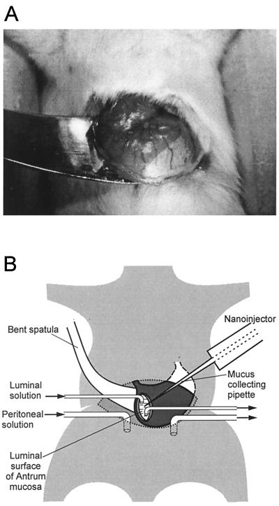
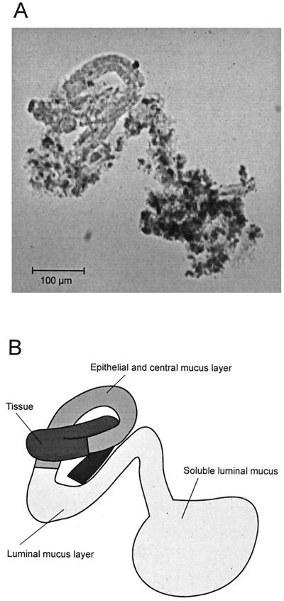
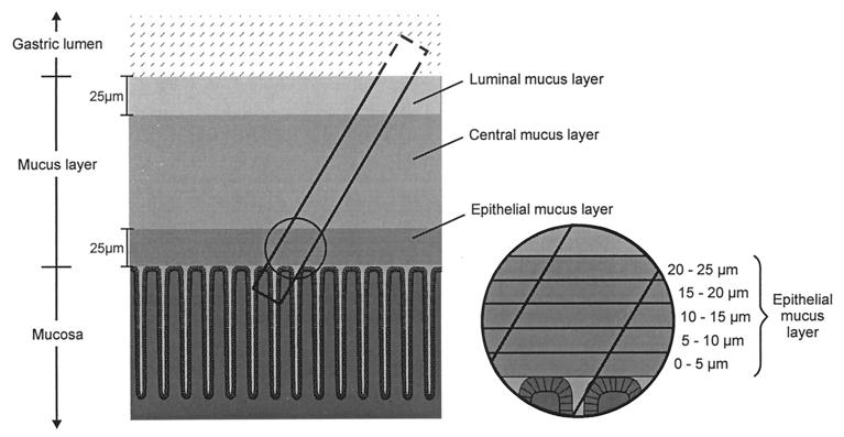
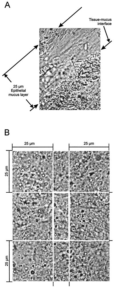
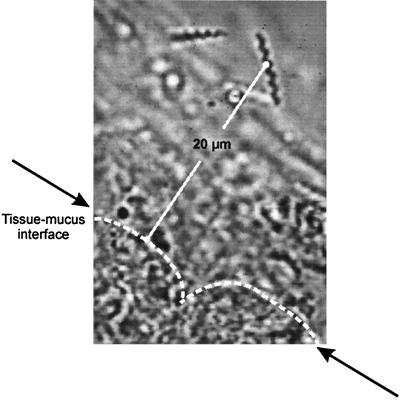
We describe a method that permits the collection of very small samples (2 nl) from precisely defined positions within the gastric mucus of anesthetized mice. This method was used to study the in vivo local distribution of bacteria within the mucus of Helicobacter felis-infected mice. A total of 200 samples from 40 mice were analyzed. Each sample was microscopically analyzed, within less than 1 min, as a native preparation. To avoid changes in bacterial location within the mucus after collection and to improve the counting accuracy, bacterial motility was blocked by adjusting the pH inside the collecting pipette to 4.5. The mucus in a collected sample was subdivided into three layers, an epithelial layer (the first 25 micron of mucus from the tissue-mucus interface), a luminal layer (the last 25 micron to the mucus-lumen interface), and the remaining central mucus layer. The volume of the analyzed segments in the sample was between 4 and 9 pl. The concentration of bacteria inside the epithelial mucus layer was 3,400 per nl, but it was only 50 per nl inside the central mucus layer. The mean distance of H. felis to the epithelial surface was 16 microm. A total of 75% of all H. felis bacteria resided in the mucus zone between 5 and 20 micron from the tissue surface, with no bacteria closer than 5 micron to the epithelial surface. This method permits the study of factors determining the density of colonization and distribution of bacteria along chemical gradients with a high precision.


Figures

FIG. 1
FIG. 1
Experimental setup. (A) Photograph of the stomach, carefully lifted through a ventral incision, and glued with tissue adhesive to the surface of a bent spatula. There is a sharp borderline between the white surface of the forestomach and the darker (red) surface of the corpus. (B) Schematic drawing of the nanoinjector setup. The collecting pipette is inserted through a small incision in the ventral antrum wall. The sample is collected from the dorsal antrum mucosa penetrating from the luminal side. The abdominal cavity is perfused with a solution similar to blood plasma; the gastric lumen is perfused with a CO2-saturated isotonic salt solution (pH 3.8).
FIG. 2
FIG. 2
Collected and ejected sample of gastric mucus. (A) Cylindrical sample (100-fold magnification). (B) Schematic drawing of the sample.
FIG. 3
FIG. 3
Schematic drawing of the pipette inside the mucus and epithelial layer and of the collected sample. The cylindrical samples contained superficial tissue and the overlying mucus. The three layers were as follows: an epithelial mucus layer (the first 25 μm of mucus from the tissue-mucus interface), a luminal mucus layer (the last 25 μm of mucus to the mucus-lumen interface), and a central mucus layer between the two. The insert shows the further subdivision of the epithelial mucus layer into five sublayers of 5 μm each.
FIG. 4
FIG. 4
Micrograph of regions analyzed under the microscope. (A) Section of a sample collected from the epithelial mucus 970-fold magnification). The photograph shows the main criteria for definition of the epithelial mucus layer: distance of less than 25 μm from the tissue surface, high transparency, and tidy fibrillar structure. (B) Magnification of the central mucus and the bordering 25-μm layers of luminal mucus and epithelial mucus.
FIG. 5
FIG. 5
Measurement of the distance from H. felis inside the epithelial mucus to the epithelial surface.

References

    1. Allen A, Hutton D A, Leonard A J, Pearson J P, Sellers L A. The role of mucus in the protection of the gastroduodenal mucosa. Scand J Gastroenterol. 1986;21(Suppl. 125):71–77. - PubMed
    1. Andrutis K A, Fox J G, Schauer D B, Marini R P, Li X, Yan L, Josenhans C, Suerbaum S. Infection of the ferret stomach by isogenic flagellar mutant strains of Helicobacter mustelae. Infect Immun. 1997;65:1962–1966. - PMC - PubMed
    1. Covacci A, Falkow S, Berg D E, Rappuoli R. Did the inheritance of a pathogenicity island modify the virulence of Helicobacter pylori? Trends Microbiol. 1997;5:205–208. - PubMed
    1. Crabtree J E, Farmery S M, Lindley I J D, Figura N, Peichl P, Tompkins D S. CagA/cytotoxic strains of Helicobacter pylori and interleukin-8 in gastric epithelial cell lines. J Clin Pathol. 1994;47:945–950. - PMC - PubMed
    1. Dunn B E, Cohen H, Blaser M J. Helicobacter pylori. Clin Microbiol Rev. 1997;10:720–741. - PMC - PubMed

Publication types