

Acid production by vaginal flora in vitro is consistent with the rate and extent of vaginal acidification
- PMID: 10496892
- PMCID: PMC96867
- DOI: 10.1128/IAI.67.10.5170-5175.1999
Acid production by vaginal flora in vitro is consistent with the rate and extent of vaginal acidification
Abstract
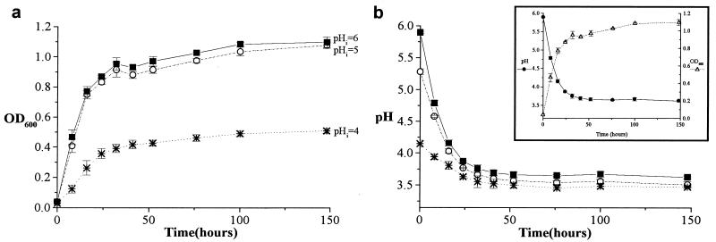
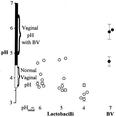
Perinatally, and between menarche and menopause, increased levels of estrogen cause large amounts of glycogen to be deposited in the vaginal epithelium. During these times, the anaerobic metabolism of the glycogen, by the epithelial cells themselves and/or by vaginal flora, causes the vagina to become acidic (pH approximately 4). This study was designed to test whether the characteristics of acid production by vaginal flora in vitro can account for vaginal acidity. Eight vaginal Lactobacillus isolates from four species-L. gasseri, L. vaginalis, L. crispatus, and L. jensenii-acidified their growth medium to an asymptotic pH (3.2 to 4.8) that matches the range seen in the Lactobacillus-dominated human vagina (pH 3.6 to 4.5 in most women) (B. Andersch, L. Forssman, K. Lincoln, and P. Torstensson, Gynecol. Obstet. Investig. 21:19-25, 1986; L. Cohen, Br. J. Vener. Dis. 45:241-246, 1969; J. Paavonen, Scand. J. Infect. Dis. Suppl. 40:31-35, 1983; C. Tevi-Bénissan, L. Bélec, M. Lévy, V. Schneider-Fauveau, A. Si Mohamed, M.-C. Hallouin, M. Matta, and G. Grésenguet, Clin. Diagn. Lab. Immunol. 4:367-374, 1997). During exponential growth, all of these Lactobacillus species acidified their growth medium at rates on the order of 10(6) protons/bacterium/s. Such rates, combined with an estimate of the total number of lactobacilli in the vagina, suggest that vaginal lactobacilli could reacidify the vagina at the rate observed postcoitally following neutralization by the male ejaculate (W. H. Masters and V. E. Johnson, Human sexual response, p. 93, 1966). During bacterial vaginosis (BV), there is a loss of vaginal acidity, and the vaginal pH rises to >4.5. This correlates with a loss of lactobacilli and an overgrowth of diverse bacteria. Three BV-associated bacteria, Gardnerella vaginalis, Prevotella bivia, and Peptostreptococcus anaerobius, acidified their growth medium to an asymptotic pH (4.7 to 6.0) consistent with the characteristic elevated vaginal pH associated with BV. Together, these observations are consistent with vaginal flora, rather than epithelial cells, playing a primary role in creating the acidity of the vagina.
Figures
References
-
- Andersch B, Forssman L, Lincoln K, Torstensson P. Treatment of bacterial vaginosis with an acid cream: a comparison between the effect of lactate-gel and metronidazole. Gynecol Obstet Investig. 1986;21:19–25. - PubMed
-
- Bartlett J G, Onderdonk A B, Drude E, Goldstein C, Anderka M, Alpert S, McCormack W M. Quantitative bacteriology of the vaginal flora. J Infect Dis. 1977;136:271–277. - PubMed
-
- Caillouette J C, Sharp C F, Zimmerman G J, Roy S. Vaginal pH as a marker for bacterial pathogens and menopausal status. Am J Obstet Gynecol. 1997;176:1270–1275. - PubMed
-
- Ciba-Geigy Corporation. Geigy scientific tables. 1. Units of measurement, body fluids, composition of the body, nutrition. West Caldwell, N.J: Medical Education Division, Ciba-Geigy Corporation; 1981. pp. 185–186.
Publication types
MeSH terms
Substances
Grants and funding
LinkOut - more resources
Full Text Sources
Other Literature Sources
Molecular Biology Databases
Miscellaneous
